Downloaded 23 times


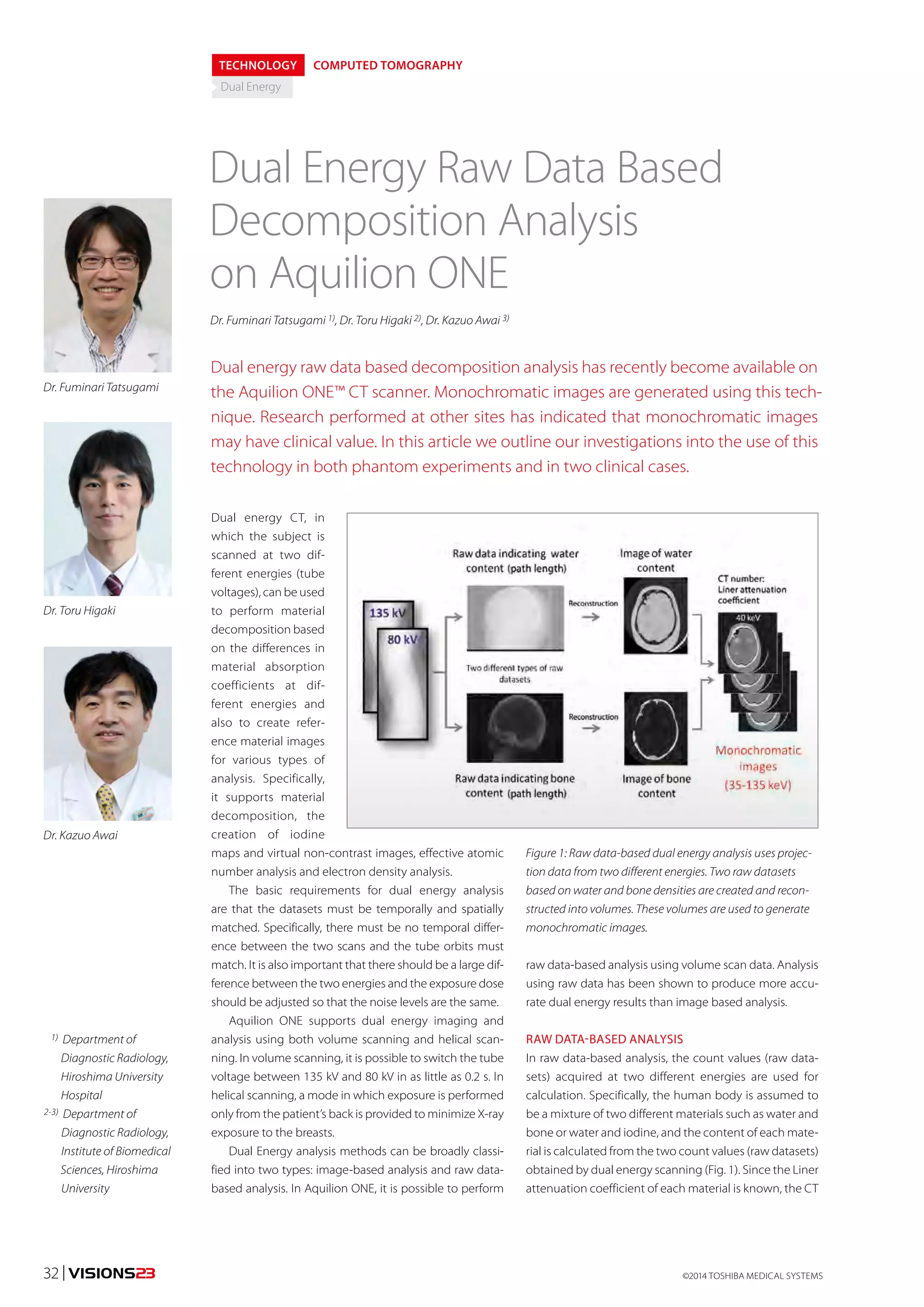


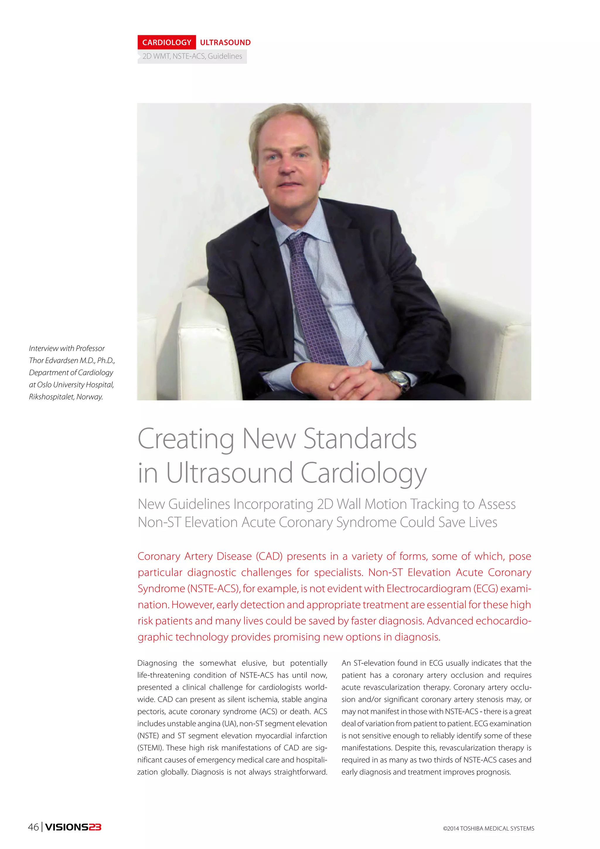


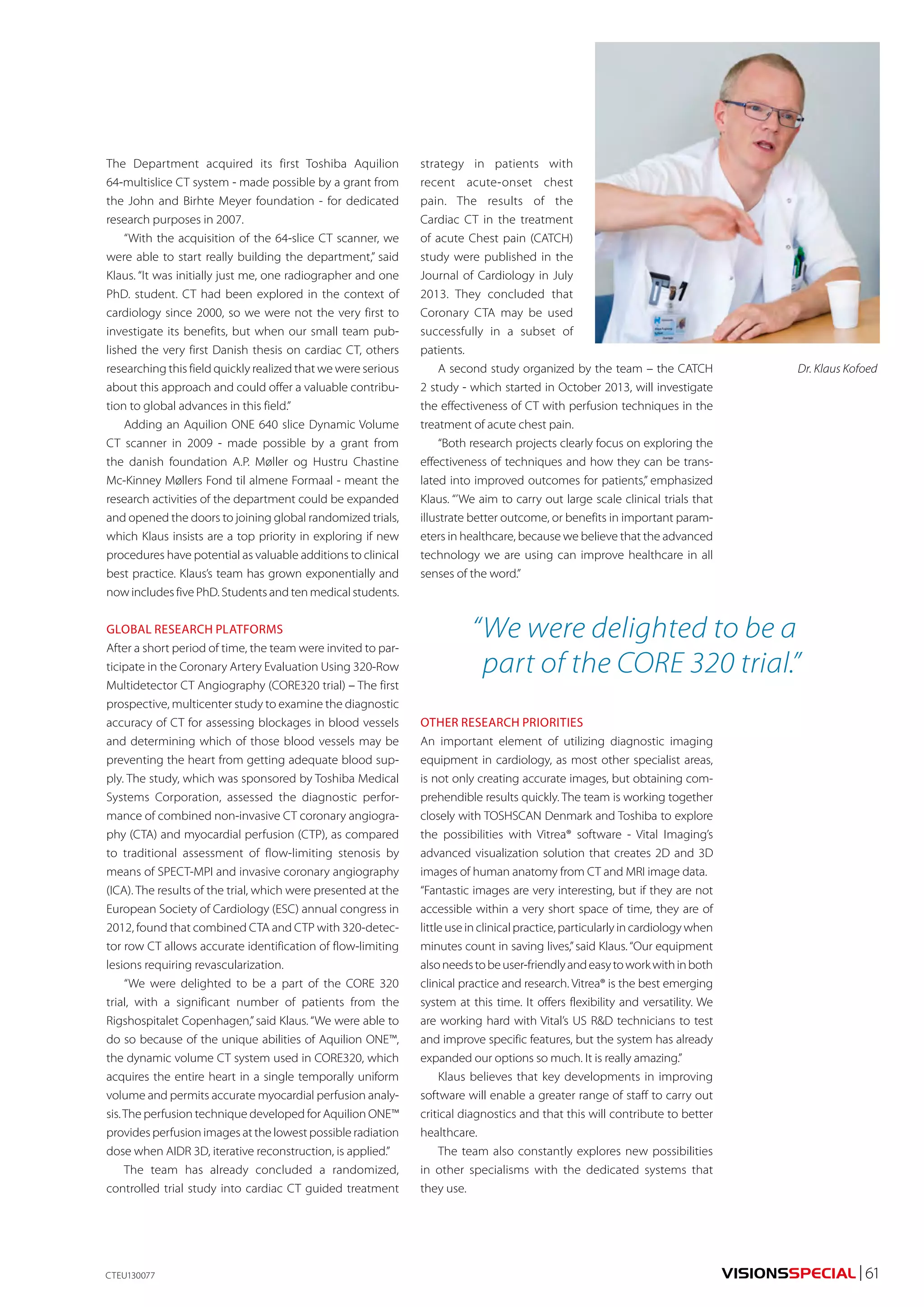


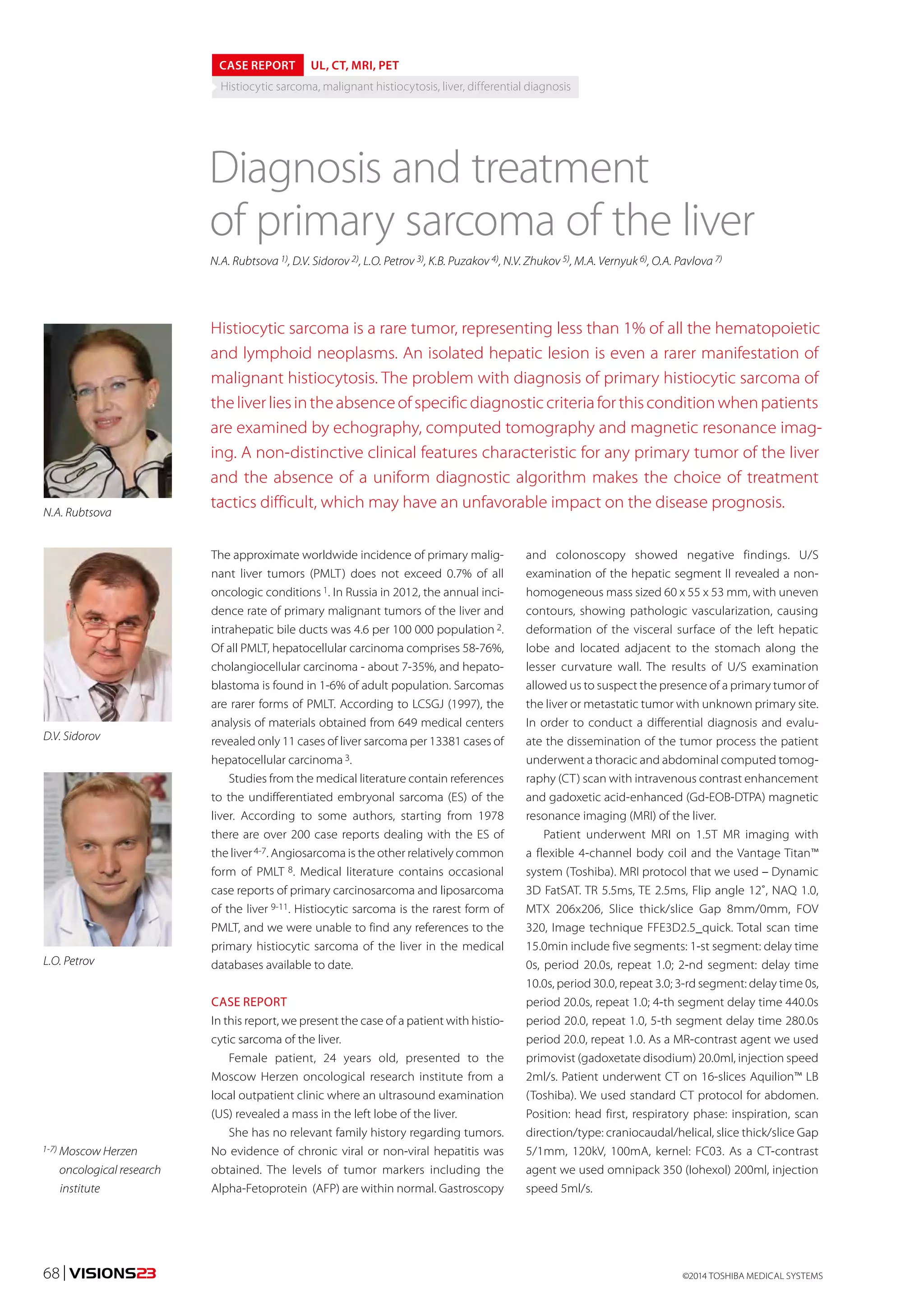

![the presence of osseous changes we performed a
CT-guided targeted biopsy of one of the foci in the body
of the L4 lumbar vertebra (Fig. 7).
The histological and immunohistochemical (IHC)
examination of the biopsied mass obtained from the left
hepatic lobe revealed the following. Most of the hepatic
tissue was replaced by the polymorphocellular lymphoid
infiltrate predominantly composed of small lympho-cytes
with an admixture of plasma cells and eosinophils.
Against the backdrop of the infiltrate, there are occasional
giant cells resembling Hodgkin cells and Beresovsky-
Sternberg-Reed cells.
Leukocytes are positive for LCA, B-lymphocytes - for CD20,
T-lymphocytes for CD3, occasional large cells - for CD30,
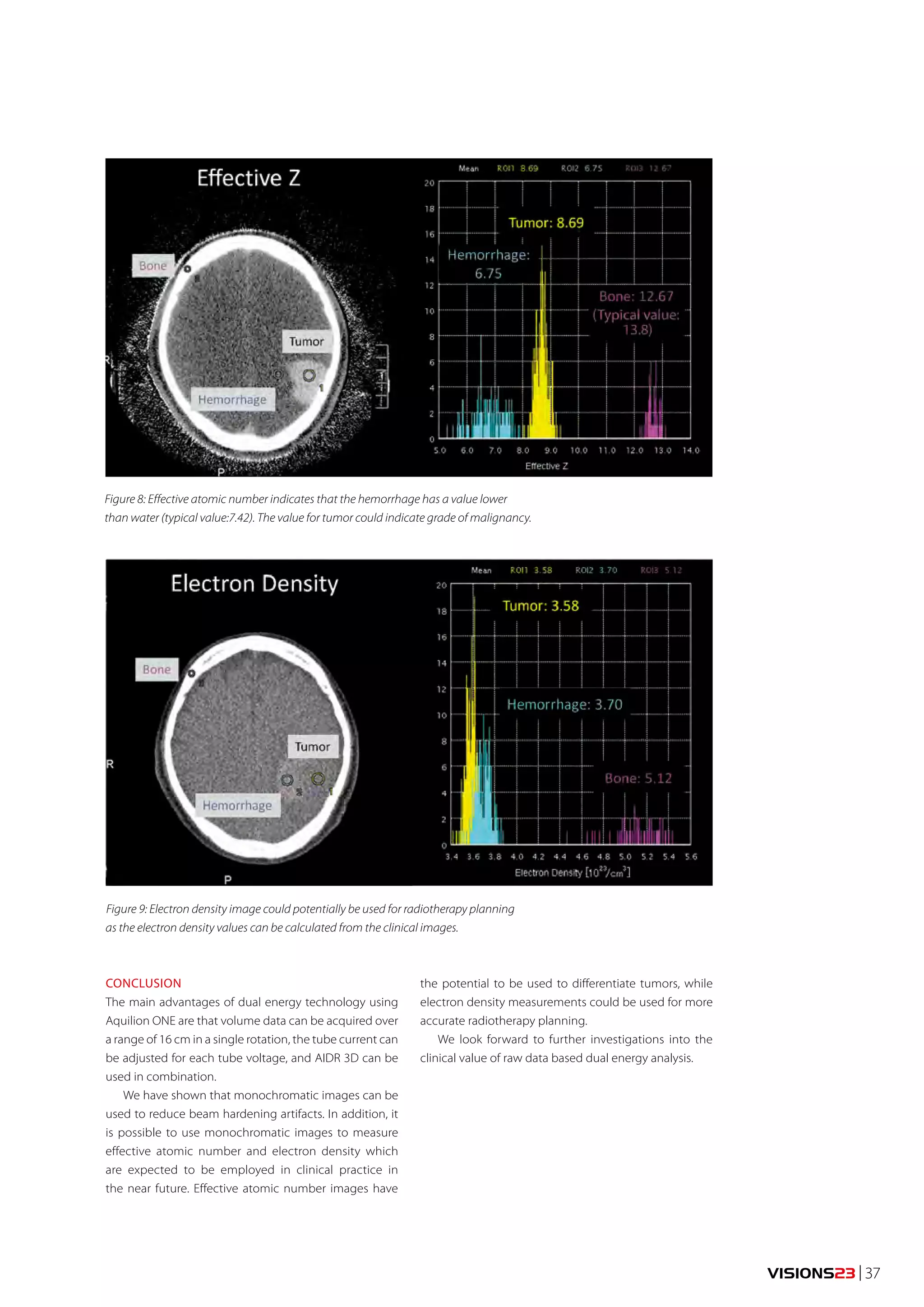
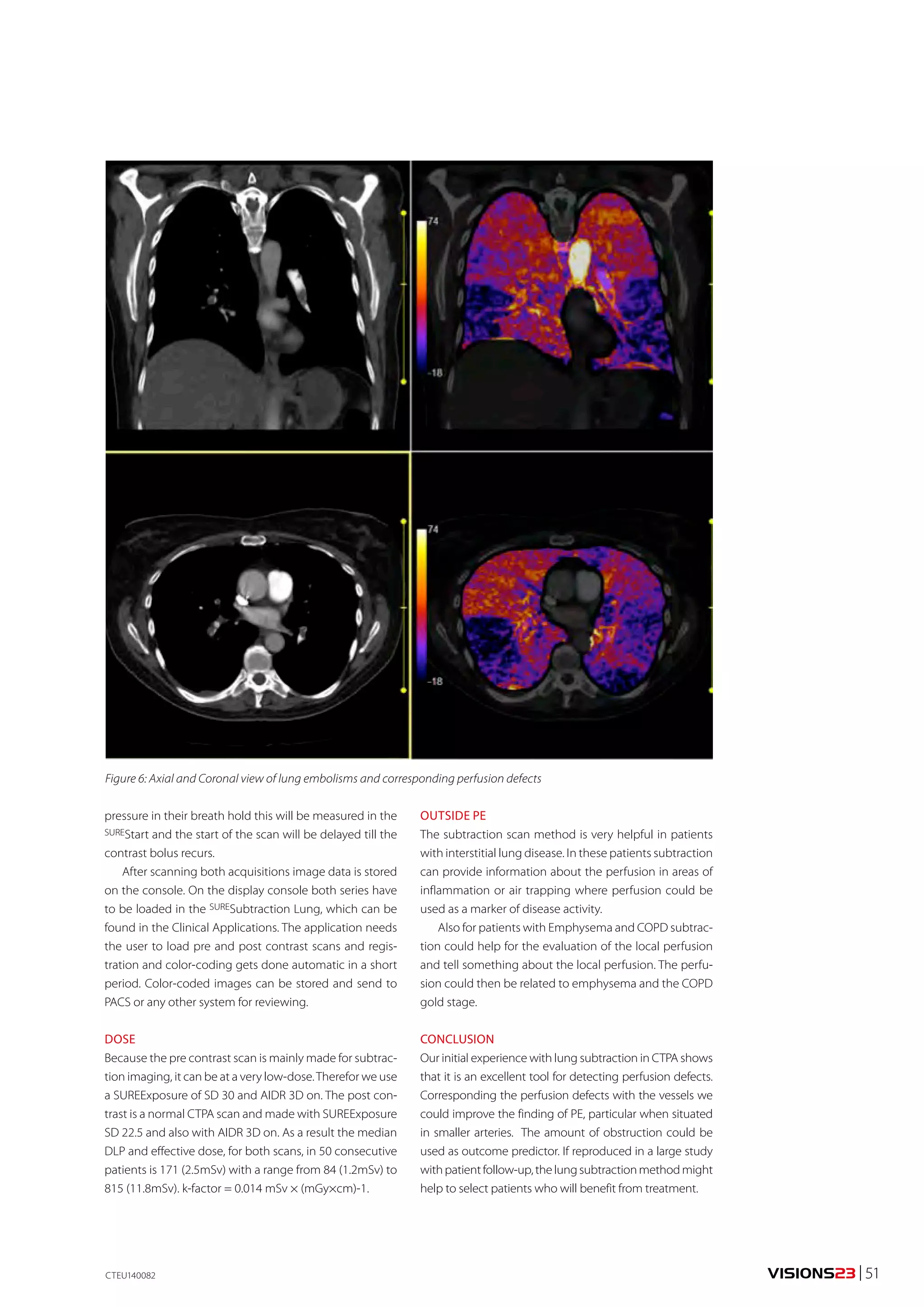
Figure 4: Contrast Enhanced T1 WI MR images of the lower thoracic and lumbar vertebrae pre-sent
consecutive axial slices. Low-signal intensity foci that do not accumulate contrast material
after dynamic intravenous contrast enhancement are encircled by a broken line and arrowed.
Figure 5: Axial CT scans of abdominal organs (tumor nodule is arrowed): а – native phase,
b – post-contrast image, hepatic arterial phase, c – arteriovenous phase, d – 3D model of
the liver highlighting the tumor, unchanged parenchyma of the right and left lobes and
branches of the hepatic artery and hepatic portal vein.
granulocytes and occasional large cells – for CD15, plasma
cells - for ЕМА, B-lymphocytes - for РАХ-5 (also, less intense
staining in occasional large cells). Approximately 30% of
tumor cells express Ki67 antigen. The morphological pat-tern
and immune phenotype are characteristic for classi-cal
Hodgkin’s lymphoma. Its variant can not be defined
because of the insufficient amount of tissue provided.
The additional ICH examination was carried out using
cell type-specific antibodies. Large and partly small cells
were positive for CD68, small fraction of cells was positive
for Bсl 6, plasma cells were positive for CD138 and nega-tive
for CD1a, CD30 and glycoforin. These findings indicate
an histiocytic sarcoma of the liver (non-langerhans type),
ICD-0 code 9755/3.
Results of histological examination of the material
obtained from the L4 vertebra biopsy: the biopsy material
is represented by a fragment of compact bone tissue with
a mosaic pattern, fragmentation and dystrophic changes
of the bone plates but with no signs of tumor growth.
Given the results of histological examination of the
hepatic mass puncture biopsy and the equivocal results
of the chest CT scan regarding the nature of foci in lungs
and bones, the patient was subjected to F-18 fluorodeoxy-glucose
(FDG) positron emission tomography (PET). PET
scan revealed a solitary focus of pathological metabolic
changes in the left lobe of the liver parenchyma. There
was no evidence of the presence of tumor in other organs
examined.
After discussing the case at the board of doctors it
was decided to conduct surgical treatment, namely, the
anatomical liver resection (bisegmentectomy SII-SIII) for
liver tumor (Fig. 8).
The dissection of the liver parenchyma was performed
using the water-jet dissector and harmonic scalpel. The
Pringle maneuver was not applied. The duration of sur-gical
intervention was 115 min., and the intra-operative
blood loss was 150 mL.
The post-operative period ran smoothly, and the
wound healed by primary intention. The patient was
discharged from hospital on day 8 of the post-operative
period. According to the post-operative histological
examination, the removed tumor nodule is the histiocytic
sarcoma of the liver, and the resection edges are intact.
Control examination performed 8 weeks post-operation
revealed no signs of tumor progression.
DISCUSSION
Histiocytic sarcoma (HS) is a rare tumor, representing
less than 1% of all the hematopoietic and lymphoid
neoplasms [12, 13]. Malignant histiocytosis or HS occurs
mostly in the mononuclear phagocytic system organs,
which is manifested in the clinical symptoms of the dis-ease.
The most common HS manifestations are lymphad-enopathy
and skeletal lesions while liver, spleen, lungs,
pleura, skin, kidneys, bone marrow and gastrointestinal
tract are less frequently affected 14.
©2014 70 | VISIONS23 TOSHIBA MEDICAL SYSTEMS](https://image.slidesharecdn.com/visions-23-140821172829-phpapp01/75/Toshiba-Visions-23-70-2048.jpg)


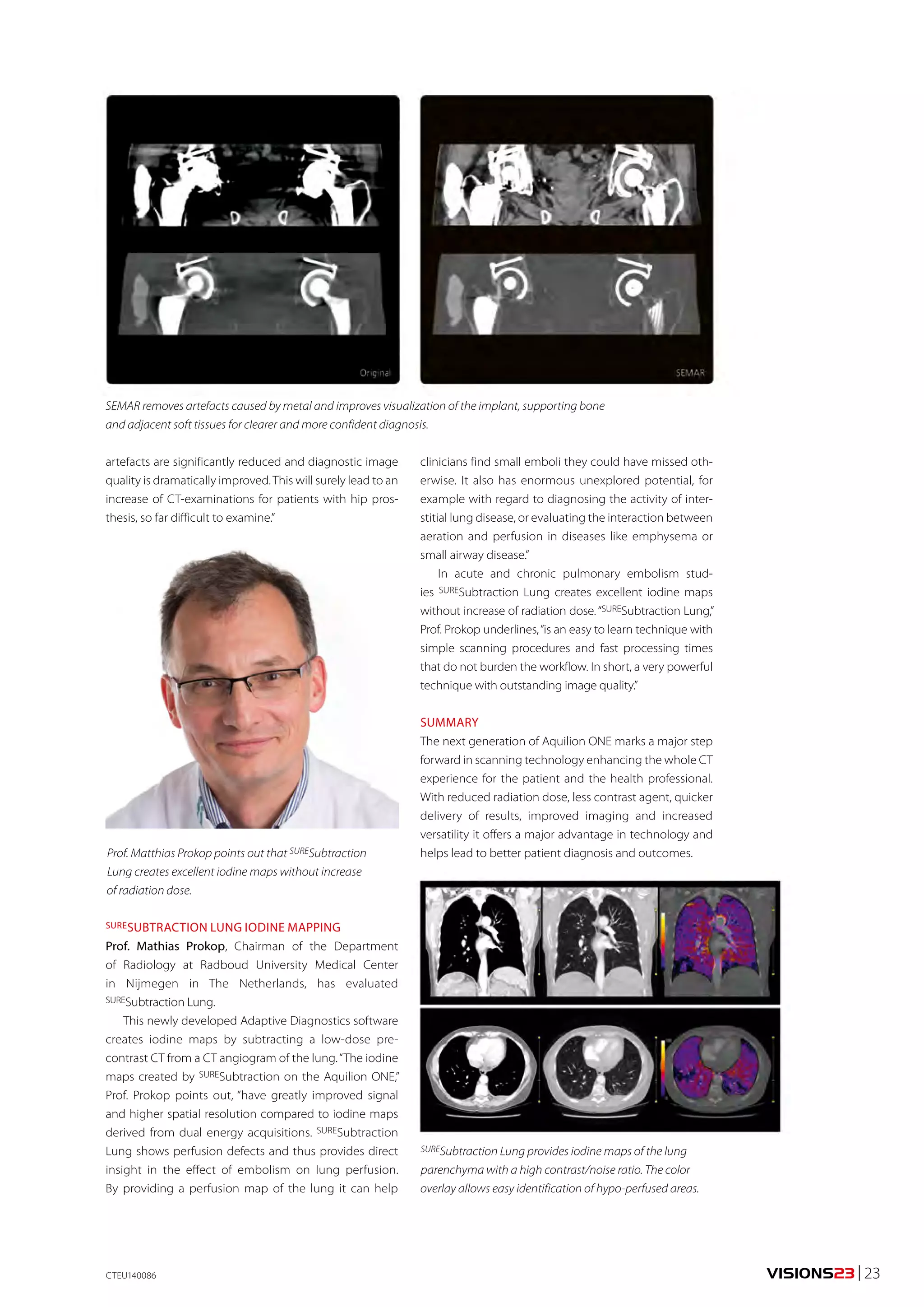
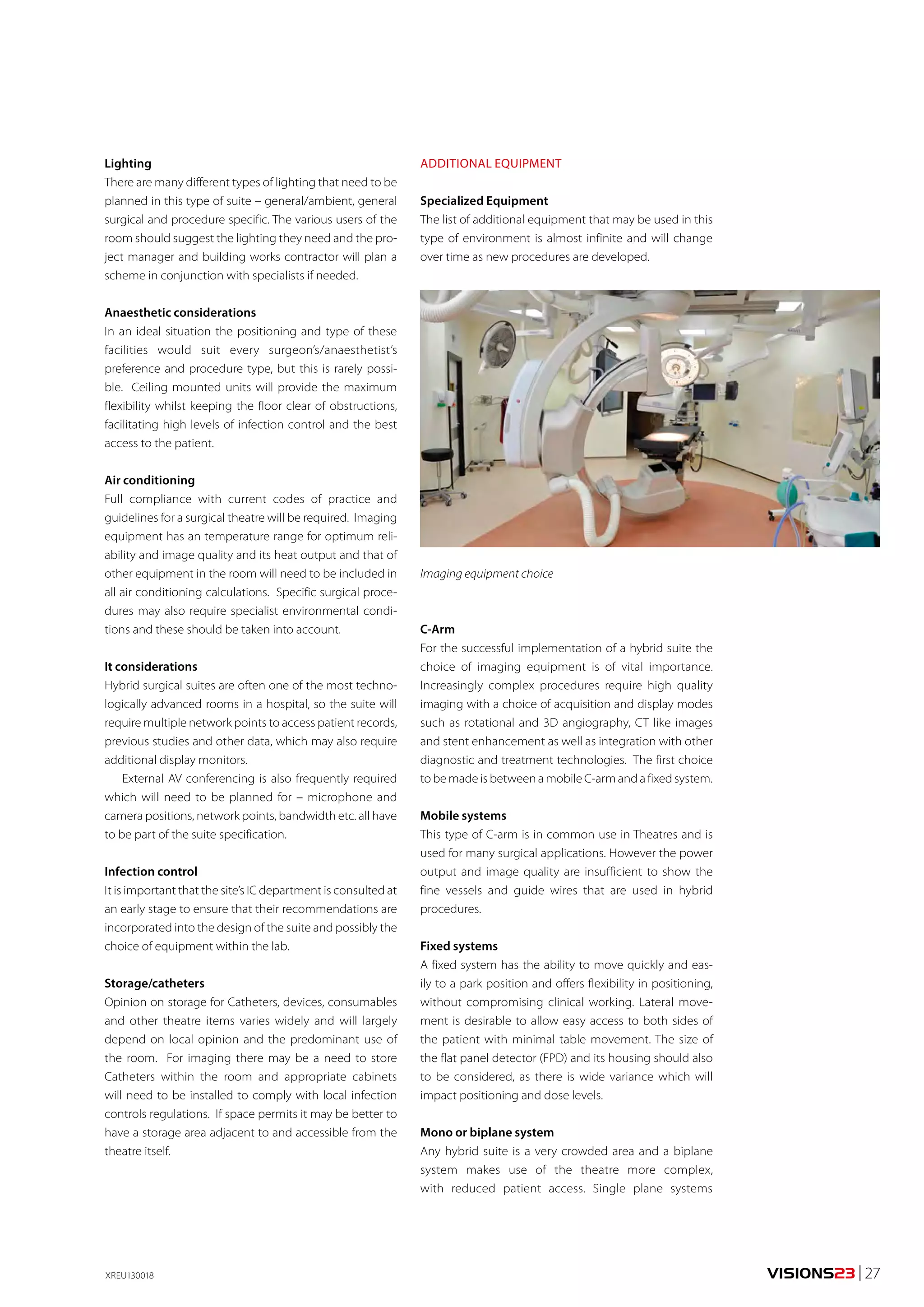
![REPORT
COMPUTED TOMOGRAPHY
Myocardial Perfusion, Coronary Angiography
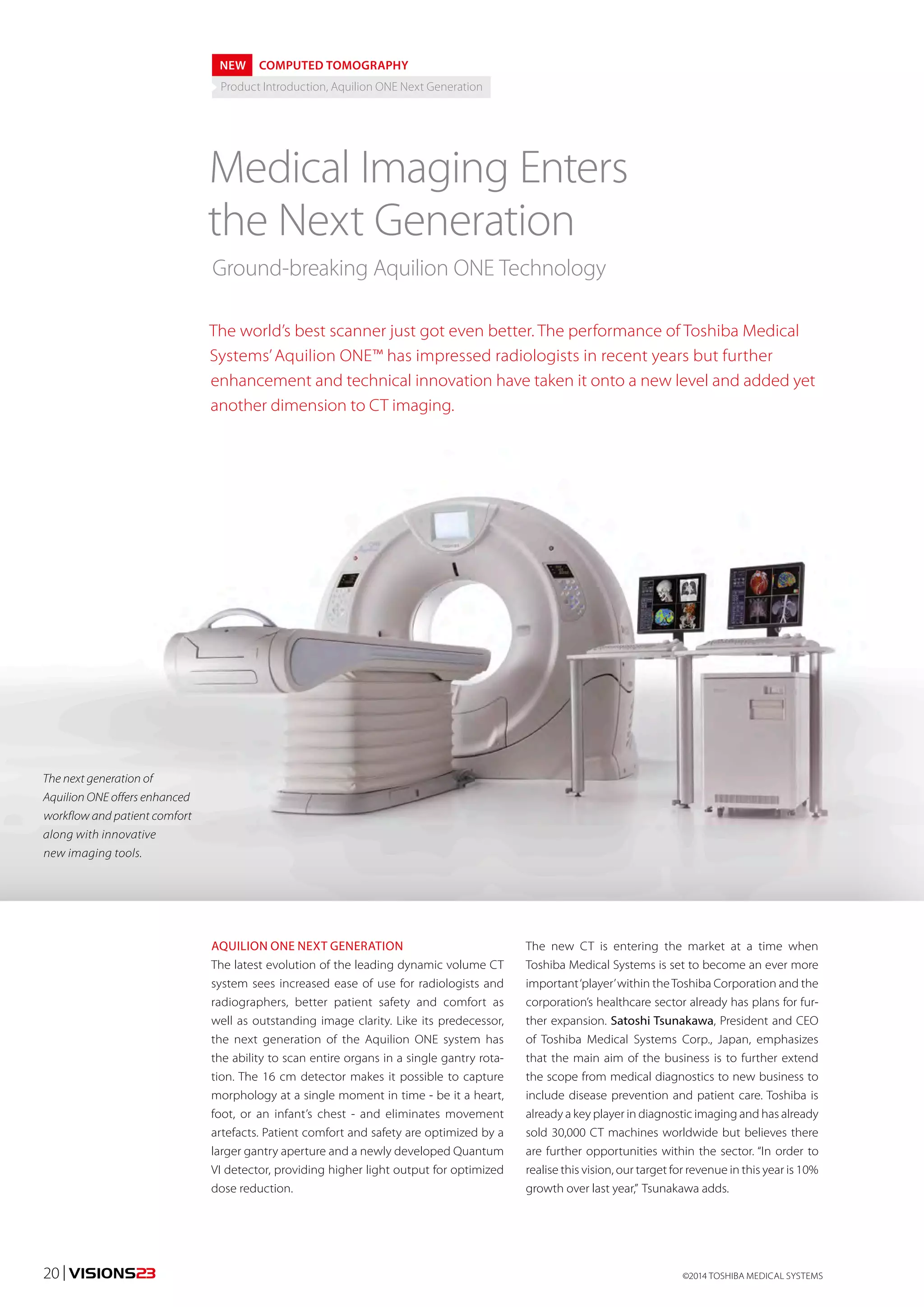
320-row CT as a single and effec-tive
platform for anatomical and
functional evaluation of coronary
artery disease: the CORE320 trial
Joanne D. Schuijf, PhD 1); Chloe Steveson, MMRS 2)
A single 320-detector row CT examination that combines coronary angiography and
myocardial perfusion is effective in the detection of haemodynamically significant
coronary stenoses, according to the international, multicenter trial CORE320. The results
have recently been published in the European Heart Journal1.
CLINICAL NEED FOR BOTH ANATOMICAL
AND FUNCTIONAL ASSESSMENT
In the diagnosis and management of coronary artery
disease (CAD), imaging plays an increasingly important
role. In particular, non-invasive CT coronary angiography
(CTA) has emerged as an attractive tool for initial evalua-tion
of patients presenting with symptoms or other signs
suggestive of CAD. Yet an important limitation of this
particular approach is that it only provides information on
the anatomical severity of disease. In contrast, knowledge
whether detected coronary stenoses result in reduced
myocardial perfusion is crucial for further management,
such as assessing the need for coronary intervention. This
notion has fuelled the development of CT myocardial
perfusion (CTP).
While initial studies have shown the feasibility of
combined CTA and CTP, these experiences were limited
to single-center evaluations in small patient cohorts.
However, for a test to be accepted in the clinical arena,
more robust, prospective data are needed. Recently, the
results from the first large, international, multicenter trial
on this topic, the CORE320 trial, have become available,
confirming the value of combined CTA and CTP with
320-detector row CT.
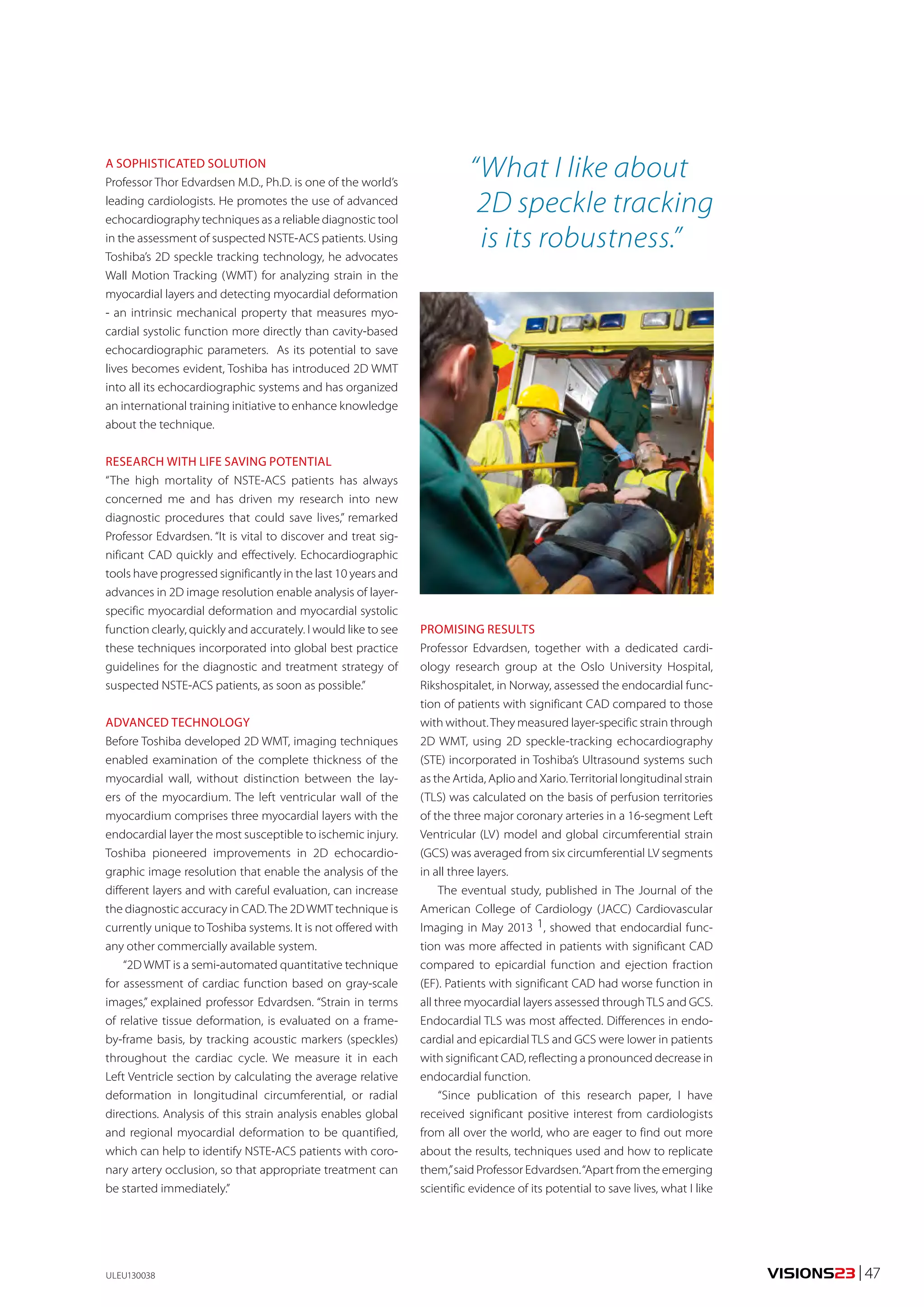
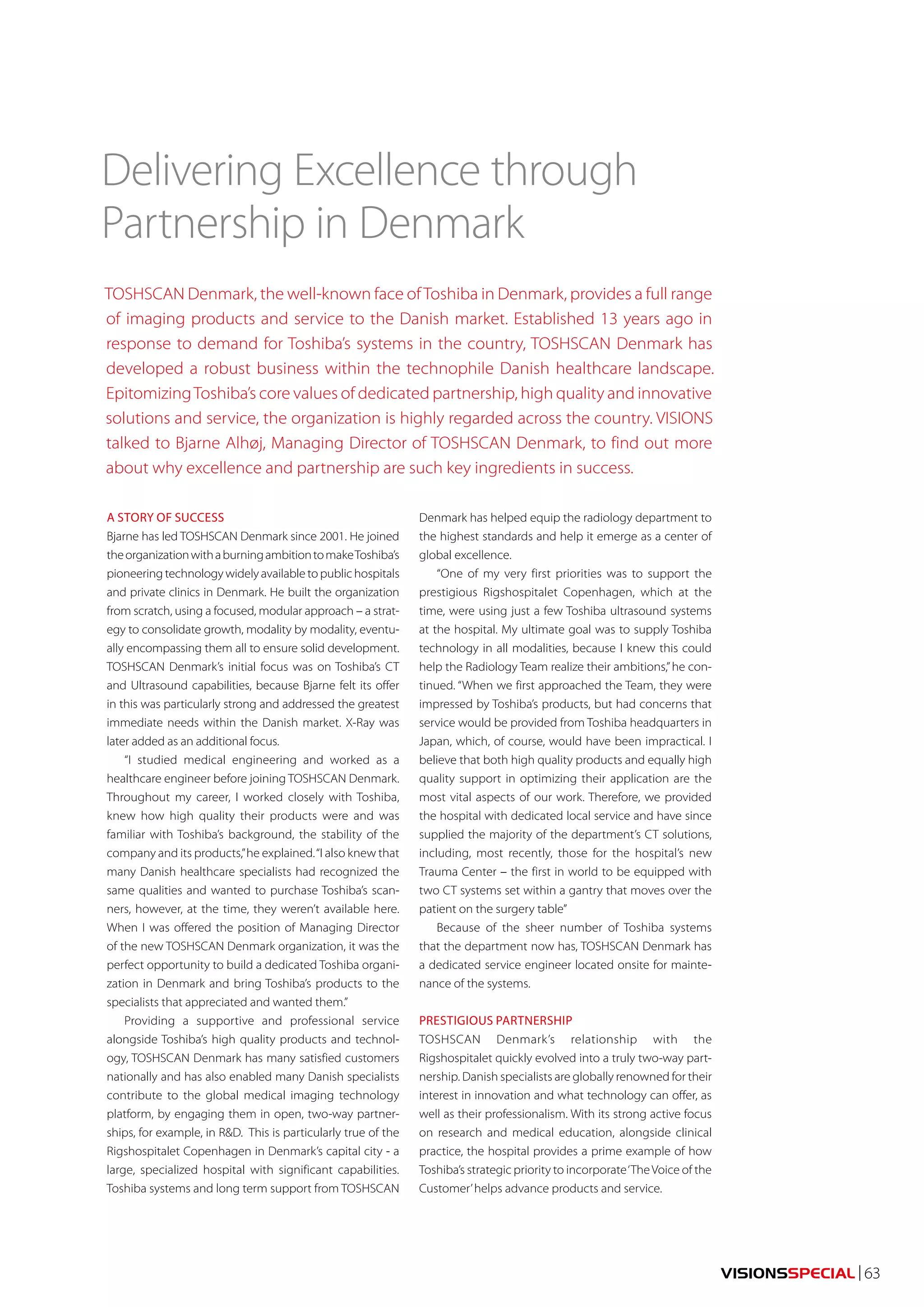
STUDY DESIGN AND METHODS
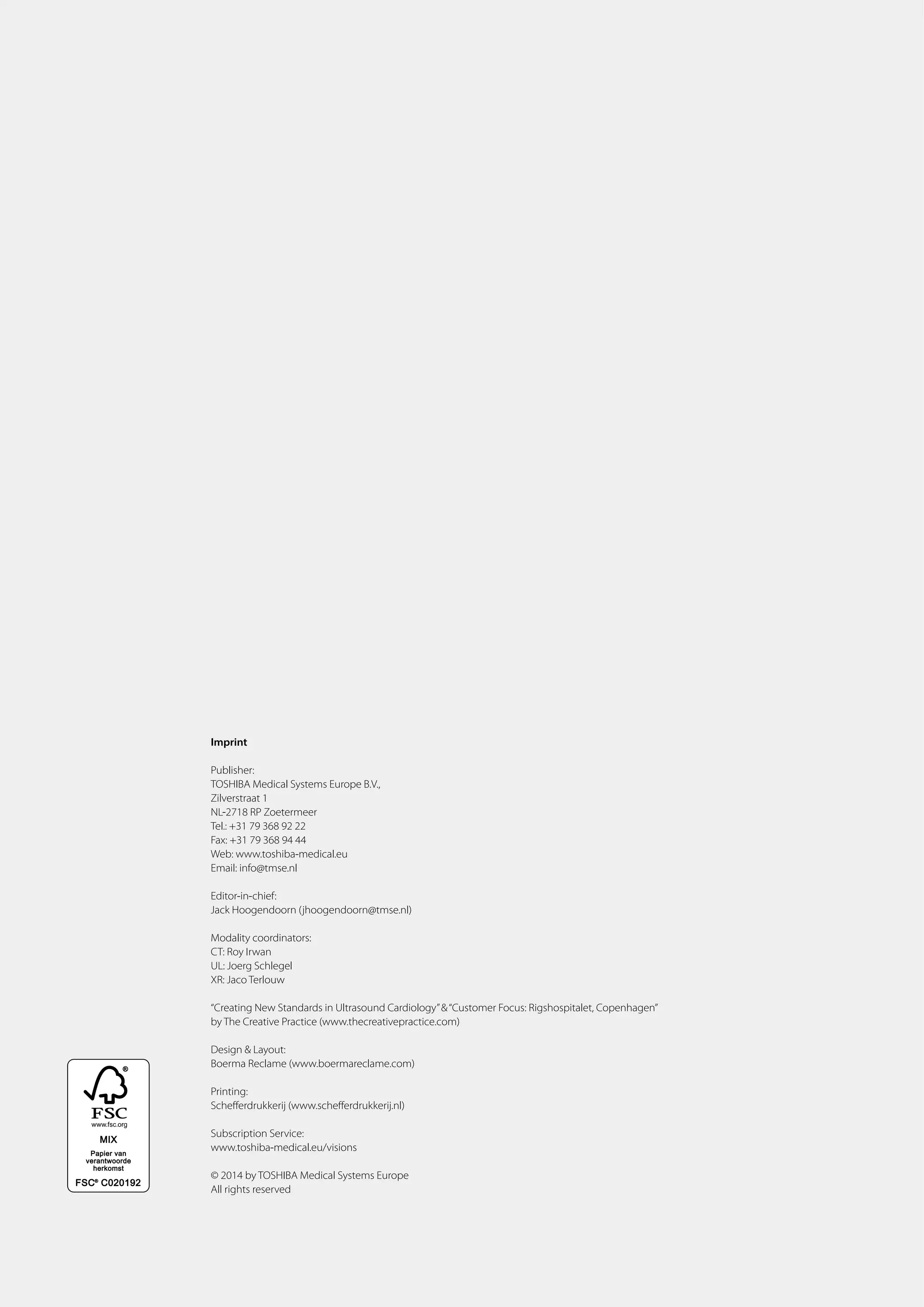
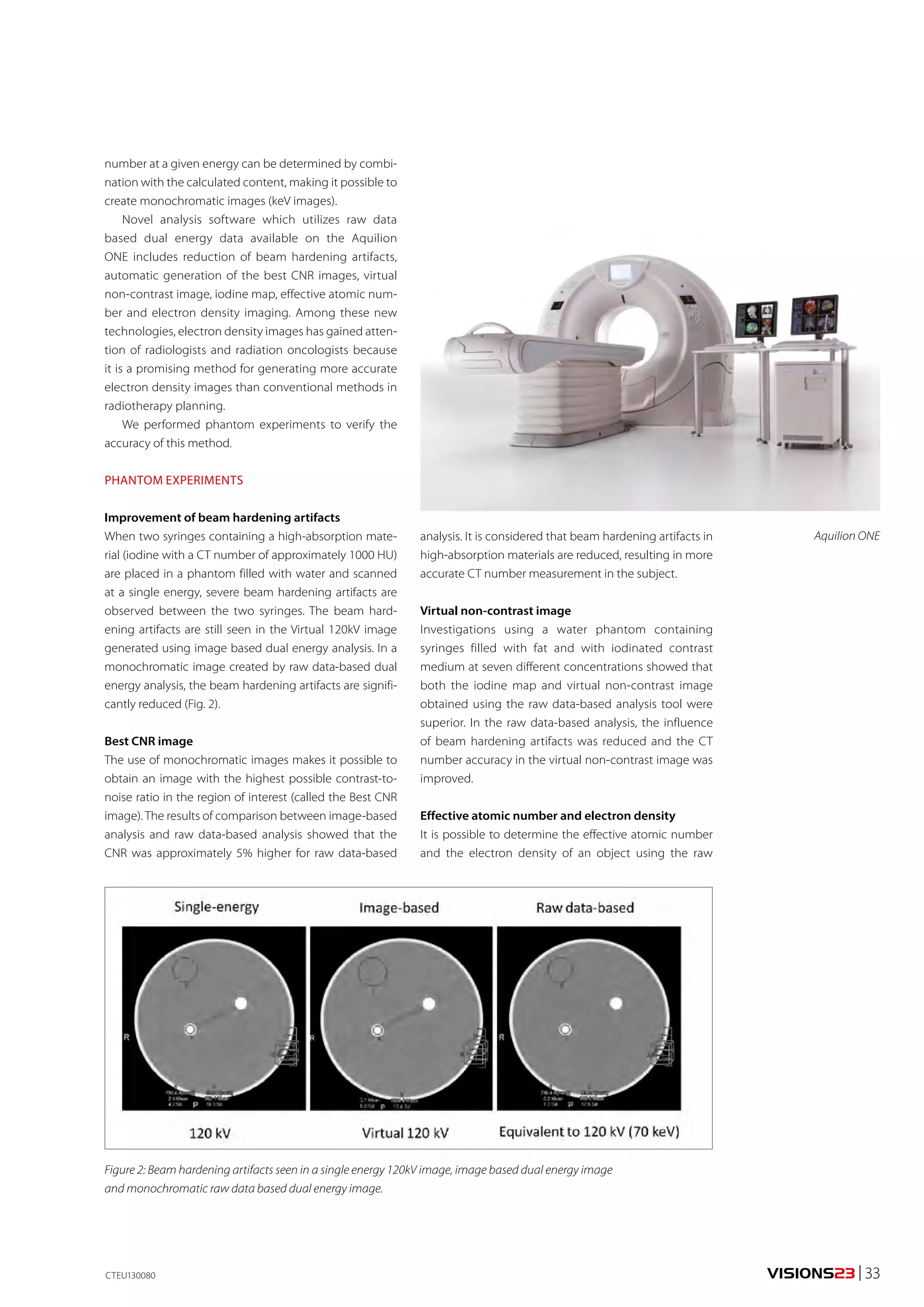
Researchers from 16 sites in eight countries enrolled
381 patients who were referred for cardiac evaluation
because of suspected or known CAD (Fig. 1). All patients
underwent a comprehensive imaging protocol consist-ing
of combined CTA and adenosine stress CTP as well as
myocardial perfusion imaging (MPI) by means of SPECT
prior to conventional invasive coronary angiography
(ICA). Imaging data were evaluated by four independent
blinded core laboratories.
Figure 1: Participating countries.
Joanne D. Schuijf
Chloe Steveson
1) Toshiba Medical
Systems Europe,
Zoetermeer,
The Netherlands
2) Toshiba Medical
Systems Corporation,
Otawara, Japan
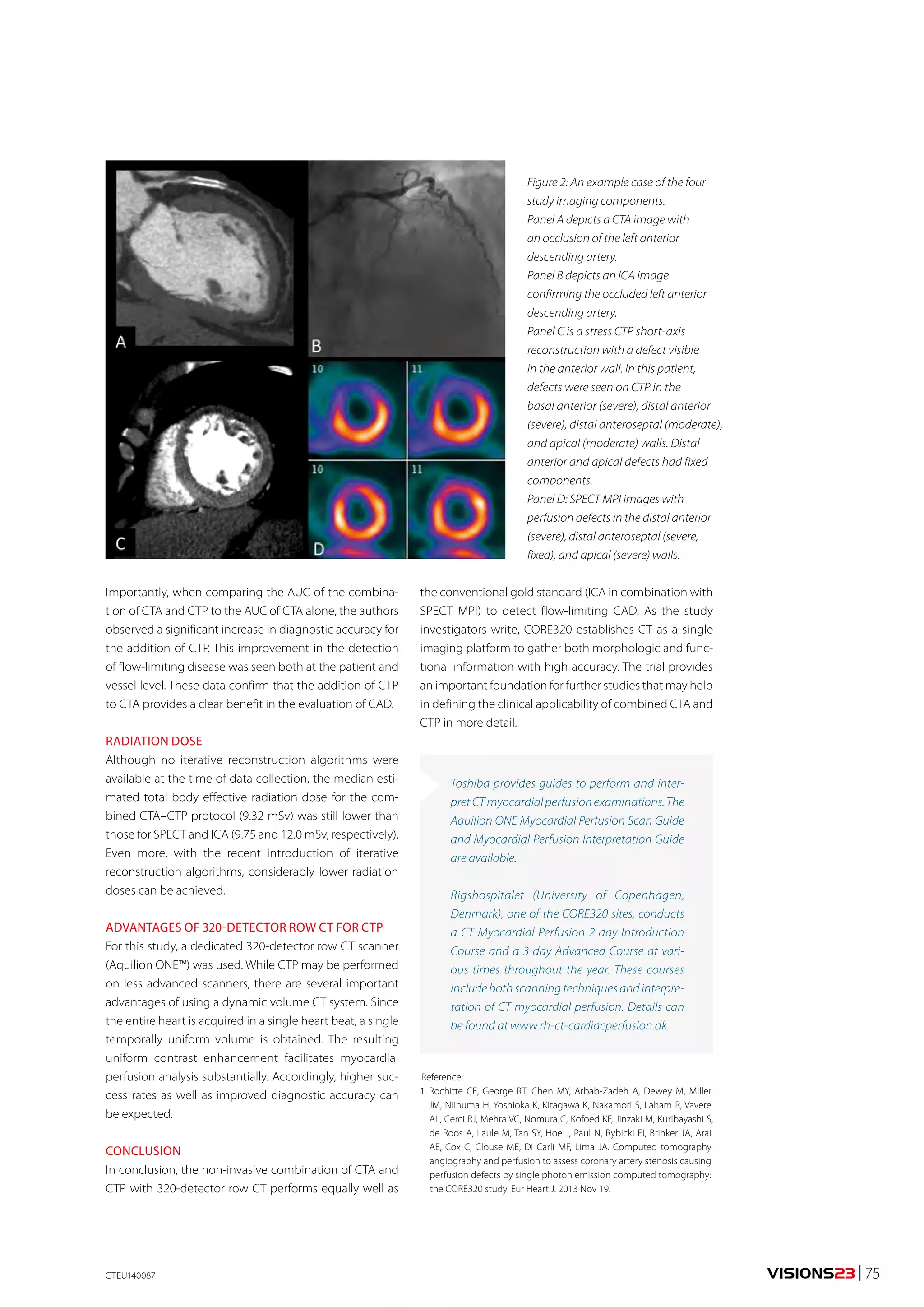
For the reference standard, each patient and vessel was
classified as normal or having CAD, defined as ≥ 50%
coronary stenosis by ICA with an associated perfusion
defect by SPECT MPI in the corresponding territory. An
example dataset of the four imaging components is pro-vided
in Fig. 2.
CLEAR BENEFIT WHEN ADDING CTP TO CTA
Based on the gold standard, 38% of patients were positive
for CAD. The patient-based diagnostic accuracy defined
by the area under the receiver operating characteristic
curve (AUC) of integrated CTA–CTP for detecting or
excluding flow-limiting CAD was 0.87 [95% confidence
interval (CI): 0.84–0.91]. In a next step, the analysis was
repeated in patients without prior CAD, revealing an even
higher AUC of 0.93 (95% CI: 0.89–0.97). For the combina-tion
of a CTA stenosis ≥50% and a CTP perfusion defect
(summed stress score ≥4), the sensitivity, specificity, posi-tive
predictive, and negative predicative values (95% CI)
were 80% (72–86), 74% (68–80), 65% (58–72), and 86%
(80–90), respectively.
74 | VISIONS23 ©2014 TOSHIBA MEDICAL SYSTEMS](https://image.slidesharecdn.com/visions-23-140821172829-phpapp01/75/Toshiba-Visions-23-74-2048.jpg)


This document provides information on several medical imaging topics and technologies: - It highlights abstracts from the ECR 2014 conference focusing on adaptive diagnostics and how they can solve clinical challenges. Abstracts discuss the latest 3T MRI experience, a new metal artifact reduction algorithm, and lung subtraction versus dual energy CT. - It also provides information on Toshiba's new products and technologies including the next generation Aquilion ONE CT scanner, planning for hybrid labs, dual energy CT applications, and dose reduction techniques for interventional procedures. - Additional sections profile customer sites and applications of Toshiba ultrasound, CT, and MRI systems.