Downloaded 86 times


![How-I-do-it
MAGNETOM Flash · 2/2011 · www.siemens.com/magnetom-world 7
Challenges of
scanning children
Safety
MRI of children poses a number of spe-cific
safety issues with patient heating
being the primary concern. Neonates
and infants in particular have immature
thermoregulation mechanisms, and
higher core body temperatures making
them particularly sensitive to RF heating
effects [1]. These mechanisms are fur-ther
affected by sedation and anesthesia
common in pediatric imaging [2], or
when babies are swaddled for imaging
[1]. Children also have a greater surface
area to weight ratio than adults. This
means for a given weight we often need
to expose a greater surface area of the
patients to the RF field. This can lead to
increased heating in children, and
decrease their ability to dissipate this
heat. There is intrinsic uncertainty in
current specific absorption rate (SAR)
predictions based on extrapolated data
from phantom models [3] particularly
due to factors such as body shape, size,
composition, and position within the MR
scanner. While definitive data on safety
risks are not yet available, close monitor-ing
of children, particularly critically ill
or compromised infants, is desirable
when using higher field strengths and
high SAR scan techniques [1].
Anesthesia is an important safety con-sideration
in pediatric MRI. While serious
complications such as death are rare,
there are significantly higher rates of
morbidity, particularly amongst neonates,
when compared to adult anesthesia [2].
Aside from adverse events there are a
number of common side effects includ-ing
nausea, vomiting, drowsiness and
agitation upon awakening, which affect
about one third of pediatric patients [2].
The challenge of monitoring patients in
the MR environment coupled with the
reduced ability for the patient to commu-nicate
adverse events creates significant
additional risks [4]. If sedation is required,
the associated risks need to be taken
into account when deciding to image
young children.
Anatomy
Normal structures in children are smaller
than in the average adult. This creates
a challenge both in terms of the available
signal, and the limits of our scan resolu-tion.
Anatomy is further complicated by
congenital anomalies and malformations
as well as developmental changes [5].
At birth we are about 75% water and we
dry out as we age to about 55–65%
water for an average adult. This is best
appreciated in the neonatal brain. The
high water content, and lack of fatty
myelin, requires an increase in TE on
T2-weighted imaging to around 150–
160 ms to improve contrast. With so
much of the available hydrogen in loosely
bound water, there often is not much
to influence relaxation. The use of fast
recovery (restore) pulses at the end of
the echo train improves the signal-to-noise
ratio (SNR) while allowing for
shorter TRs to be used (Fig. 1).
T1 contrast can be particularly flat requir-ing
an increase in TR to around 1,200 ms
at 1.5 Tesla. The use of inversion recovery
techniques, and magnetization prepared
3D imaging such as MPRAGE, are evident
at many institutions, particularly at
higher field strengths [5].
Pathology
Children are notoriously poor reporters
of symptomatology, and their often
vague and non-specific symptoms can
belie the seriousness of their condition.
Clinical examination is often very diffi-cult,
so MRI requests are seldom specific.
There are many transient appearances
on MR images that can be considered
normal at some stages of development
and abnormal at others. Recognizing the
appearance of normal from abnormal
development on MR images and deter-mining
the optimal sequences and factors
to best display them presents a challenge
to technologists with little pediatric
experience.
An awareness of the conditions that
are commonly found in the pediatric
population is necessary to tailor scans
appropriately.
Physiology
Pulse rate, blood flow, and respiration
rates are considerably faster in children,
with normal heart rates that can be in
excess of 140 bpm and respiratory rates
of 40/min [5]. Children typically find it
difficult to satisfactorily hold their breath,
creating significant challenges in cardiac,
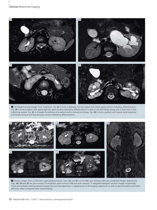
2
2 Coronal PD-weighted image of an osteo-chondral defect (OCD) of the distal phalynx of the
right toe.](https://image.slidesharecdn.com/magnetomflashissue47-00090255-140821160532-phpapp02/85/Pediatric-Imaging-Issue-47-7-320.jpg)
![How-I-do-it
chest and abdominal imaging. Increased
flow rates lead to artifacts from blood
vessels and cerebrospinal fluid (CSF)
pulsations, creating difficulties with
spine and Time-of-Flight (TOF) vessel
imaging [5]. Differences and evolution
in pediatric physiology may also lead
wto changes in the mechanism of injury,
or the types of injuries that occur in
children, such as growth plate injuries
and osteo-chondral defects (OCD) [6].
Behavioral
Sedation or anesthetic is commonly
required for younger children or those
with significant behavioral problems.
Factors such as temperament, stress,
pain, and illness play an important role
in patient compliance, creating difficul-ties
in establishing definitive age limits
for identifying which children will require
these procedures [7]. Encouraging chil-dren
to co-operate for an MRI examina-tion
and identifying those who cannot
are arguably the most significant chal-lenges
in pediatric MRI.
Techniques in scanning
children without sedation
Preparation
At our institution we begin scanning
without sedation from about five years
of age, although some positive out-comes
have been obtained with patients
as young as three years. Adequate prep-aration
of children for the MRI procedure
has been vital in achieving these results.
Our facility employs the services of edu-cational
play therapists who use a range
of resources to assist children to comply
with the procedure, such as brochures,
MRI toys and storybooks, discussions with
parents, and, most importantly, the
‘mock MRI’ procedure.
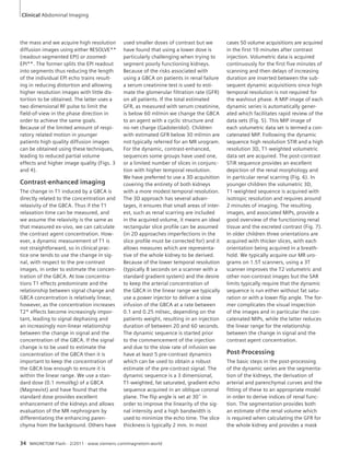
Simulation
The ‘mock MRI’ procedure involves chil-dren
undergoing a simulated scan with
the assistance of a play therapist prior
to the actual diagnostic scan. It acts as
both a screening tool, to assist in identi-fying
children who are likely to be able
to comply with the MRI procedure,
and also helps to prepare these children,
by familiarizing them with the environ-ment,
sounds, and equipment, while
teaching them skills (such as breathing,
relaxation, or distraction) to cope with
the actual procedure (Fig. 2). Use of
the ‘mock’ magnet has led to a marked
reduction in the numbers of patients
who have required anesthetic [7] and
reduced the time required for the diag-nostic
scan [8]. Several pediatric facili-ties
in various countries have introduced
a mock procedure in their facilities in
recent years [9].
Communication
Specialist staff and equipment are clearly
helpful in assisting children to comply
with an MR scan. However, for technolo-gists,
an awareness of how to talk to chil-dren
and adolescents at different stages
of development and the use of psycho-logical
techniques, such as distraction and
relaxation, can be the critical factor deter-mining
whether a young person is will-ing,
or able, to carry out the procedure.
Many children are withdrawn or uncom-municative
when nervous about a medi-cal
procedure, and taking the time to
help the child to feel safe and secure in
the environment is important. Compli-ance
with preschool children may be
facilitated by engaging in pretend play,
where the child can be encouraged to
frame the experience in familiar and non-threatening
ways [10]. Nonverbal com-munication
comprises a significant pro-portion
of a child’s interaction with the
world at this stage, and young children
can pick up on their parents’ anxiety or
the technologist’s impatience through
nonverbal clues. They may not under-stand
these feelings and can interpret
them as anger or fear of the examination.
Professionals who work with children
typically take steps to ensure that both
their verbal communication and body
3 Mock MRI simulator – this procedure identifies patients that are able to comply with
the requirements of an MRI examination, as well as prepare them for the clinical scan,
saving unnecessary appointments and valuable scanner time.
8 MAGNETOM Flash · 2/2011 · www.siemens.com/magnetom-world
3](https://image.slidesharecdn.com/magnetomflashissue47-00090255-140821160532-phpapp02/85/Pediatric-Imaging-Issue-47-8-320.jpg)
![language are reassuring and convey calm-ness
and confidence (Table 1). Positive
reinforcement, where the child is praised
for their efforts at each step, can be very
helpful.
School age children are able to engage
more actively in the procedure, and may
respond well to efforts to increase their
perceived control. Medical examinations
often take the locus of control away
from the patient, and this is particularly
true in pediatrics where someone else
usually makes the decisions for the
patient. Empowering children by offer-ing
some choice in how they can have
the scan can be helpful. This is particu-larly
important during adolescence; a
period of rapid social and physical
changes [10], when increased autonomy
is important, yet can be hampered by
serious illness. Adolescents are less likely
than children or adults to blindly follow
instructions, and may be reluctant to
accept or comply with the scan in the
absence of a flexible approach, where the
technologist is sensitive to their concerns.
Distraction and relaxation
Distraction can be a powerful tool for
reducing anxiety and increasing patient
compliance. Distraction techniques
can be either active or passive. Passive
techniques such as audiovisual aids are
useful during the scan when patients are
required to lie still in the bore. Having
a point of interest (such as a parent or
video screen) is helpful in maintaining
the patient in one position. Active tech-niques
which require patient participa-tion
such as relaxation breathing, guided
imagery, or complex puzzle tasks, are
useful in relaxing children before MRI or
performing interventions such as intra-venous
cannulation and general anes-thetic
(GA) inductions.
Successful use of
intravenous (IV) contrast
IV cannulation is a major cause of anxi-ety
in young patients presenting for
MRI examination. Limiting the use of IV
contrast in pediatric examinations can
often mean the difference between a
successful awake scan and a rebook for
sedation. This requires the support of
the radiologists to make decisions regard-ing
whether the benefits of contrast are
worth the potential distress to the patient.
Where contrast is necessary, it is often
helpful to separate the procedures of
IV placement and the MR exam by either
placing the cannula before the examina-tion
or offering a break between the
pre and post contrast scans. Many
children respond well to being able to
How-I-do-it
choose an IV site. Active distraction
techniques can be helpful, and there are
several aids available to assist with the
pain, such as local anesthetic creams,
ice, or nitrous oxide.
Protocols and sequences
Protocol based scanning can be difficult
in presenting pediatric patients, as the
required sequences differ dramatically
depending upon pathology, patient age,
compliance, and the clinical questions
being asked. It is often necessary for the
technologist or radiologist to screen the
examination as it progresses and tailor
the sequences for the patient and pathol-ogy.
A wide field-of-view scan can be
helpful to obtain an overview to screen
for other pathologies, particularly in
children who are difficult to examine
clinically. Children can be unpredictable
in how long they will remain still, so it
is important to prioritize sequences with
the highest diagnostic yield such as T2,
FLAIR, and diffusion. Scanning in multi-ple
planes or using 3D sequences can
help delineate disorders as well as mini-mize
the chance of pathology being
missed through partial voluming or inter-slice
gap.
Often it is necessary to modify a proto-col
or sequence when imaging children
of different sizes or capabilities. It is
important to strike a balance between
optimum image resolution and scan time.
MAGNETOM Flash · 2/2011 · www.siemens.com/magnetom-world 9
Table 1: Communicating with children
Engage with the child Get down on their level Use simple language Maintain eye contact
Frame the experience Help them verbalize Involve the child’s past Smile
their experience experiences / play
Empower the child Offer limited choices Praise good behavior Be positive “I know you can do this”](https://image.slidesharecdn.com/magnetomflashissue47-00090255-140821160532-phpapp02/85/Pediatric-Imaging-Issue-47-9-320.jpg)


![in reviewing and diagnosing complex
congenital conditions, and may reduce
the number of 2D sequences performed.
It allows for high resolution, no gap
imaging which can be used to accurately
measure lesion size, and monitor changes
in follow up imaging (Fig. 5).
BLADE
Rotating k-space techniques are utilised
in pediatric MR imaging to reduce arti-facts
from physiological motion in the
brain, as well as other body areas such
as the shoulder, chest, abdomen, and
pelvis. It is particularly useful with
younger patients scanned at 3T where
complex and turbulent flow artifacts can
mask pathology [11]. Recent studies
show improvement in lesion conspicuity
in the posterior fossa through reduction
in pulsation artifacts [12]. Disadvan-tages
of BLADE include increased scan
time, altered image contrast, increased
SAR, and reduction in sensitivity to some
pathology, particularly haemorrhage
[13]. Motion reduction with propeller
sequences can be utilized to obtain lim-ited
diagnostic information in moving
patients; however, their limitations
restrict widespread use for correcting
voluntary patient motion in pediatric
patients (Fig. 6).
Parallel imaging
The advent of parallel imaging tech-niques
and multiple element, phased
array coils has transformed pediatric
imaging in recent years, providing a
boost in either signal or speed. Parallel
imaging techniques combine signals
from several coil elements to produce an
image with increased SNR, or allow par-tial
sampling to reduce scan time. The
use of parallel image acceleration and
multiple acquisitions can be used to aver-age
motion artifacts in pediatric imag-ing.
Parallel imaging techniques can also
be exploited to reduce the duration of
breathhold imaging, allowing dynamic
capture of fast moving pediatric
anatomy. Parallel imaging also reduces
inhomogeneity artifacts such as seen in
diffusion-weighted imaging [14].
Time resolved angiography
When imaging arterio-venous malforma-tions
and vascular shunts it is important
for treatment and management to iden-tify
feeder vessels as well as the direction
of blood flow. Rapid heart rates and high
flow rates in children often make imag-ing
of complex vasculature difficult with
traditional MR angiography techniques.
Time resolved contrast-enhanced MR
angiography (MRA) techniques can pro-vide
anatomical as well as functional
assessment of these vascular conditions
[14].
High fi eld strength
imaging (3T)
Higher field strengths offer the opportu-nity
to address many of the difficulties
encountered with pediatric MR imaging.
The increased SNR allows for smaller vox-els
and increased resolution, or reduced
averages for increased speed. Parallel
imaging factors can be increased further
reducing scan time. Prolonged T1 times
facilitate better background suppression
for MRA and improved visualization of
paramagnetic contrast agents [5]. The
advantages offered by higher field
strengths have lead to the viability of
several new techniques in pediatric MRI.
Unfortunately, higher field strengths
can also present a number of challenges.
The increased field strength leads to
greater RF deposition, resulting in
increased heating (SAR), which can cause
sequence limitation in pediatric imag-ing.
B1-field inhomogeneities, chemical
shift, motion artifacts and susceptibility
artifacts are more pronounced at higher
field strengths. However, there are a
number of new techniques, which offer
potential to mitigate against these diffi-culties.
Prolonged T1 relaxation at higher
field strengths creates challenges in
image contrast, particularly in the neo-natal
brain [5].
How-I-do-it
Emerging techniques
in pediatric MRI
SWI
Susceptibility-weighted imaging is being
increasingly utilized in pediatric patients
for imaging trauma, vascular disease such
as haemorrhage, telangiectasia, and cav-ernous
and venous angiomas, tumors and
epilepsy imaging, as well as investigating
metabolic disorders (Figs. 7, 8). The use
of the phase images can be used to differ-entiate
calcification from haemhorrage in
lesions [15].
Parallel transmit technology
The use of multiple coil elements to
transmit part of the RF pulse results in
shorter pulse durations, reductions in
SAR, and corrections of patient-related
inhomogeneities [16]. This addresses
some major challenges of pediatric MRI,
particularly at higher field strengths.
Diffusion Tensor Imaging
DTI has provided insights into connectiv-ity
and plasticity in the developing brain.
It is now entering the clinical realm in
the assessment of traumatic brain injury,
epilepsy and white matter disease [14].
Arterial Spin Labeling
ASL provides functional information of
blood perfusion by magnetically tagging
inflowing blood upstream from the region
of interest. Persistence of the ‘tag’ limits
its use in adults; however, this is of less
concern in pediatric patients, due to fast
flows and relatively short perfusion dis-tances
[5]. This technique offers the
potential to investigate regions of hypo-and
hyper-perfusion, in conditions such
as stroke or tumors, without the use of
intravenous contrast media; however,
further validation is required to demon-strate
the clinical utility of this technique
in pediatric patients [17].
MR urography
Magnetic resonance urography provides
both anatomical and functional assess-ment
of the kidneys and urinary collect-ing
system. The multi-planar capabilities
MAGNETOM Flash · 2/2011 · www.siemens.com/magnetom-world 13](https://image.slidesharecdn.com/magnetomflashissue47-00090255-140821160532-phpapp02/85/Pediatric-Imaging-Issue-47-13-320.jpg)


![in this population. MRI in children
can be extremely challenging physically,
mentally, and emotionally, even for a
seasoned pediatric technologist; however,
these very challenges are also what make
pediatric imaging such an interesting
and rewarding field for MR technologists.
Acknowledgments
I would like to thank the patients and
staff of the Royal Children’s Hospital,
Melbourne, for their inspiration, advice,
and support in compiling this paper.
References
1 Machata AM, Willshke H, Kaban B, Prayer D,
Marhofer P (2009) “ Effect of Brain MRI on Body
Core Temperature in Sedated Infants and Children”
British Journal of Anaesthesia 102(3):385-9.
2 Cohen MM, Cameron Cal B, Duncan PG (1990)
“Pediatric Anesthesia Morbidity and Mortality
in the Perioperative Period” Anesthesia and
Analgesia 70”160-7.
3 Homman H, Bornert P, Eggers H, Nehrke K, Dossel
O, Graesslin I (2011) “Toward individualised SAR
models and in vivo validation” Magnetic reso-nance
in Medicine doi: 10.1002/mrm.22948.
4 Serafini G, Ongaro C, Mori A, Rossi C, Cavalloro F,
Tagliaferri C, Mencherini S, Braschi A (2005)
“Anesthesia for MRI in the Pediatric Patient”
Minerva Anesthesiology Jun 71(6) 361-6.
5 Dagia C, Ditchfield M. (2008) “3T MRI in
paediatrics: Challenges and clinical applications”
European Journal of Radiology doi:10.1016/j.
ejrad.2008.05.019.
6 Hussain HM, Barnes CE (2007) “Pediatric skeletal
trauma- Plain film to MRI” Applied Radiology
36(8):24-33.
7 Carter AJ, Greer ML, Gray SE, Ware, RS (2010)
“Mock MRI: reducing the need for anaesthesia in
children” Pediatric Radiology Aug;40(8):1368-74.
8 de Amorim e Silva,C.T.J., Mackenzie,A.,
Hallowell,L.M., Stewart, S.E., Ditchfield, M.R.
(2006) “Practice MRI: Reducing the need for se-dation
and general anaesthesia in children under-going
MRI” Australasian Radiology 50(4),319-323.
9 Hallowell, L.M., Stewart, S.E., de Amorim e Silva,
C.T., Ditchfield, M.R. (2008) “Reviewing the
process of preparing children for MRI” Pediatric
Radiology 38(3), 271-279.
10 Gable S (2010) “Communicating effectively
with children” University of Missouri, Columbia,
Outreach and Extension information sheet.
11 Vertinsky AT, et al. (2009) “Performance of
PROPELLER relative to standard FSE T2-weighted
imaging in pediatric brain MRI” Pediatric
Radiology. Oct;39(10):1038-47.
12 Von Kalle T, et al. (2010) “Diagnostic Relevant
Reduction of Motion Artifacts in the Posterior
Fossa by syngo BLADE Imaging” MAGNETOM
Flash 43(1/2010):6-11.
13 Forbes KP, et al. (2003) “Brain Imaging in the
Unsedated Pediatric Patient: Comparison of
Periodically Rotated Overlapping Parallel Lines
with Enhanced Reconstruction and Single-Shot
Fast Spin-Echo Sequences” American Journal of
Neuroradiology 24:794-798.
14 Shenoy-Bangle A, Nimkin K, Gee MS (2010)
“Pediatric Imaging: Current and Emergent
Trends” Journal of Postgrad Medicine 56:98-102.
15 Zhen Wu, Sandeep Mittal, Karl Kish, Yingjian Yu,
J. Hu, Mark Haake (2009) “Identification of Calci-fication
with Magnetic Resonance Imaging Using
Susceptibility-Weighted Imaging: A Case Study”
Journal of Magnetic Resonance Imaging 29(1):
177-182.
16 Wald LL, Adalsteinsson E (2009) “Parallel Trans-mit
Technology for High Field MRI” MAGNETOM
Flash 40(1/2009):124-135.
17 Chen J, et al. (2009) “Arterial Spin Labelling
Perfusion MRI in Pediatric Arterial Ischemic
Stroke- Initial Experiences” Journal of Magnetic
Resonance Imaging February; 29(2) 282-290.
18 Grattan-Smith JD, Jones R (2008) “Magnetic
resonance urography in Children” Magnetic
Resonance Imaging Clinics of North America
16 515-531.
19 Sinha R (2009) “Role of MRI in Chron’s disease”
Clinical Radiology 64 (4):341-352.
Contact
Glenn Cahoon
Royal Children’s Hospital
Melbourne
Australia
Glenn.Cahoon@rch.org.au
*MR scanning has not been established as safe for
imaging fetuses and infants under two years of age.
The responsible physician must evaluate the benefit
of the MRI examination in comparison to other
imaging procedures.
How-I-do-it
of MRI are ideal for displaying complex
congenital anomalies of the genito-urinary
tract. This information can be
used to predict outcome and select
patients that are most likely to benefit
from surgical intervention [18].
MR enterography
Crohn’s disease is a serious and lifelong
condition affecting the digestive system.
It affects primarily the ileum and colon
causing inflammation, ulceration and
can lead to abscess formation or fistulae
to other organs. Approximately 30% of
patients with Crohn’s disease will pres-ent
before the age of 20. MRI provides
the ability to diagnose and monitor this
condition as well as complications such
as peri-anal fistulae without exposing
the patient to radiation [19].
Conclusion
Magnetic resonance imaging referrals
for children continue to rise. The lack of
ionizing radiation coupled with the high
level of detail afforded by MR imaging
has made it the examination of choice for
a growing number of pediatric presenta-tions.
Most children who require mag-netic
resonance imaging will be examined
in specialist pediatric centers or hospi-tals;
however, heightened pressure on
specialist centers has lead to many chil-dren
being scanned in non-specialist
facilities where technologists may have
little experience in pediatric imaging.
These referrals need to be treated differ-ently
to standard adult imaging requests
in order to ensure diagnostic imaging
with minimal distress and intervention
to the patient. There is no ‘one size fits
all’ approach to imaging children and
pediatric MRI requires dedicated special-ist
knowledge, flexibility, and expert
input from the technologist. An aware-ness
of the challenges in pediatric MRI
and experience in pediatric imaging
techniques is vital to successful exami-nation
16 MAGNETOM Flash · 2/2011 · www.siemens.com/magnetom-world](https://image.slidesharecdn.com/magnetomflashissue47-00090255-140821160532-phpapp02/85/Pediatric-Imaging-Issue-47-16-320.jpg)


![Oncology Clinical
son to viable tumor tissue. We postu-lated
that the water mobility within a
tumor would increase over time after
treatment and that the magnitude of
change would be related to the effective-ness
of therapy which results in mem-brane
damage and subsequent reduction
in cell density. Based on a clinical feasi-bility
study in eight patients [1], we
found a correlation between the increase
of signal in ADC maps and the amount
of necrotic tumor-cells in treated human
osteogenic sarcomas, indicated by the
histological Salzer-Kuntschik-index.
Chemotherapy is considered satisfactory
if more than 90% of tumor volume
becomes necrotic (Salzer-Kuntschik grades
1, 2 or 3). In these cases, the increase in
ADC value was more than 0.33 x10⁻³
mm2/sec despite steady tumor volumes
during chemotherapy. Calculation of
ADC was based on a three scan-trace DWI
EPI MR sequence with b-values up to
700 s/mm2; a linear decay of the signal
was assumed. These preliminary results
reveal that the calculation of an ADC
value seems a promising quantitative
and sensitive substitute for monitoring
the response to chemotherapy in osteo-sarcoma.
The value of DWI for determin-ing
therapy response in case of osteo-sarcoma
could also be shown in animal
studies e.g. we observed a significant rela-tionship
between dosages of treosulfan
applied in a Ewing sarcoma mice model,
and effect on ADC as well as delayed
tumor volume response [4].
Since the cellularity of osteosarcomas is
heterogeneous, especially post-chemo-therapy,
the variations in the ADC values
are large. For this reason, Kiyoshi [2]
considered that the average ADC did not
necessarily reflect the higher cellularity
component in osteosarcomas. He pre-sumed
that the areas with the highest
cellularity within heterogeneous tumors
best reflect the characteristics of the
tumors. According to Kiyoshi, we are
now using the minimum ADC in the solid
tumor components. A significant differ-ence
can be demonstrated between the
patients with a good response to chemo-therapy
and those with a poor response.
We can therefore assume that the mini-mum
ADC, which reflects a higher cellu-lar
component in the tumor, provides
more valuable information for patients
with osteosarcoma.
DWI as a prognostic tool
Based on the results of Kiyoshi’s study,
the use of the minimum ADC for evalu-ating
the histological response to chemo-therapy
in osteosarcoma is considered
preferable to the use of the average ADC.
Calculating the ADC ratios ([ADCpost –
ADCpre] / ADCpre) using the minimum
ADC, patients with a good response had
a significantly higher minimum ADC
ratio than those with a poor response
(1.01 ± 0.22 and 0.55 ± 0.29 respec-tively,
p < 0.05).
Since the high tumor necrosis rate after
chemotherapy is an important prognos-tic
factor, the minimum ADC ratio might
be the most useful prognostic factor in
imaging for patients with osteosarcoma
[3]. The COSS-study group (European
Cooperative Osteosarkoma study group)
is currently prospectively testing this fas-cinating
new method. Final results show-ing
the significance of DWI-monitoring
of osteosarcomas are expected in 2014.
MAGNETOM Flash · 2/2011 · www.siemens.com/magnetom-world 19
only low impact on the mineralized
matrix of the osteogenic sarcoma. How-ever,
neo-adjuvant chemotherapy here
does have a significant favorable impact
on event-free survival. It is essential to
monitor the response to chemotherapy
to determine whether the prescribed
treament regime is effective. Treatment
response is considered successful if
more than 90% of the tumor cells histo-logically
show necrosis. However, histo-logical
assessment of tumor necrosis dur-ing
the course of chemotherapy would
require repeated biopsy [1].
Diffusion-weighted imaging
The use of water diffusion as a surrogate
marker to probe tumor necrosis is com-pelling
because the parameter is strongly
affected by membrane permeability
between intra- and extracellular com-partments,
active transport and direc-tionality
of cellular structures that impede
water mobility. Therefore, diffusion-weighted
imaging (DWI) could be used
to characterize highly cellular regions
of tumors versus regions which change
their cellularity (quantifiable by calculat-ing
the apparent diffusion coefficient
(ADC)) within the tumor over time (acel-lular
and necrotic regions). It could also
detect treatment response, which mani-fests
itself as a change in cellularity within
the tumor over time [1]. Subtle changes
in the degree of restriction of diffusion
by, for example, an alteration in cell mem-brane
integrity or permeability to water,
are reflected in changes in the diffusion-weighted
MR signal. Therefore, we expect
a signal change in diffusion-weighted
images in necrotic tumour in compari-](https://image.slidesharecdn.com/magnetomflashissue47-00090255-140821160532-phpapp02/85/Pediatric-Imaging-Issue-47-19-320.jpg)


![Contact
PD Dr. Dr. Günther Schneider
Dept. of Diagnostic and Interventional
Radiology
Saarland University Hospital
Kirrberger Strasse
66421 Homburg/Saar
Germany
dr.guenther.schneider@uks.eu
MRI often is also able to differentiate
the origin of soft tissue masses [6].
Regarding availability of MRI, at least in
maximum-care centres, MRI is available
on an emergency basis. In addition, by
applying sequences such as HASTE or
using motion artifacts reducing k-space
acquisition schemes like BLADE, MRI can
provide a high and robust image quality
even in less compliant patients, where
conventional MR techniques might fail.
We present two cases from our daily
routine that probably would not have
undergone MRI ten years ago. These
cases clearly demonstrate that MRI can
play an important role in imaging of
inflammatory complications and infection
outside the brain in pediatric patients.
These new possibilities using todays state
of the art imaging techniques in MRI are
asking for a re-evaluation of a daily
practise, where CT is often considered
the one and only emergency imaging
modality beyond ultrasound imaging.
General remarks on applied
imaging protocols
All images shown were acquired at
1.5 Tesla (MAGNETOM Aera); for the
body trunk (pelvis / abdomen to the
upper mediastinum), a combination of
the 18-channel body coil(s) with the
integrated 32-channel spine coil was
used; for imaging of the neck and skull
base, the standard 20-channel head /
neck coil was added. Administration of
contrast media is in our view essential in
cases of inflammation and its potential
complications. We also apply routinely
diffusion-weighted imaging (DWI) for
abdominal scans. According to our expe-rience
DWI allows excellent visualization
of inflamed tissue. However, its relevance
for differentiating inflammation versus
tumor or therapy response to anti-inflammatory
drugs, is still subject of
clinical research.
Clinical Abdominal Imaging
a higher clinical efficiency in small chil-dren
and adolescents compared to adults.
This is mainly due to the reduced depth
to be imaged allowing higher freqeuency
probes and better spatial resolution in
pediatric ultrasound examinations com-pared
to its application in older and
especially obese patients. Furthermore,
manoeuvres to displace disturbing air
and alternative approaches to image rel-evant
structures e.g. assessment of the
appendix via a dorsolateral subrenal view
instead of the left fronto-lateral window,
are more often successful in younger
patients.
But what if ultrasound fails or because
of the invasiveness of the therapeutic
decision further imaging is required to
strengthen the diagnostic confidence
and therapy planning?
In most of these cases, computed tomog-raphy
(CT) is used as a robust, fast and
nowadays 24/7 available imaging modal-ity.
But despite all attempts to reduce radi-ation
exposure, the utilization of imag-ing
modalities like x-ray, fluoroscopy and
CT [1], still use ionizing radiation with
potentially harmful effects. This is espe-cially
true for children, who are more
susceptible to ionizing radiation as com-pared
to adults [NRPB (1999). “Guidelines
on patient dose to promote the optimiza-tion
of protection for diagnostic medical
exposure.” Radiologial Protection Board
10(1).].
In this clinical scenario, MRI offers two
advantages as compared with CT:
First a superior soft tissue contrast and
second a complete lack of ionizing radia-tion.
Consequently, MRI is very often
used in clinical settings, when inflamma-tion
is considered a differential diagno-sis,
e.g. juvenile dermatomyositis, juve-nile
idiopathic arthritis, meningitis and
encephalitis [2-5] are clinical questions
where CT cannot provide the required
information offered by MRI. In addition,
References
1 image gentlySM – The Alliance for Radiation Safety
in Pediatric Imaging
http://www.pedrad.org/associations/5364/ig/
2 Damasio MB, Malattia C, Martini A, Tomà P.
Synovial and inflammatory diseases in childhood:
role of new imaging modalities in the assessment
of patients with juvenile idiopathic arthritis.
Pediatr Radiol. 2010 Jun;40(6):985-98. Epub
2010 Apr 30. Review.
3 Tzaribachev N, Well C, Schedel J, Horger M.
Whole-body MRI: a helpful diagnostic tool for
juvenile dermatomyositis case report and review
of the literature. Rheumatol Int. 2009
Oct;29(12):1511-4. Epub 2009 Mar 20. Review.
4 Bonthius DJ, Karacay B., Meningitis and encepha-litis
in children. An update. Neurol Clin. 2002
Nov;20(4):1013-38, vi-vii. Review.
3 Faingold R, Oudjhane K, Armstrong DC,
Albuquerque PA. Magnetic resonance imaging
of congenital, inflammatory, and infectious
soft-tissue lesions in children. Top Magn Reson
Imaging. 2002 Aug;13(4):241-61. Review.
6. Turkington JR, Paterson A, Sweeney LE,
Thornbury GD. Neck masses in children.
Br J Radiol. 2005 Jan;78(925):75-85. Review.
30 MAGNETOM Flash · 2/2011 · www.siemens.com/magnetom-world](https://image.slidesharecdn.com/magnetomflashissue47-00090255-140821160532-phpapp02/85/Pediatric-Imaging-Issue-47-30-320.jpg)


![Clinical Abdominal Imaging
cessing algorithms permit the evaluation
of the split renal function by generating
time-intensity curves representative for
the renal function, as well as many other
parameters. The use of MRU for the
assessment of urolithiasis, vesicoureteral
reflux, renal trauma, and fetal urinary
tract abnormalities is still partially
limited and technical refinements are
required. Judicious use of gadolinium-based
(Gd) contrast agents in children
at risk for nephrogenic systemic fibrosis
(NSF) should be employed with atten-tion
so as to avoid new occurrences.
Purpose
The aim of our work is to promote the
use of MRU in pediatrics trying to pres-ent
a safe and reliable MRI protocol for
anatomic imaging that is generally
accepted. Defining imaging and proce-dural
recommendations in pediatric uro-radiology
is an important task, not only
in standardization of pediatric uroradio-logic
imaging protocols but also in
reducing invasiveness and radiation
dose. The lack of standardization for
quantitative renal functional evaluation
and urinary excretion assessment
requires new studies and extensive inter-disciplinary
consultations.
Patient preparation
The adequate preparation is a prerequi-site
for good image quality [1–2]. We do
not routinely place a bladder catheter
in order to reduce invasiveness of the
procedure, although catheterization of
small children is recommended in case
of megaureter (with or without reflux).
The intravenous hydratation and admin-istration
of furosemide are crucial for
reducing the concentration of Gd [1].
Diuretics are recommended in both
static urography and dynamic urography
15 minutes before contrast administra-tion.
In this context, we adopted the
F-15 protocol and we administered stan-dardized
hydratation and diuretics prior
to Gd, in order to reduce artifacts and
shorten the examination time [3]. In
children younger than six years of age
40 MAGNETOM Flash · 2/2011 · www.siemens.com/magnetom-world
and those who are non-cooperative for
breathhold techniques, sedation should
be performed with ketamine (Ketalar)
and midazolam (Dormicum) according
to the department’s standard sedation
protocol [4–5]. Blood pressure, respira-tion,
heart rate, and oxygen saturation
are continuously monitored throughout
MR imaging in all patients.
Imaging protocol
MRU examinations are performed on a
1.5T MAGNETOM ESSENZA, using a
4-channel Body Matrix coil. The MRU
protocol should allow a correct evalua-tion
of the renal parenchyma, upper
urinary tract, bladder, renal vessels and
surrounding structures. Such a compre-hensive
‘one-stop-shop’ protocol must
be able to provide the required morpho-logical
and functional information
within 30–35 minutes. Our MRU proto-col
consists of a native MRI examination
with T2w coronal, T1w and T2w axial
sequences, administration of diuretic
followed by a dynamic study with gado-
Table 1: Sequence parameters – 1.5T MAGNETOM ESSENZA
t2_tru-fi_
loc
haste_
16sl_sag
t2_blade_
tra_fs
t2_
blade_
cor_fs
t2_tse
3d_cor
t1_sin-gle_
vibe_
cor
t1_
vibe_
dyn_cor
t1_tse
3d_sag
vibe_
fs_cor_
bh
single_
cor_CE
TE 2.09 59 123 123 627 0.83 0.83 9 2.38 1.09
TR 4.17 700 2280 1800 1600 2.62 2.62 696 4.38 3.11
FA 50 160 150 150 150 10 10 150 8,0 25
BW 501 789 362 362 219 1090 1090 219 610 520
FOV 40 38 30 32 34 38 38 34 40 38,5
ST 8.0 8.0 5,0 5,0 1,5 5,0 3.35 1,5 4,0 1,5
TA 0:13 0:36 0:48 0:43 2:48 0:04 13:51 1:13 0:11 0:14
SpS 7 – – – 60 32 32 60 80 56
Legend: TA = time acquisition in sec., TE = echo time, TR = repetition time, FA = flip angle, FOV = field-of-view, ST = slice thickness,
bh = breathhold, SpS = slices per slab, BW = bandwidth, VIBE = Volume Interpolated GRE](https://image.slidesharecdn.com/magnetomflashissue47-00090255-140821160532-phpapp02/85/Pediatric-Imaging-Issue-47-40-320.jpg)

![3A 3B 3C 3D
3 Ureteropelvic junction (UPJ) obstruction leading to hydronephrosis with dilatation of the right pyelocalyceal system in a 9-year-old girl. Coronal
T2w syngo BLADE image (3A). MR angiography following Gd administration (Single COR CE) demonstrates accessory right lower pole renal artery
(arrows), compressing the ureteropelvic junction – Thin MIP images (3B, 3C). Right side hydronephrosis demonstrated on 3D coronal T2w TSE maximum
intensity projection (3D). The girl was treated successfully by pyeloplasty.
linium (Gd) injection and 3D reconstruc-tions.
This MRU protocol allows the
combination of the whole information,
provided by conventional exams, ultra-sound
and scintigraphy in a single
examination, with or without minimal
drug toxicity – furosemid and Gd.
We commence the MRU examination
with a 3-plane localizer and we use a
sagittal view to depict the kidneys and
bladder in order to obtain an oblique
coronal plan angled parallel to the long
axis of the kidneys, including ureters
and bladder. We select a large field-of-view
(FOV) above both hemidiaphragms
so as to avoid artifacts from aliasing or
post-contrast signal intensity decline in
the upper renal poles. Following the cor-onal
T2w plane, we perform axial T2w
and T1w sequences. Fat-suppression
techniques are used in T1 and T2 hyper
intense findings and in cases of suspi-cion
of tumor formation – in and out of
phase sequences for contour delinea-tion.
An important pre-contrast
sequence is 3D T2w urogram with fat-suppression.
T1-weighted gradient-echo
sequence with fat-saturation (3D VIBE
dynamic) is used for the post-contrast
scan. The dynamic scan is repeated
within 15 minutes, following Gd injec-tion
with increasing intervals between
acquisitions, for the need of post-processing.
We inject the standard dose
of 0.1 mmol/kg of gadolinium. Regard-less
of the patient’s size, placement of
the coils is crucial. In newborns and
small children we use the smaller Flex
coil. Otherwise we use the Body Matrix
coil to scan older children. It is recom-mended
to adjust the Body Matrix coil
anteriorly to assure proper signal distri-bution
throughout the volume. The
technical parameters of our sequences
are presented in Table 1.
Discussion
MR urography is a feasible method for
evaluation of urinary tract pathology in
neonates and infants [6]. It overcomes
the limitations of the conventional imag-ing
techniques and is a superior tool in
many aspects, especially in the evalua-tion
of parenchymal kidney diseases and
poorly functioning systems, assessment
of ureteral anatomy, renal vasculature
and tumors. The method is particularly
helpful for further therapeutic decisions,
planning of surgical intervention and
future diagnostic work-up.
The most common MRU techniques,
used to visualize the urinary tract, are
the static (T2w) MRU and excretory
(T1w) MRU [7–9]. Static MRU uses the
long relaxation times of the liquids by
T2w sequences (Fig. 1). Three-dimen-sional
(3D) respiratory-triggered
sequences are used to obtain thin-sec-tion
data sets that can be further post-processed
to create volume rendered
(VR) or maximum intensity projection
(MIP) images of the entire urinary tract
42 MAGNETOM Flash · 2/2011 · www.siemens.com/magnetom-world
(Fig. 2) [10–11]. Excretory (T1w) MRU is
similar to CT urography and intravenous
urography and is performed after intra-venous
injection of Gd. The use of con-ventional
dose – 0.1 mmol/kg and in
some occasions low-dose Gd opacifica-tion
– 0.01 mmol/kg allows to maintain
the linearity between signal and Gd con-centration,
which is essential for quanti-tative
measurements and analysis.
Administration of diuretics improves the
quality of MRU by increasing the quan-tity
of the urine and therefore lead to
better dilution and appropriate distribu-tion
of Gd in the urinary tract [12–13].
The most important sequence of excre-tory
MRU is 3D gradient-echo. Fat-sup-pression
is recommended for better
demonstration of the ureters. MR angi-ography,
in addition to excretory MR
urography, demonstrates excellently the
renal vasculature (Fig. 3). Modern MR
units scan simultaneously in one volume
the kidneys, the ureters and the bladder,
using 3D gradient-echo sequences in
one breathhold [14–15]. Sometimes
segmental scanning of the kidneys or
bladder separately is recommended for
more detailed image visualization. An
advantage of static MRU over dynamic,
T1w MRU is the demonstration of the
ureters and the ureterovesical junction
in cases of obstruction or impaired renal
function [6, 16].
There is a growing number of publica-tions
concerning the criteria for assess-](https://image.slidesharecdn.com/magnetomflashissue47-00090255-140821160532-phpapp02/85/Pediatric-Imaging-Issue-47-42-320.jpg)
![Abdominal Imaging Clinical
Contact
Georgi Hadjidekov, MD
MC “Pro-Vita”
Montevideo str. N66
Sofia 1632
Bulgaria
jordiman76@yahoo.com
MAGNETOM Flash · 2/2011 · www.siemens.com/magnetom-world 43
ment of the renal function in children
by dynamic MRU, but the achievement
of consensus requires more and deeper
investigations. Functional criteria
include renal transit time, differential
renal function, and time–signal intensity
curves. The technique is challenging and
is best performed in specialized centers.
However, this single MRI examination
could avoid the use of multiple modali-ties
(e.g. US, conventional urography,
renal scintigraphy) that individually help
assess various morphologic features,
renal function and excretion and there is
an expanding use of MRI. Current limita-tions
for widespread distribution of the
technique are understaffed pediatric
radiologic departments and time-con-suming
post-processing.
Alongside the advantages of MRU, men-tioned
above, it is necessary to note
some limitations. Sometimes it requires
a placement of bladder catheter, admin-istration
of furosemide and Gd, sedation
and even anesthesia (for newborns and
younger children), which is a comple-mentary
risk [16]. Breathhold tech-niques
could not be applied in neonates
and small infants. We use syngo BLADE
to minimize motion artifacts. Patient
preparation and the examination itself
are time-consuming; post-processing
and calculation of functional curves and
differential renal function require an
additional 30 minutes.
Until recently it was believed that the
extracellular Gd-based contrast agents
are safe. However, in 2006, it was dem-onstrated
that some Gd-based contrast
agents may provoke the development of
NSF and/or a generalized fibrotic disor-der
in renal failure patients [17]. Gd-ions,
released from Gd-based MR con-trast
agents, are the likely etiologic
agent of NSF [18]. The guidelines of the
European Society of Urogenital Radiol-ogy
(ESUR) suggest a very careful
administration of Gd in children with
renal failure. Absolute contraindications
are high levels of creatinine and a glomer-ular
filtration under 30 ml/min/1.73 m2
and in cases of glomerular filtration
between 30 and 60 ml/min/1.73 m2 dis-cussions
with pediatric nephrologists are
mandatory. Written consent should be
obtained in spite of the fact that most
cases of NSF occur in adults and the
reported cases of NSF without Gd
administration. In all patients at high
risk of developing NSF and in the pediat-ric
group, cyclic Gd-helators should be
used due to their higher stability [19].
Conclusion
MRU has the potential to become the
leading diagnostic modality in the near
future of a wide range of pathological
conditions affecting the urinary tract in
newborns and children. It integrates
exquisite anatomical information with a
variety of functional data and avoids
ionizing radiation. MRU is increasingly
employed as a problem solver when con-ventional
imaging studies remain incon-clusive
and its growing application will
likely improve availability and reduce
cost in the future. The introduction of
parallel imaging, higher field strength
systems, multielement coil technology,
and gadolinium-based contrast agents
with increased T1 relaxivity will further
improve the overall signal-to-noise ratio
(SNR) and spatial resolution of the
images. The advances of molecular
imaging techniques will provide new
insights about the nature of hereditary
diseases in pediatric nephrology and
urology. Potential future applications
include also virtual endoscopy and MRU-guided
procedures.
*MR scanning has not been established as safe for
imaging fetuses and infants under two years of age.
The responsible physician must evaluate the benefit of
the MRI examination in comparison to other imaging
procedures.
References
1 Grattan-Smith, J.D., S.B. Little, and R.A. Jones,
MR urography in children: how we do it. Pediatr
Radiol, 2008. 38 Suppl 1: p. S3-17.
2 Leyendecker, J.R., C.E. Barnes, and R.J. Zagoria,
MR urography: techniques and clinical applica-tions.
Radiographics, 2008. 28(1): p. 23-46;
discussion 46-7.
3 Grattan-Smith, J.D., et al., MR imaging of kid-neys:
functional evaluation using F-15 perfusion
imaging. Pediatr Radiol, 2003. 33(5): p. 293-
304.
4 Cengiz, M., Z. Baysal, and S. Ganidagli, Oral
sedation with midazolam and diphenhydramine
compared with midazolam alone in children
undergoing magnetic resonance imaging.
Paediatr Anaesth, 2006. 16(6): p. 621-6.
5 Lin, T.F., et al., Antiemetic and analgesic-sparing
effects of diphenhydramine added to morphine
intravenous patient-controlled analgesia. Br J
Anaesth, 2005. 94(6): p. 835-9.
6 Riccabona, M., et al., Feasibility of MR urography
in neonates and infants with anomalies of the
upper urinary tract. Eur Radiol, 2002. 12(6):
p. 1442-50.
7 Nolte-Ernsting, C.C., et al., MR urography today.
Abdom Imaging, 2003. 28(2): p. 191-209.
8 Garcia-Valtuille, R., et al., Magnetic resonance
urography: a pictorial overview. Br J Radiol,
2006. 79(943): p. 614-26.
9 Nolte-Ernsting, C.C., et al., Diuretic-enhanced
gadolinium excretory MR urography: comparison
of conventional gradient-echo sequences and
echo-planar imaging. Eur Radiol, 2001. 11(1):
p. 18-27.
10 Roy, C., et al., MR urography in the evaluation of
urinary tract obstruction. Abdom Imaging,
1998. 23(1): p. 27-34.
11 O’Malley, M.E., et al., MR urography: evaluation
of a three-dimensional fast spin-echo technique
in patients with hydronephrosis. AJR Am J
Roentgenol, 1997. 168(2): p. 387-92.
12 Szopinski, K., et al., Magnetic resonance uro-graphy:
initial experience of a low-dose Gd-DT-PA-
enhanced technique. Eur Radiol, 2000.
10(7): p. 1158-64.
13 Hughes, J., et al., MR urography: evaluation of
different techniques in non-dilated tracts.
Clin Radiol, 2002. 57(11): p. 989-94.
14 Nolte-Ernsting, C.C., G.B. Adam, and R.W.
Gunther, MR urography: examination techniques
and clinical applications. Eur Radiol, 2001.
11(3): p. 355-72.
15 Sudah, M., et al., MR urography in evaluation of
acute flank pain: T2-weighted sequences and
gadolinium-enhanced three-dimensional FLASH
compared with urography. Fast low-angle shot.
AJR Am J Roentgenol, 2001. 176(1): p. 105-12.
16 Riccabona, M., Pediatric MRU-its potential and
its role in the diagnostic work-up of upper
urinary tract dilatation in infants and children.
World J Urol, 2004. 22(2): p. 79-87.
17 Thomsen, H.S. and P. Marckmann, Extracellular
Gd-CA: differences in prevalence of NSF. Eur J
Radiol, 2008. 66(2): p. 180-3.
18 Abraham, J.L., et al., Dermal inorganic gado-linium
concentrations: evidence for in vivo
transmetallation and long-term persistence in
nephrogenic systemic fibrosis. Br J Dermatol,
2008. 158(2): p. 273-80.
19 Martin, D.R., et al., Individual kidney blood flow
measured with contrast-enhanced first-pass
perfusion MR imaging. Radiology, 2008. 246(1):
p. 241-8.](https://image.slidesharecdn.com/magnetomflashissue47-00090255-140821160532-phpapp02/85/Pediatric-Imaging-Issue-47-43-320.jpg)


![Neurology Clinical
2 Contrast-enhanced axial T1-weighted images demonstrate an increased pachymeningeal and leptomeningeal enhancement in a 13-year-old
girl with viral meningitis.
MAGNETOM Flash · 2/2011 · www.siemens.com/magnetom-world 61
tions in the basal ganglia and in the
periventricular region. The children may
suffer from hydrocephalus, but also from
microcephaly. In contrast to congenital
CMV infection, disorders of cortical
development do not usually occur [1].
Generally, the consequences tend to be
less serious the later the infection occurs
during gestation.
Due to widespread immunization cam-paigns,
congenital rubella infections
have become an exceedingly rare occur-rence
in the western world. The severity
of the infections depends on the gesta-tional
age – infections have an especially
severe course when they occur during the
first trimester. The MR imaging appear-ance
can resemble that of connatal toxo-plasmosis.
Microcephaly and calcifica-tions
are observed [2].
Cytomegalovirus (CMV) infections are
by far the most common connatal infec-tion
in the western world. CMV infections
during pregnancy are estimated to be ten
times more common than congenital
toxoplasma infections – occurring in up
to 1% of live births. However, only 5 to
10% of connatal CMV infections affect
the brain. A common presenting symp-tom
in children is hearing loss. It is not
unusual for children to be referred for
MR imaging prior to surgery for a cochlear
implant to assess the morphology of the
inner ear. The MR imaging appearance
varies depending on the gestational age
at the time of infection. CMV infections
may lead to polymicrogyria and / or
lissencephaly, especially if they occur
during the second trimester of pregnancy.
Very commonly, patchy white matter
lesions with hyperintense signal on
T2-weighted and FLAIR sequences are
present (Fig. 1). These are often conflu-ent
and may occur in the absence of
abnormalities of cortical development [3].
Herpes simplex virus (HSV) infections
of the brain in the neonate are usually of
type II and are acquired during vaginal
birth. MR imaging usually demonstrates
rapidly progressive signal alterations of
the white matter eventually leading to
cystic encephalomalacia with ensuing
atrophy of the brain [4].
Other congenital infections include lym-phocytic
choriomeningitis (LCM) virus
infection, syphilis, congenital HIV infec-tion
and congenital varicella zoster
infection.
Generally, connatal infections should
always be taken into consideration when
imaging a child with developmental delay
and / or epilepsy. Children with congeni-tal
hearing loss usually undergo MR imag-ing
prior to surgery for a cochlear implant.
Connatal infections – especially connatal
CMV infections – are a rather common
cause for congenital hearing loss and the
MR imaging should always be scrutinized
for possible signs of connatal infections
such as white matter lesions and / or dis-orders
of cortical development.
Meningitis, cerebritis
and abscess
Meningitis is the most common neuro-infection
throughout childhood. The
infectious agent may reach the meninges
by different routes [5]:
■ hematogeneously
■ via an adjacent infection,
e.g. mastoiditis
■ through the choroid plexus
■ via a penetrating injury
■ by rupture of a cerebral abscess.
Most commonly, the infectious agent is
viral. Viral meningitis is not a classic
imaging diagnosis, but imaging may be
performed to rule out complications
and / or to rule out contraindications to
perform a diagnostic lumbar puncture.
2A 2B](https://image.slidesharecdn.com/magnetomflashissue47-00090255-140821160532-phpapp02/85/Pediatric-Imaging-Issue-47-61-320.jpg)
![Clinical Neurology
3 Contrast-enhanced axial T1-weighted images (3A), diffusion-weighted images (3B) and the concomitant ADC-map (3C) show an intracranial
abscess with diffusion restriction.
MR imaging may show a thickening of
the meninges with an increased
enhancement after the administration of
contrast agent – this can affect the lep-tomeninges
and / or the pachymeninges
(Fig. 2). However, the MR imaging
appearance may also be completely nor-mal
in viral meningitis. A normal MR
appearance generally does not rule out
viral meningitis. Complications of viral
meningitis include concomitant enceph-alitis,
hydrocephalus and vasculitis.
Bacterial meningitis usually progresses
more rapidly with a more severe clinical
course than viral meningitis. Again, MR
imaging is mostly performed to investi-gate
complications and / or to rule out
contraindications for lumbar puncture.
The thickening of the meninges is usually
more pronounced in bacterial meningitis
compared to viral meningitis. Subdural
empyemas are a potential complication
of bacterial meningitis, the occurrence
of which leads to a pronounced worsen-ing
of the prognosis [6]. Hemophilus influ-enzae
may, however, lead to sterile sub-dural
fluid collections that need to be
differentiated from empyema.
Other complications of bacterial menin-gitis
include hydrocephalus, venous
infarctions due to thromboses, and arte-rial
infarctions due to arteritis / vasculi-tis.
Moreover, bacterial meningitis may
lead to cerebritis and abscess formation,
as well as to ventriculitis.
Tuberculous meningitis may occur in
children or in adults. Tuberculosis prefer-entially
affects the basal meninges,
which show a marked thickening and
enhancement on MR imaging. Focal
tuberculomas may also be seen, which
are often hypointense on T2-weighted
MR imaging. Common complications
include hydrocephalus and infarctions
in the basal ganglia and thalami.
A focal infection of the brain is called
cerebritis. At the stage of a cerebritis,
the focal infection has not yet become
centrally necrotic. MR imaging usually
62 MAGNETOM Flash · 2/2011 · www.siemens.com/magnetom-world
reveals a contrast enhancement of the
entire lesion with a perifocal edema.
At a later stage, cerebritis may become
a cerebral abscess, especially if left
untreated. Cerebral abscesses in children
may be a result of a sinusitis or mastoid-itis
and are then commonly located in
close proximity to either the paranasal
sinuses or the petrous bones. MR imaging
reveals one of the hallmark features of
cerebral abscesses: a centrally restricted
diffusion, which aids in differentiating
cerebral abscess formation from primary
brain tumors [7]. On conventional MR
imaging, the central portion of a cerebral
abscess is usually hyperintense on
T2-weighted imaging and hypointense
on T1-weighed imaging, while the rim
of the lesion shows a pronounced
enhancement after the administration
of contrast agent (Fig. 3).
Especially in small children, ventriculitis
frequently occurs as a complication of
meningitis. This may lead to periventric-ular
and intraventricular abscesses and
the prognosis is rather unfavorable.
3A 3B](https://image.slidesharecdn.com/magnetomflashissue47-00090255-140821160532-phpapp02/85/Pediatric-Imaging-Issue-47-62-320.jpg)
![Neurology Clinical
MAGNETOM Flash · 2/2011 · www.siemens.com/magnetom-world 63
MR imaging typically shows a pronounced
enhancement of the ventricular epen-dyma
and dilatation of the ventricles.
Later in the disease process, loculations
with septae commonly ensue.
Viral encephalitis
Viral encephalitis is another important
neuro-infectious differential diagnosis
both in children and in adults. There is a
plethora of viral infectious agents that
may cause encephalitis. In the following,
this report focuses on HSV I encephalitis,
SSPE, tick-borne encephalitis and Ras-mussen
encephalitis.
HSV encephalitis is a crucial diagnosis
not to be missed on MR imaging. The
earlier treatment is initiated, the better
the diagnosis is. About one third of HSV
encephalitis occurs in children, most of
which are HSV type I infections. Initial
symptoms may be rather unspecific;
therefore, the imaging diagnosis can be
crucial for the patient’s further progno-sis.
HSV preferentially involves the limbic
system, and so an involvement of the
temporal lobes with a signal hyperinten-sity
on T2-weighted and FLAIR sequences
especially in the temporomesial portion
is a common hallmark feature of the
disorder (Fig. 4). The insular / peri-Sylvian
regions are commonly involved as well.
However, the MR imaging appearance
can also be atypical. An asymmetrical
involvement of the limbic structures is
rather common. In addition, the pattern
of involvement can also be unspecific
and does not necessarily demonstrate
a preferential involvement of the limbic
system, especially in small children [8].
If in any doubt, the suspicion of HSV
encephalitis should be raised and treat-ment
should be swiftly initiated, until HSV
infection is ruled out by cerebrospinal
fluid (CSF) diagnostics.
Varicella-zoster-encephalitis is a rare
complication of chicken pox. Affected
children usually present with cerebellar
signs such as ataxia or dysarthria. On
MR imaging there is usually pronounced
edema of the cerebellum. Supratentorial
lesions may also be present, e.g. in the
cortex or in the basal ganglia.
Subacute sclerosing panencephalitis
(SSPE) is a devastating and eventually
lethal disorder of the brain. It is consid-ered
a late complication of measles
infection. Affected children suffer from
a progressive neurodegeneration with
dementia and pareses. MR imaging ini-tially
shows patchy signal alterations in
the cortical and subcortical regions with
hyperintensities on T2-weighted and
FLAIR sequences. In the further course,
pronounced atrophy ensues [9].
Other brain manifestations associated
with measles infection include acute
postinfectious encephalitis, which is prob-ably
an autoimmune disorder in relation
to the measles virus, and progressive
infectious encephalitis, which is a persis-tent
infection of the brain.
Tick-borne encephalites usually have
a peak occurrence in early summer,
when ticks are most prevalent. Depend-ing
on the region, the symptoms and
imaging appearance may vary. Tick-borne
encephalitis most commonly starts with
flu-like symptoms, which may be followed
by a cerebral and / or spinal affectation.
On MR imaging, edema of the basal gan-glia
and thalami with pronounced signal
hyperintensities on T2-weighted and
FLAIR sequences is the most common
imaging pattern. However, both the cor-tical
ribbon and the spinal cord may be
involved as well.
Rasmussen encephalitis is a progres-sive
disorder of the brain, the cause of
which is still unknown. Both viral agents
and autoimmune processes are discussed
as potential causes. Affected children
usually suffer from epilepsy and progres-sive
hemiparesis with the epilepsy com-monly
being refractory to medical ther-apy.
The disorder starts focally and
subsequently spreads throughout the
cerebral hemisphere. MR imaging shows
cortical and subcortical edema and later
progressive atrophy. It is important to
remember that MR imaging can be com-pletely
normal in the early stages of the
disease [10].
Parasitic infections
Parasites are among the most common
infectious agents affecting the brain
world-wide, even though they are rather
rare in the western world.
Neurocysticercosis is caused by the
tapeworm taenia solium [11]. Affected
children commonly complain of head-
3C](https://image.slidesharecdn.com/magnetomflashissue47-00090255-140821160532-phpapp02/85/Pediatric-Imaging-Issue-47-63-320.jpg)


![How-I-do-it
Optimized Examination Technique
Reduces Examination Time for Children
with Retinoblastoma by Half
Anton S. Quinsten, RT; Sebastian Blex, RT; Nicola Keiper, RT; Sophia L. Goericke, MD
University Hospital Essen, Diagnostic and Interventional Radiology and Neuroradiology, Essen, Germany
Introduction
Retinoblastoma is the most common
(intraocular) eye tumor of childhood
with an incidence of approx. one affected
child per 18,000 live births. If the tumor
is detected and treated at an early stage,
more than 95% of patients will survive
[1]. Tumors that are present in one eye
only are referred to as unilateral retino-blastoma.
Approx. 2/3 to 3/4 of affected
children experience unilateral retinoblas-toma
[3]. When the tumors are present
in both eyes, they are referred to as
bilateral retinoblastoma. Because most
patients are children, the examination
usually has to be performed under gen-eral
anesthesia.
The most common symptoms include
leukocoria, strabismus, glaucoma and
loss of vision. In 30% of cases both eyes
are affected.
The diagnosis is usually made on the
basis of a clinical examination, ophthal-moscopy
and ultrasound. Magnetic reso-nance
imaging* (MRI) is the procedure of
choice for visualizing the local ophthal-mic
findings, the extent of the tumor
and possible associated brain changes,
which is essential for treatment planning.
An MRI examination of retinoblastoma
poses a major challenge even for the
most experienced examiner. Suspected
retinoblastoma requires far more than a
standard examination of the orbita.
Producing high resolution images with
optimized sequences and MR hardware
is crucial for treatment planning and
ultimately for its success. As Technolo-gists
we play a significant role in this
process.
Examination
Apart from examining the orbits, the
brain also needs to be visualized. The
MRI examination can provide informa-tion
on whether there is extraocular
tumor growth and/or whether there
is a tumor of the bones or brain tissue.
Because the patients are children, the
examination is performed mostly under
general anesthesia, and special care is
required before, during and after the
examination.
Since in a third of cases both orbits are
affected, it is advisable to examine both
eyes. Previously we used to examine each
68 MAGNETOM Flash · 2/2011 · www.siemens.com/magnetom-world
eye separately. This used to take about
two hours. Now with the new technique
the whole examination including both
orbits takes approximately 57 minutes.
In the following we would like to explain
our examination technique, now used
routinely, which allows the brain and
both eyes to be simultaneously visual-ized
at high resolution in a considerably
shorter examination time.
* MR scanning has not been established as safe for
imaging fetuses and infants under two years of age.
The responsible physician must evaluate the benefit of
the MRI examination in comparison to other imaging
procedures.
References
1 http://www.uk-essen.de/augenklinik/
retinoblastom.html
2 http://www.onmeda.de/krankheiten/
retinoblastom-definition-1779-2.html
3 http://www.uk-essen.de/augenklinik/
retinoblastom.html
4 http://www.kinderkrebsinfo.de/e9031/e10591/
e77086/e63970/index_ger.html
5 http://www.krebsgesellschaft.de/
pat_ka_retinoblastom_definition,108207.html
6 http://www.dr-gumpert.de/
html/retinoblastom.html](https://image.slidesharecdn.com/magnetomflashissue47-00090255-140821160532-phpapp02/85/Pediatric-Imaging-Issue-47-68-320.jpg)


![Clinical Thorax
Case Report: MRI of the Lung in a Young
Child with Abscess Pneumonia Caused by
H1N1 Infection
Migle Sumkauskaite1; Jens-Peter Schenk, PD2; Olaf Sommerburg, PD3; Michael Puderbach, MD1,4;
H.-P. Schlemmer, MD, PD1; Monika Eichinger, MD1
1 German Cancer Research Center (DKFZ), Dept. of Radiology, Heidelberg, Germany
2 University Hospital Heidelberg, Div. of Pediatric Radiology, Dept. of Diagnostic and Interventional Radiology,
Heidelberg, Germany
3 University Hospital Heidelberg, Div. of Pediatric Pulmonology and Cystic Fibrosis Center, Dept. of Pediatrics III,
Heidelberg, Germany
4 Chest Clinic Heidelberg, Dept. of Diagnostic and Interventional Radiology, Heidelberg, Germany
74 MAGNETOM Flash · 2/2011 · www.siemens.com/magnetom-world
Introduction
Influenza is an infectious disease caused
by RNA viruses of the family Orthomyxo-viridae
[1]. The most common symp-toms
of the infection are: fever, rhinor-rhoea,
sore throat, cough, muscle pain,
frontal/retro-orbital headache, weak-ness/
fatigue [1–3]. Pneumonia is a com-mon
complication of influenza in young
children. [1]. We present a case of
abscess pneumonia caused by H1N1
infection in a young child and discuss
the MRI findings of the lungs during
the course of the disease.
Patient history
A 5-year-old girl with a history of rhini-tis,
cough and fever over a period of 2–3
weeks, with proven H1N1 infection but
unsuccessfully treated with antibiotics
was admitted to our hospital because of
progressive dyspnoea and fever of
39.7°C. A chest radiograph showed a
consolidation of the right lower lung
field (Fig. 1).
Laboratory tests demonstrated nonspe-cific
findings of systemic inflammatory
illness:
CRP of 203.9 mg/l (normal < 5 mg/l),
erythrocyte count 4.0 /pl (normal
3.9–5.3/pl),
haemoglobin 10.8 g/dl (normal
11–14.5 g/dl),
1 Chest-X-ray at admission: homogenous opacification of the right lower lung field and
obscured costo-diaphragmatic angle (arrow).
1](https://image.slidesharecdn.com/magnetomflashissue47-00090255-140821160532-phpapp02/85/Pediatric-Imaging-Issue-47-74-320.jpg)

![2A
Clinical Thorax
2B
2C
3
1
2
2
76 MAGNETOM Flash · 2/2011 · www.siemens.com/magnetom-world
Discussion
In clinical practice, as a widely available
imaging modality, chest x-ray is essential
for primary diagnoses of lung diseases.
Actually CT is considered to be the “gold
standard” for the assessment of lung
parenchyma and the majority of lung
pathology. However, repeated examina-tions
to evaluate the progress of the dis-ease
are frequently necessary in severe
illness. This leads especially in small
children to cumulative radiation doses,
which can be avoided if radiation-free
imaging modalities are used.
Magnetic resonance imaging (MRI) as a
radiation-free technique was already
proposed as a potential alternative for
lung imaging in the late 80s [4]. At that
time, MRI technology was not able to
produce results comparable to CT [5].
New technologies and strategies were
able to overcome the inherent difficul-ties
of MRI of the lung [6]. With the
introduction of parallel imaging in clini-cal
practice, faster image acquisition
became possible and thus substantial
improvement in temporal and/or spatial
resolution [7–9]. MRI has a lower spatial
resolution than CT, but its major advan-tage
is the possibility to characterize dif-ferent
aspects of tissue based on differ-ent
contrasts in T1w and T2w, as well as
enhancement after contrast media
administration. Additionally, MRI is able
to visualize different regional functional
aspects of the lung parenchyma (pulmo-nary
hemodynamics, perfusion, ventila-tion).
Today, lung MRI is considered a sensitive
detector of infiltrative and solid lung
pathology. It is therefore the ideal fol-low-
up assessment of pneumonia, pleu-ral
disease or chronic lung patients with
pulmonary exacerbations. In children
with cystic fibrosis it is more and more
used as a monitoring strategy for lung
disease progression [10].
In this report we present a child with a
severe abscess pneumonia caused by
H1N1 virus infection. In addition to the
areas of consolidation, abscesses were
found in the right lower lobe accompa-nied
by bilateral pleural effusion.
For assessment of lung parenchyma,
T2-weighted images as well as
3
3
2 First MRI: axial T2-weighted HASTE (2A), syngo BLADE (2B) and
T1-weighted VIBE image post contrast (2C) showing abscess pneumonia
of the right lower lobe. Areas of a homogenous increase in T2w signal
(consolidation) in the right lower lobe (1) and multiple inclusions of in
T2w hyper and in T1w hypo intense areas with circular peripheral contrast
media enhancement (abscesses, 2) are demonstrated. Additionally a small
bilateral pleural effusion is visualized (3).](https://image.slidesharecdn.com/magnetomflashissue47-00090255-140821160532-phpapp02/85/Pediatric-Imaging-Issue-47-76-320.jpg)
![T1-weighted images before and after
contrast media injection are performed.
T1-weighted sequences pre- and post-contrast
are used for the detection of
lymph nodes especially in the mediasti-num
and tumor infiltration into the
thorax wall. T2-weighted sequences are
nowadays the method of choice for the
assessment of lung parenchyma, and
additionally enable visualization of the
pathology of mediastinum and thoracic
wall.
Ex vivo experiments and in vivo experi-ence
have shown that solid lung pathol-ogy
can be successfully detected with
fast T1-weighted gradient echo
sequences (T1-GRE). For lung nodules
larger than 4–5 mm, 3D gradient echo
sequences reach the detection rates
of conventional helical CT with a single
row detector technique [11].
The T2-weighted ‘Half-Fourier single-shot
turbo spin-echo’ (HASTE) sequence
has a sensitivity of 85%–90% for lung
nodules larger than 4 mm and a sensitiv-ity
of 100% for lung lesions larger than
8 mm [12, 13]. Post-contrast imaging is
necessary for differentiation of pleural
processes, such as empyema, abscesses
or metastatic spread of malign tumors,
as well as for the evaluation of solid
intraparenchymal benign/malign tumor
masses [12].
In airways disease the reflex of hypoxic
vasoconstriction leads to impaired perfu-sion
in less ventilated lung areas. To
detect lung perfusion defects, an injec-tion
of contrast media at the same time
that a repeated 3D GRE sequence with
high temporal resolution is acquiring
MRI data is necessary.
In the presented case, MRI examinations
were performed on a 1.5 Tesla MR scan-ner
(MAGNETOM Avanto, Siemens Erlan-gen,
Germany). Orientated on a previ-ously
published standard protocol for
lung image examination [11], a naviga-tor
triggered T2w BLADE and T2w HASTE
sequence as well as a T1w VIBE
sequence pre- and post-contrast media
in inspiratory breath hold were used.
For functional assessment a 3D GRE
sequence with high temporal resolution
with parallel contrast media injection
was performed.
3A
3B
2
2
2
1
1
1
3
3
3 First follow-up MRI: axial T2-weighted HASTE (3A), syngo BLADE (3B)
and T1-weighted VIBE image post contrast (3C): progressive pleural effusion
on the right side (1) and unchanged right lower lobe consolidation with
abscesses (2). Bronchial wall thickening is visible in the left lower lobe (3).
MAGNETOM Flash · 2/2011 · www.siemens.com/magnetom-world 77
3C](https://image.slidesharecdn.com/magnetomflashissue47-00090255-140821160532-phpapp02/85/Pediatric-Imaging-Issue-47-77-320.jpg)

![References
1 Delaney JW, Fowler RA. 2009 influenza A
(H1N1): a clinical review. Hosp Pract (Minneap).
2010:38(2):74–81.
2 Smith SM, Gums JG. Antivirals for influenza:
strategies for use in pediatrics. Paediatr Drugs.
2010 Oct 1;12(5):285–99.
3 Nelson Textbook of Pediatrics Elsevier 18th
edition 2011, editors: Robert M. Kliegman, MD,
Richard E. Behrman, MD, Hal B. Jenson, MD and
Bonita M.D. Stanton, MD.
4 Fiel SB, Friedman AC, Caroline DF et al. Magnetic
resonance imaging in young adults with cystic
fibrosis. Chest. 1987:91:181–184.
5 Carr DH, Oades P, Trotman-Dickenson B et al.
Magnetic resonance scanning in cystic fibrosis:
comparison with computed tomography.
Clin Radiol. 1995:50:84–89.
6 Kauczor HU, Heussel CP, Schreiber WG et al.
[New developments in MRI of the thorax].
Radiologe. 2001:41:279–287.
7 Swift AJ, Woodhouse N, Fichele S et al.
Rapid lung volumetry using ultrafast dynamic
magnetic resonance imaging during forced vital
capacity maneuver: correlation with spirometry.
Invest Radiol. 2007:42:37–41.
8 Heidemann RM, Griswold MA, Kiefer B et al.
Resolution enhancement in lung 1H imaging
using parallel imaging methods. Magn Reson
Med. 2003:49:391–394.
9 Fink C, Bock M, Puderbach M et al. Partially
parallel three-dimensional magnetic resonance
imaging for the assessment of lung perfusion-initial
results. Invest Radiol. 2003:38:482-488.
10 M. Puderbach, M. Eichinger, J. Haeselbarth, A.
Niemann, M.Mall, H.-U. Kauczor Magnetic
Resonance Imaging (MRI) for early diagnosis of
lung changes in Children with Cystic Fibroisis
AJRCCM, 2008:177:773, Abstract issue.
11 Puderbach M, Hintze C, Ley S, Eichinger M,
Kauczor H-U, Biederer J. MR imaging of the
chest: A practical approach at 1,5 T. Eur J of
Radiology, 2007: 64: 345–355.
12 MRI of the lung, Springer-Verlag Berlin Heidel-berg,
1st edition 2009, editor: Kauczor H.-U.
13 Biederer J, Hintze C, Fabel M MRI of pulmonary
nodules: technique and diagnostic value. Cancer
Imaging, 2008: 8(1): 125–130.
Contact
Monika Eichinger, MD
Department of Radiology, E010
German Cancer Research Center
Im Neuenheimer Feld 280
69120 Heidelberg
Germany
Phone: +49 6221 422498
m.eichinger@dkfz-heidelberg.de
1, 2
1, 2
5B
5C
5 MRI after clinical restitution: T2-weighted axial syngo BLADE image (5A)
and T2-weighted axial HASTE image (5B) demonstrate the residual infiltrates
in the right lower lobe with dysatelectasis (1) and pleural thickening (2).
Subtraction image of MR perfusion from a dorsal slice shows homogenous
perfusion (5C).
* MR scanning has not been established as safe for
imaging fetuses and infants under two years of age.
The responsible physician must evaluate the benefit of
the MRI examination in comparison to other imaging
procedures.
MAGNETOM Flash · 2/2011 · www.siemens.com/magnetom-world 79
5A](https://image.slidesharecdn.com/magnetomflashissue47-00090255-140821160532-phpapp02/85/Pediatric-Imaging-Issue-47-79-320.jpg)
![Clinical Thorax
Case Report: Virtual MR
Broncoscopy in the Fetus*
Heron Werner1,4; Pedro Daltro1,4; Thomas M. Doring1,2,4; Marcio Bernardes1,2; Jorge R. Lopes dos Santos3;
Ricardo Fontes3; Simone Belmonte2; Romeu Cortes Domingues1,2
1Clínica de Diagnóstico por Imagem (CDPI, DASA), Rio de Janeiro, Brazil
2Clínica Multiimagem (MI, DASA), Rio de Janeiro, Brazil
3Instituto Nacional de Tecnologia (INT), Rio de Janeiro, Brazil
4Department of Radiology, Universidade Federal do Rio de Janeiro (UFRJ), Rio de Janeiro, Brazil
Objective
To show magnetic resonance imaging
(MRI) with virtual bronchoscopy (VB) in
the evaluation of fetal airway patency.
Introduction
‘Virtual bronchoscopy’ is a technique
that involves the creation and evaluation
of representations of the bronchial tree
and surrounding structures using spatial
information derived from imaging
modalities other than the bronchoscope
itself [1]. Initially, these two-dimen-sional
1A 1B 1C
80 MAGNETOM Flash · 2/2011 · www.siemens.com/magnetom-world
and later three-dimensional (3D)
bronchial tree representations were
generated from computed tomography
images. However, VB can now also be
performed using data from MRI. There
are several software programs available
to help generate accurate and realistic
1 Post-processing sequence TrueFISP 3D (1A), a volume rendering (thin VRT) thickness of 1.8 mm. 3D view of the lungs with airway paths
(sagittal view, 1B) and virtual broncoscopic image of normal carina and mainstem bronchi (1C).
* MR scanning has not been established as safe for imaging fetuses and infants under two years of age. The responsible physician must evaluate the benefits of the MRI
examination compared to other imaging procedures.](https://image.slidesharecdn.com/magnetomflashissue47-00090255-140821160532-phpapp02/85/Pediatric-Imaging-Issue-47-80-320.jpg)
![Thorax Clinical
used. Its high-resolution with excellent
contrast fetal imaging allows visualiza-tion
of internal tissues to be realized [5].
In this article we describe how VB was
performed in both a normal fetus that
underwent MRI at 28 weeks gestation
due to placenta previa with suspicion of
placenta accreta, and also in a 37 week
fetus with cervical teratoma.
Methods
MRI examination was performed using
a 1.5T scanner (MAGNETOM Avanto,
Siemens Healthcare, Erlangen, Germany).
The protocol involved a T2-weighted sin-gle
shot Turbo Spin Echo sequence in
the three planes of the fetal body
(HASTE; shortest repetition time (TR);
echo time (TE) 140 ms; field-of-view
300–200 mm; matrix 256×256; slice
thickness 4 mm; acquisition time 17 s;
40 slices). In addition, we applied a
3D T2-weighted balanced steady state
sequence (TrueFISP) in the sagittal plane
(TRUFI; TR 3.02 ms; TE 1.34 ms; voxel
size 1.6×1.6×1.6 mm3; flip angle 70°;
parallel acquisition PAT factor 2; acquisi-tion
time, 0.26 s). The entire examina-tion
time was less than 20 minutes. The
3D MRI datasets were reconstructed to
produce an interactive surface model
of the fetal respiratory tract that could
be viewed from any angle. First, a 3D
model of the fetal airway was created
from the overlapping image layers gen-erated
by MRI using the Mimics software
(Materialise, Leuven, Belgium), which
allowed the surface of the airway to be
delineated using contrast detection in
the relevant areas of interest. The 3D
model thereby generated was exported
using the standard triangular language
file format and then converted into an
OBJ file using the MAYA 3D modeling
2 3A 3B
MAGNETOM Flash · 2/2011 · www.siemens.com/magnetom-world 81
renderings of the bronchial tree [3, 4].
MR images are a prerequisite to obtain
good quality images of the airway paths
of the fetus. MRI is used to generate a
3D model of the airway; in most cases
volume rendering techniques (VRT) are
used and do also allow interactive fly
through e.g. for intervention planning.
In addition, VB based on MRI provides
additional information about anatomy
beyond the airway wall is important for
therapy planning e.g. surgery.
The development of imaging technology
has led to vast improvements in fetal
evaluation. The primary method of fetal
assessment is the ultrasound examina-tion
because it is effective, patient-friendly,
cost-efficient and considered to
have no collateral effects [2]. When
ultrasound cannot provide sufficiently
high-quality images or further (func-tional)
information is required, MRI is
2 T2-weighted sequence (coronal view)
of the fetus (28 weeks) with teratoma.
3 (3A) 3D reconstruction showing the teratoma and its relationship to the upper airway, trachea
and lung as well as virtual fly-through (3B).](https://image.slidesharecdn.com/magnetomflashissue47-00090255-140821160532-phpapp02/85/Pediatric-Imaging-Issue-47-81-320.jpg)
![Clinical Thorax
82 MAGNETOM Flash · 2/2011 · www.siemens.com/magnetom-world
References
1 Ferguson JS, McLennan G. Virtual bronchoscopy.
Proc Am Thorac Soc. 2005; 2: 488–491.
2 Frates M, Kumar AJ, Benson CB, Ward VL,
Tempany CM. Fetal anomalies: comparison of MR
imaging and US for diagnosis. Radiology 2004;
232: 398–404.
3 Lam WW, Tam PKH, Chan FL, Chan K, Cheng W.
Esophageal atresia and tracheal stenosis: Use of
three-dimensional CT and virtual bronchoscopy in
neonates, infants, and children. AJR 2000; 174:
1009–1012.
4 Merritt SA, Gibbs JD, Yu KC, Patel V, Rai L, Cornish
DC, Bascom R, Higgin WE. Image-guided bron-choscopy
for peripheral lung lesions. Chest 2008;
134: 1017–1026.
5 Prayer D, Brugger PC, Prayer L. Fetal MRI:
techniques and protocols. Pediatr Radiol 2004;
34: 685–693.
6 Werner H, Dos Santos JRL; Fontes R, Daltro P,
Gasparetto E, Marchiori E, Campbell S. Virtual
bronchoscopy in the fetus. Ultrasound Obstet
Gynecol 2011; 37: 113–115.
7 Werner H, Dos Santos JRL; Fontes R, Daltro P,
Gasparetto E, Marchiori E, Campbell S. Additive
manufacturing models of fetuses built from three-dimensional
ultrasound, magnetic resonance
imaging and computed tomography scan data.
Ultrasound Obstet Gynecol 2010; 36: 355–361.
Contact
Heron Werner
Clínica de Diagnóstico
por Imagem (CDPI, DASA)
Rio de Janeiro
Brazil
heron.werner@gmail.com
software (Autodesk, San Rafael, CA,
USA) [6, 7]. This program allows the vir-tual
positioning of observation cameras
while working with multiple on-screen
windows. Since the development of the
3D model, the software has allowed the
user to determine the best positioned
viewpoints for visualization of the 3D
model and also facilitated the adjust-ment
of lighting parameters to improve
contrast resolution. Using the navigation
mode it is also possible to perform
virtual bronchoscopy to visualize the
upper respiratory tract from the pharynx
downwards through the tracheobron-chial
tree with a quality similar to that
obtainable by videotaped bronchoscopy.
Results
There were two main outcomes: the
possibility of creating 3D virtual airway
paths from fetuses with cervical tumors,
and the carrying out of VB based on
those images.
After delivery of the child and based on
VB, a complete resection of the teratoma
was conducted.
Conclusion
It was demonstrated that MRI data can
be used to create a 3D model of the
respiratory tract in a normal and abnor-mal
fetus. We believe that this technique
could become a useful tool for the
assessment of fetal airway patency.
4 3D view of the fetus obtained by
TrueFISP 3D.
4](https://image.slidesharecdn.com/magnetomflashissue47-00090255-140821160532-phpapp02/85/Pediatric-Imaging-Issue-47-82-320.jpg)


![Cardiology Clinical
MAGNETOM Flash · 2/2011 · www.siemens.com/magnetom-world 85
up to accommodate anomalous cardiac
axes for functional imaging and across
great vessels for flow quantification.
Methods
When performing cardiac MR imaging
in children regardless of the indication,
consideration must be given to respira-tory
motion suppression and cardiac gat-ing.
While older children can breathhold
and have heart rates similar to those of
adults, imaging of infants and younger
children requires sequence modification
to adapt to free-breathing and higher
heart rates.
Respiratory motion
suppression
Static bright and black blood sequences:
Respiratory motion suppression can be
performed using several methods other
than breathhold, depending upon the
cardiac sequence. Static steady state free
precession (TrueFISP) bright blood
sequences as well as black-blood half
Fourier single shot turbo-spin echo
(HASTE) can be performed as single shot
techniques, with all lines of k-space
acquired in a single heartbeat. TrueFISP
techniques with inversion recovery can
also be performed for delayed contrast
enhanced imaging using single shot
acquisition of k-space. While disadvan-tages
include lower spatial resolution,
single shot sequences, because of their
fast acquisition, are relatively insensitive
to both respiratory motion and arrhyth-mias
or variations in heart rate.
Cine sequences:
Bright blood cine steady state free pre-cession
(TrueFISP) or gradient recalled-echo
(TurboFLASH) sequences can be
performed at multiple (usually 3) aver-ages
to reduce respiratory motion artifact.
Alternatively, non-triggered real-time
TrueFISP cines may be performed, with
the R-wave providing time-stamp infor-mation
for ventricular size and function
parameter calculation. While real-time
techniques are quick and useful for func-tional
parameter calculation, a disadvan-tage
is their low resolution making them
a poor option for simultaneous anatomy
assessment.
TrueFISP coronary/anatomy assessment:
Bright blood TrueFISP volumetric imag-ing
of the coronaries and cardiovascular
anatomy can be performed without
added intravenous contrast agent using
navigators for respiratory motion supres-sion.
These navigators, which are set
up to monitor diaphragmatic motion and
to acquire data in end expiration, are
ideal for children who often have regular
respiratory patterns and are sedated.
Cardiac gating
EKG-gating for cardiac MR in children
can often be easier than EKG-gating for
adults. While it is true that some chil-dren
with cardiomyopathies and congen-ital
heart disease have arrhythmias, they
are less likely to have the irregular heart
rate of atrial fibrillation so common in
older adults. Their fast heart rates, in
general, tend to shorten the MR acquisi-tion
and, therefore, the overall examina-tion
time. Difficulties arise when the
heart rates are too fast to permit ade-quate
T2-weighting, (such as with
T2-weighted Turbo Spin Echo sequences)
or are too short to permit adequate
recovery of the inversion recovery pulse
used to null the myocardium in post
contrast sequences [1]. In these cases,
the acquisition can be set to occur over
multiple heartbeats to maintain adequa-teT2-
weighing or to permit adequate
recovery of the inversion recovery pulse.
Arrhythmias can be addressed with single
shot techniques in static data acquisition
as described above or through real-time
cine data acquisition, again with the
caveat that for both, the spatial resolu-tion
will be relatively poor.
Suggested pediatric protocols
for 3T MAGNETOM Trio, A Tim
system, and 1.5T MAGNETOM
Avanto
Standard 6-channel body array coils may
be used for toddlers and older children.
Infants may require placement in a head
coil.
The principal difference between the
protocols as optimized for the 1.5T and
3T systems is the more frequent use of
gradient recalled echo (TurboFLASH)
sequences at 3T, particularly, in patients
with MR-compatible metallic implants in
whom MR imaging is desired. While True-
FISP sequences can be performed at 3T,
ideal conditions for TrueFISP imaging,
such as high flip angle and short TR may
not be met because of increased specific
absorption rate (SAR) and frequency off-resonance
effects. For instance, in order
to keep the SAR within limits at 3T, the
flip angle must be reduced for TrueFISP
sequences [2]. This causes a drop in blood
pool signal and decreases the contrast
between the blood pool and myocardium.
Consequently, while blood-pool/myocar-dial
contrast-to-noise (CNR) with gradi-ent
recalled echo TurboFLASH sequences
may be less than in TrueFISP imaging at
1.5T, TrueFISP CNR on 3T will be lower
secondary to lower flip angle require-ments
secondary to SAR. In addition,
increased sensitivity to flow-induced
noise, known as the magneto-hydrody-namic
effect, can interfere with EKG-gat-ing
at 3T. Adjusting frequency offsets on
a slice orientation basis may decrease
off-resonance effects, but increases over-all
study time. Thus, because of these
limitations for TrueFISP imaging at 3T,
TurboFLASH sequences are frequently
substituted in pediatric cardiac MR imag-ing
protocols.
Conversely, contrast-enhanced MR angi-ography
usually performs well at 3T. The
time-resolved MR angiography sequence,
syngo TWIST, can be very useful at both
3T and 1.5T for demonstrating the pres-ence
of intracardiac shunts, leaks across
baffles, or conduits status post repair,
using a relatively low gadolinium-based
intravenous contrast dose [3].
Delayed contrast-enhanced MR imaging,
in general, is used infrequently in con-genital
heart imaging, however, it may
have utility in assessing patients followed
post-operatively. Its presence may have
predictive value in some patient popula-tions,
such as those patients status post
Fontan, where enhancing ventricular
fibrosis is associated with increased
arrhythmias and downstream ventricular
dysfunction [4].](https://image.slidesharecdn.com/magnetomflashissue47-00090255-140821160532-phpapp02/85/Pediatric-Imaging-Issue-47-85-320.jpg)

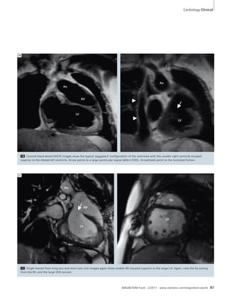


![Cardiology Clinical
1C 1D
Surgical repair is done in the first year
of life for symptomatic AV canal defects.
Large defects may require a Dacron
patch or suture closure.
Reference
1 Kafka H, Mohiaddin RH. Cardiac MRI and pulmo-nary
MR angiography of sinus venosus defect
and partial anomalous pulmonary venous con-nection
in cause of right undiagnosed ventricular
enlargement. AJR Am J Roentgenol. Jan 2009;
192(1):259-66.
MAGNETOM Flash · 2/2011 · www.siemens.com/magnetom-world 93
Discussion
ASD is the second most common con-genital
heart defect after ventricular
septal defect (VSD), and the most com-mon
to become symptomatic in adult-hood.
Ostium secundum defects account
for majority of the ASDs. Females are
affected more often than males.
ASDs can be associated with syndromes,
which include Holt Oram Syndrome
(in which an ASD is accompanied by
upper extremity anomalies) and Trisomy
21. A majority of the children with
Trisomy 21 have an ostium primum and
atrioventricular septal defect.
Common signs and symptoms include
dyspnea, accentuation of right precor-dial
thrust, systolic ejection murmur,
and wide splitting of P2.
Adults may develop pulmonary hyper-tension
from unknown cases of ASD.
Radiology fi ndings
Primary diagnosis is made by echocar-diography
in infants and children.
Chest radiographs can show cardiomeg-aly.
Often, an electrocardiogram can
show right axis deviation.
A cine MRI, has a sensitivity and specific-ity
of over 90% in delineating septal
defects, and can be particularly useful
when other tests are inconclusive [1].
MRI is very useful for depiction of func-tion,
flow, and anatomy in older children.
Treatment
Spontaneous closure occurs in many
children with secundum ASD. A trans-catheter
percuteneous closure device is
the treatment of choice, if ncessecary.
A primum ASD is not amenable to a
closure device and patients are operated
on at 3–5 years of age.
Contact
Harnoor Singh, MD
University of Texas-Medical Branch
Galveston, TX
USA
ha3singh@utmb.edu
The cine images for
this case are available
at our MAGNETOM
World internet page.](https://image.slidesharecdn.com/magnetomflashissue47-00090255-140821160532-phpapp02/85/Pediatric-Imaging-Issue-47-93-320.jpg)
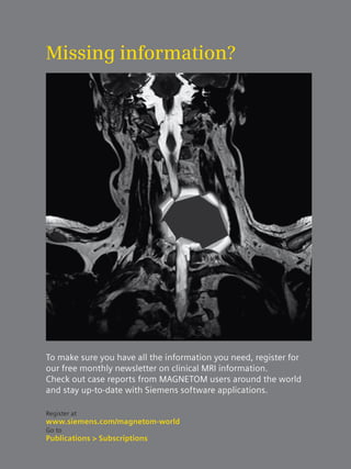


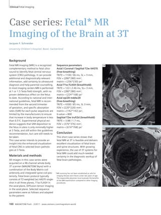


![Clinical Fetal Imaging
Case series:
MR Imaging of the Fetus*
Nguyen Thi Thu Trang, MD1; Ha To Nguyen, MD1; Gregor Kasprian, MD2; Lisa Chuah, PhD3
1Department of Diagnostic Radiology, Maternal and Obstetrics Tu Du Hospital, Ho Chih Min City, Vietnam
2Department of Radiology, Division of Neuroradiology, Medical University of Vienna, Vienna, Austria
3Siemens Healthcare, Regional Headquarters, Singapore
Background
Fetal MRI is increasingly being used as
an adjunct to prenatal ultrasound (US)
in the clinical setting. This case series
features a number of fetal imaging cases
at Tu Du Hospital, a specialized obstetrics
and gynecology hospital in Ho Chi Minh
City, Vietnam. Since the installation of a
MAGNETOM Espree in April 2011 (the first
MR system in the hospital), approximately
20 fetal MR imaging cases were per-formed
to date. As noted in Aaron Flam-mang’s
How-I-do-it article in this issue of
MAGNETOM Flash, ultrasound can resolve
most clinical diagnostic questions. Never-theless,
beyond situations related to
ultrasound-associated limitations (e.g.,
obese mother, fetal position/condition,
smaller, sector-shaped field-of-view), MRI
can, in some cases, provide additional,
complementary findings, improve diag-nosis
and modify the treatment approach
across a number of conditions (e.g.,
cerebral malformations and acquired
pathologies, pulmonary malformations,
renal abnormalities, detection of cleft lip
and palate) [1, 2, 3]. These cases were
selected to highlight situations where MR
imaging had provided additional informa-tion
in fetuses with sonographically diag-nosed
or suspected pathologies.
Case 1
34-week-old fetus with hemor-rhage
in the left cerebellum
and heart defects
The ultrasound on the 28-year-old
mother had suggested the presence
of vermian hypoplasia and MRI was
requested for confirmation.
The size and shape of the vermis looked
normal on MR images (Fig. 1A). MR
imaging additionally revealed a left
cerebellar hemispheric lesion as the
result of hemorrhage (Figs. 1B, 1C, 1D).
1A
108 MAGNETOM Flash · 2/2011 · www.siemens.com/magnetom-world
Materials and methods
All patients reported in the case series
were scanned on a 1.5T MAGNETOM
Espree (software version syngo MR B17)
using a combination of two 6-element
Body Matrix coils and the 24-element
Spine Matrix coil. In all cases, the mother
was scanned in a left lateral decubitus
position, which can be achieved com-fortably
within the 70 cm bore of the
MAGNETOM Espree. For all the cases
reported below, ultrasound was first per-formed,
with MR imaging requested to
provide additional information for diag-nostic
confirmation and/or prediction of
postnatal outcome.
1A The vermis appears to
be normal in shape and size as
seen on sagittal T2w HASTE
images of the fetal brain.
HASTE: 18 slices, SL 4 mm
(no gap), TR/TE 2600/102 ms;
FOV (280*280) mm2; matrix
(230*256) px2, BW 476 Hz/px,
acquisitions 1.](https://image.slidesharecdn.com/magnetomflashissue47-00090255-140821160532-phpapp02/85/Pediatric-Imaging-Issue-47-108-320.jpg)
![Fetal Imaging Clinical
1B–D A lesion present in
the left cerebellum as seen
in the coronal T2w HASTE
image was revealed by EPI
diffusion-weighted imaging
(1C, b700) and ADC maps
(1D) to be the result of a
recent hemorrhage.
HASTE: 18 slices, SL 4 mm
(no gap), TR/TE 2600/102 ms;
FOV (280*280) mm2;
matrix (230*256) px2,
BW 476 Hz/px, acquisitions 1;
EPI diffusion: 20 slices,
SL 4 mm (no gap), TR/TE
6400/87 ms, FOV (280*280),
Matrix (112*160) px2;
BW 1157 Hz/px, PAT factor 2;
b values: 0, 700; flip angle 90°,
acquisitions 1.
1E The presence of an abnor-mally
large heart was noted
on the axial T2w HASTE images.
HASTE: 20 slices, SL 4 mm
(no gap), TR/TE 2600/102 ms,
FOV (260*260) mm2, matrix
(205*256) px2, BW 476 Hz/px,
PAT factor 2, acquisitions 1.
MAGNETOM Flash · 2/2011 · www.siemens.com/magnetom-world 109
1E
MR imaging also indicated congenital
heart disease (oversized heart) which
was not noted in the ultrasound report
(Fig. 1E). However, MR is not the typical
modality of choice for cardiac imaging in
the fetus as the fetal heart is small with
a high beat rate and cardiac triggering is
not possible in the fetal setting [4] (but
see [5]). The mother underwent another
ultrasound examination which allowed
the diagnosis of heart defects. The fetus
was brought to term but died approxi-mately
two week postpartum with death
attributed to cardiac failure.
1B 1C
1D](https://image.slidesharecdn.com/magnetomflashissue47-00090255-140821160532-phpapp02/85/Pediatric-Imaging-Issue-47-109-320.jpg)

![Fetal Imaging Clinical
c
3A–C Axial HASTE (3A), sagittal TrueFISP (3B), and coronal TrueFISP (3C) images show an intact right lung (a) with compression of the heart (b)
and the left lung (c) by the bowels (d) and spleen. HASTE: 25 slices, SL 4 mm (no gap), TR/TE 2800/102 ms, FOV (280*280) mm2, matrix (205*256)
px2, BW 476 Hz/px, PAT factor 2, acquisitions 1. Sagittal TrueFISP: 20 slices, SL 4 mm (no gap), TR/TE 4.1/2.1 ms, FOV (340*340) mm2, matrix
(384*512) px2, BW 488 Hz/px, flip angle 70°, acquisitions 1. Coronal TrueFISP: 20 slices, SL 4 mm (no gap), TR/TE 4.1/2.1 ms, FOV (300*300) mm2,
matrix (384*512) px2, BW 488 Hz/px, flip angle 70°, acquisitions 1.
The presence of an interhemispheric cyst
was noted on ultrasound and MR imag-ing
indicated that it may be an arachnoid
cyst (Figs. 4A, 4B). In addition, MR indi-normal
MAGNETOM Flash · 2/2011 · www.siemens.com/magnetom-world 111
Case 3
34-week-old fetus with
diaphragmatic hernia
Early ultrasounds (22 GW and 24 GW)
of this fetus had not noted any abnor-malities.
However, at 34 weeks, congen-ital
diaphragmatic hernia was diagnosed
with ultrasound and MR was requested
3A 3B 3C
Case 4
32-week-old fetus with a
midline arachnoid cyst and
corpus callosum agenesis
to provide prediction of postnatal out-come.
MR imaging indicated herniation
of the bowel and spleen, with hypoplasia
of the left lung and heart (Figs. 3A–C).
However, as MRI-based lung volumetry
of the right lung indicated a normal vol-ume
of 40 ml (see [6] for a summary of
4A–B Coronal TrueFISP
images which show a large
arachnoid cyst in the left
hemisphere and partial
agenesis of the corpus cal-losum
(4B). Coronal True-
FISP: 25 slices, SL 4 mm
(no gap), TR/TE 4.7/2.4 ms,
FOV (288*288) mm2,
matrix (240*320) px2,
BW 488 Hz/px, flip angle
70°, acquisitions 1.
4A 4B
fetal lung volumes), postnatal
viability was considered to be positive.
The fetus was brought to term and
immediately transferred to the pediatric
unit for treatment.
cated an associated partial agenesis of
the corpus callosum (Fig 4B). The fetus
is presently under follow-up.
a
b
d
d
a
c](https://image.slidesharecdn.com/magnetomflashissue47-00090255-140821160532-phpapp02/85/Pediatric-Imaging-Issue-47-111-320.jpg)
![5A 5B
5A–B Coronal HASTE (5A) and sagittal TrueFISP (5B) images show a large cyst in the spleen,
dorsolateral to the stomach. HASTE: 12 slices, SL 4 mm (0.4 mm gap), TR/TE 2600/102 ms,
FOV (260*260) mm2, matrix (205*256) px2, BW 476 Hz/px, PAT factor 2, acquisitions 1. True-
FISP: 13 slices, SL 4mm (no gap), TR/TE 4.3/2.1 ms, FOV (300*300) mm2, natrix (384*512) px2,
BW 488 Hz/px, flip angle 70°, acquisitions 1.
Contact
Nguyen Thi Thu Trang, MD
Department of Diagnostic Radiology
Maternal and Obstetrics Tu Du Hospital
284 Cong Quyns, District 1
Ho Chi Minh City
Vietnam
nhenconbs@gmail.com
Clinical Fetal Imaging
Conclusion
Fetal MR imaging was recently imple-mented
in Vietnam. While this is a new
service, with only a small number of
patients at this point, this case series
demonstrates that MR can offer comple-mentary
information to ultrasound, to
improve prenatal diagnosis and manage-ment.
With growing experience and expertise
and increased acceptance of this proce-dure
amongst the gynecologists and
pediatricians, it is expected that this ser-vice
will grow and will play an increas-ingly
important role in prenatal diagno-sis
and management. The information
from ultrasound and MRI are typically
complementary. However, in the future,
MR imaging may make its most signifi-cant
contributions to fetal imaging
through the provision of metabolic and
functional information such as spectros-copy,
functional MRI (fMRI), diffusion-weighted
imaging (syngo REVEAL) and
tractography [7, 8, 9, 10] and enabling
the examination of fetal anatomy at
the microstructural level.
Acknowledgements
Invaluable technical support and exper-tise
was provided by Mr Ha-Thuc Nhan,
Application Specialist, Chi Anh Med-Tech
Co Ltd, in sequence optimization, data
collection and selection of images for
this article.
9 Mitter C, Kasprian G, Brugger PC, Prayer D.
(2011). Three-dimensional visualization of fetal
white-matter pathways in utero. Ultrasound
Obstet Gynecol, 37: 252–253.
10 Story L, Damodaram MS, Allsop JM, McGuinness
A et al. (2010). Proton magnetic resonance spec-troscopy
in the fetus. Eur J Obstet Gynecol Reprod
Biol, manuscript in press.
References
1 Jelin AC, Norton ME, Bartha AI, Fick AL et al.
(2008). Intracranial magnetic resonance imaging
finds in the surviving fetus after spontaneous
monchorionic cotwin demise. Am J Obstet
Gynecol; 199: 398.e1–395.e5.
2 Nemec SF, Kasprian G, Brugger PC, Bettelheim D
et al. (2011). Abnormalities of the upper extrem-ities
on fetal MRI. Ultrasound Obstet Gynecol,
Manuscript in press.
3 Mailáth-Pokorny M, Worda C, Krampl-Bettelheim
E, Watzinger F. (2010). What does magnetic
resonance imaging add to the prenatal ultra-sound
diagnosis of facial clefts? Ultrasound
Obstet Gynecol, 36: 445–451.
4 Brugger PC (2011) MRI of the fetal heart. In:
D Prayer (ed) Fetal MRI (pp 247–258). Springer:
Heidelberg.
5 Manganaro L, Savelli S, Di Maurizio M, Perrone
et al. (2009). Assessment of congenital heart dis-ease
(CHD): Is there a role for fetal magnetic res-onance
imaging (MRI)? Eur J Radiol, 27: 172–180.
6 Kasprian G (2011) MRI of the normal fetal lung.
In: D Prayer (ed) Fetal MRI (pp 215–234). Springer:
Heidelberg.
7 Garel C. (2008). Fetal MRI: What is the future.
Ultrasound Obstet Gynecol, 31: 123–128.
8 Kasprian G, Brugger PC, Weber M, Krssak M et al.
(2008). In utero tractography of fetal white
matter development. Neuroimage, 43: 213–224.
Case 5
20-week-old fetus with cyst
in spleen
Ultrasound in this 19-week-old fetus had
suggested the presence of a cyst in the
left diaphragm. However, MR imaging
indicated the presence of a large splenic
cyst, seen on the T2w HASTE and T2w
TrueFISP images as a hyperintense mass
dorsolateral to the stomach (Figs. 5A, B).
The patient is presently under follow-up.
112 MAGNETOM Flash · 2/2011 · www.siemens.com/magnetom-world
* MR scanning has not been established as safe for
imaging fetuses and infants under two years of age.
The responsible physician must evaluate the benefit of
the MRI examination in comparison to other imaging
procedures.](https://image.slidesharecdn.com/magnetomflashissue47-00090255-140821160532-phpapp02/85/Pediatric-Imaging-Issue-47-112-320.jpg)


This document summarizes techniques for pediatric MRI. It notes that children require specialized skills and equipment due to differences from adults in disease types, sensitivity to radiation, and physiology/behavior. Longer scan times are often needed for neonates due to tissue properties. Techniques to improve image quality include using restore pulses and optimized protocols. Safety is a primary concern, especially for heating risks in neonates/infants. Patient cooperation challenges can be addressed through explanation, mock scans, distraction techniques, and anesthesia if needed. Overall the goal is high quality diagnostic images while minimizing distress.


