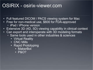
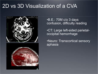
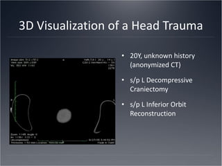
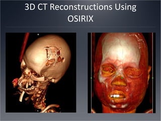
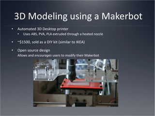
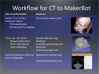
Three Dimensional Radiologic Visualization and Modeling in Clinical Practice Kaylesh Pandya, OMS-IV Nova Southeastern University College of Osteopathic Medicine
Objectives Overview of diagnostic radiology technologies, shortfalls Introduce Osirix DICOM viewer and demonstrate 3D reconstructions 3D Printing and modeling of CT using a MakerBot
Introduction Diagnostic radiology is a staple of most fields of medicine - guides both diagnosis and treatment Taught as a core competency starting in medical school Requires understanding and knowledge of anatomy, imaging modality physics,  'thinking in three dimensions while seeing in only two' Only 1.2% of US physicians are radiologists (AMA) Even fewer patients are radiologists! Communicating radiology findings between medical professionals and with patients can be difficult
DICOM Medical Imaging Format D igital  I maging and  CO mmunications in  M edicine Industry standard file format and network communication protocol for storing, retrieving, and manipulating medical imaging  Allows for centralized server storage of radiology studies PACS -  P icture  A rchiving and  C ommunication  S ystem "Mix and match" DICOM viewers depending on specialty and modality: X-ray CT MRI PET / PET-CT U/S Bone Scan
Imaging studies Radiology modalities and technologies are evolving, yet interpreting these studies has remained relatively static PACS systems 2D viewing on-screen Printed films Outside studies on CD Different viewers depending on the imaging center May not be opened on hospital-owned computers (security settings) Difficult to view radiologic imaging at point-of-care
OSIRIX - osirix-viewer.com Full featured DICOM / PACS viewing system for Mac Free for non-medical use, $600 for FDA-approved iPad / iPhone version Extensive 3D (4D, 5D) viewing capability in clinical context Can export and interoperate with 3D modeling formats Same tools used in other industries & sciences Virtual Reality CNC Mills  Rapid Prototyping MakerBot P&O?
2D vs 3D Visualization of a CVA  B.E.: 70M c/o 3 days confusion, difficulty reading CT: Large left-sided parietal-occipital hemorrhage Neuro: Transcortical sensory aphasia
3D Visualization of a Head Trauma 20Y, unknown history (anonymized CT) s/p L Decompressive Craniectomy s/p L Inferior Orbit Reconstruction
3D CT Reconstructions Using OSIRIX
3D Modeling using a Makerbot Automated 3D Desktop printer Uses ABS, PVA, PLA extruded through a heated nozzle ~$1500, sold as a DIY kit (similar to IKEA) Open source design Allows and  encourages  users to modify their Makerbot
Makerbot 101 (makerbot.com)
Workflow for CT to MakerBot Data Transformation Resource Export CT to ‘Surface Rendered’ Object Pick appropriate density level for imaging Osirix (osirix-viewer.com) “ Clean up” 3D model Reduce detail level Fill in dead spaces Add scaffolding  Blender (blender.org) MeshLab (meshlab.sourceforge.net) SketchUp (sketchup.google.com Generate Machine Code and print to MakerBot  ReplicatorG (replicat.org) MakerBot (makerbot.com
Applications and Implications Patient and family education Surgical and Rehab planning Medical education Prosthetics and Orthotics prototyping FDA – approved implantable parts in the future? HIPAA issues of manufacturing 3D models? Souvenirs?
Acknowledgements Allhandsactive.com @Ech0labs Matthew Oishii Blender.org hackerspaces.org @hackerspaces @i3detroit  i3detroit.com @makerbot wiki.makerbot.com Osirix-viewer.com

3D Visualization of DICOM datasets using a MakerBot

  • 1.
    Three Dimensional RadiologicVisualization and Modeling in Clinical Practice Kaylesh Pandya, OMS-IV Nova Southeastern University College of Osteopathic Medicine
  • 2.
    Objectives Overview ofdiagnostic radiology technologies, shortfalls Introduce Osirix DICOM viewer and demonstrate 3D reconstructions 3D Printing and modeling of CT using a MakerBot
  • 3.
    Introduction Diagnostic radiologyis a staple of most fields of medicine - guides both diagnosis and treatment Taught as a core competency starting in medical school Requires understanding and knowledge of anatomy, imaging modality physics, 'thinking in three dimensions while seeing in only two' Only 1.2% of US physicians are radiologists (AMA) Even fewer patients are radiologists! Communicating radiology findings between medical professionals and with patients can be difficult
  • 4.
    DICOM Medical ImagingFormat D igital  I maging and  CO mmunications in  M edicine Industry standard file format and network communication protocol for storing, retrieving, and manipulating medical imaging  Allows for centralized server storage of radiology studies PACS - P icture A rchiving and C ommunication S ystem "Mix and match" DICOM viewers depending on specialty and modality: X-ray CT MRI PET / PET-CT U/S Bone Scan
  • 5.
    Imaging studies Radiologymodalities and technologies are evolving, yet interpreting these studies has remained relatively static PACS systems 2D viewing on-screen Printed films Outside studies on CD Different viewers depending on the imaging center May not be opened on hospital-owned computers (security settings) Difficult to view radiologic imaging at point-of-care
  • 6.
    OSIRIX - osirix-viewer.comFull featured DICOM / PACS viewing system for Mac Free for non-medical use, $600 for FDA-approved iPad / iPhone version Extensive 3D (4D, 5D) viewing capability in clinical context Can export and interoperate with 3D modeling formats Same tools used in other industries & sciences Virtual Reality CNC Mills  Rapid Prototyping MakerBot P&O?
  • 7.
    2D vs 3DVisualization of a CVA  B.E.: 70M c/o 3 days confusion, difficulty reading CT: Large left-sided parietal-occipital hemorrhage Neuro: Transcortical sensory aphasia
  • 8.
    3D Visualization ofa Head Trauma 20Y, unknown history (anonymized CT) s/p L Decompressive Craniectomy s/p L Inferior Orbit Reconstruction
  • 9.
  • 10.
    3D Modeling usinga Makerbot Automated 3D Desktop printer Uses ABS, PVA, PLA extruded through a heated nozzle ~$1500, sold as a DIY kit (similar to IKEA) Open source design Allows and encourages users to modify their Makerbot
  • 11.
  • 12.
    Workflow for CTto MakerBot Data Transformation Resource Export CT to ‘Surface Rendered’ Object Pick appropriate density level for imaging Osirix (osirix-viewer.com) “ Clean up” 3D model Reduce detail level Fill in dead spaces Add scaffolding Blender (blender.org) MeshLab (meshlab.sourceforge.net) SketchUp (sketchup.google.com Generate Machine Code and print to MakerBot ReplicatorG (replicat.org) MakerBot (makerbot.com
  • 13.
    Applications and ImplicationsPatient and family education Surgical and Rehab planning Medical education Prosthetics and Orthotics prototyping FDA – approved implantable parts in the future? HIPAA issues of manufacturing 3D models? Souvenirs?
  • 14.
    Acknowledgements Allhandsactive.com @Ech0labsMatthew Oishii Blender.org hackerspaces.org @hackerspaces @i3detroit i3detroit.com @makerbot wiki.makerbot.com Osirix-viewer.com