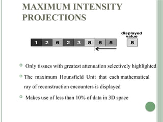
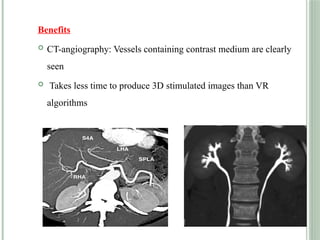
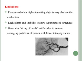
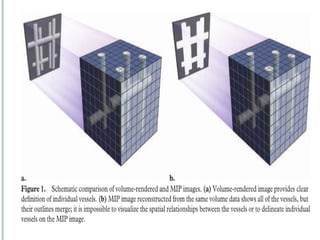
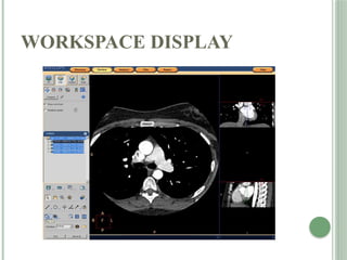
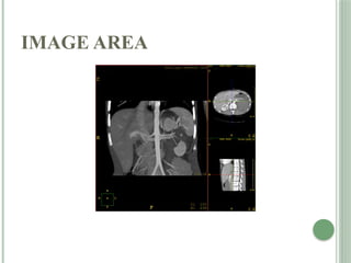
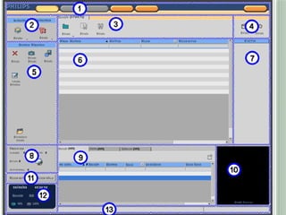
The document discusses various post-processing techniques in computed tomography (CT) including multi-planar reconstruction, 3D imaging methods such as surface and volume rendering, and maximum and minimum intensity projections. It outlines the advantages and disadvantages of these techniques, their applications in different fields such as surgical planning and radiation therapy, and details on 4D CT imaging for analyzing physiological processes over time. Additionally, it describes specialized tools and options available for advanced CT analysis, indicating the complexity and potential benefits of these technologies in medical imaging.
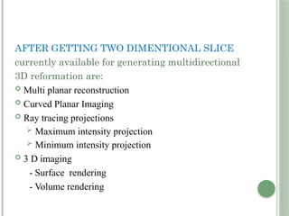


![COMPLETE BARIUM STUDIES Of GIT NAD [Adrian Dungu Niyimpa].pdf](https://cdn.slidesharecdn.com/ss_thumbnails/bastudies-gitnad-220902221043-d535eaa3-thumbnail.jpg?width=640&height=640&fit=bounds)


![Hypothalamus short notes on location, function and disorders by Dr. Neha [PT]...](https://cdn.slidesharecdn.com/ss_thumbnails/hypothalamusbydr-260124142231-2b48143d-thumbnail.jpg?width=640&height=640&fit=bounds)


