Feasibility of CT scan studies with triple split bolus intravenous contrast ...
Somatom sessions 32
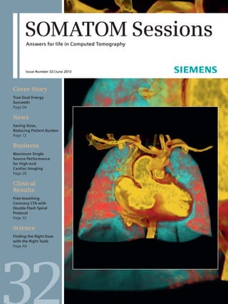
1. SOMATOM Sessions
Answers for life in Computed Tomography
Issue Number 32 / June 2013
Cover Story
True Dual Energy
Succeeds
Page 06
News
Saving Dose,
Reducing Patient Burden
Page 12
Business
Maximum Single
Source Performance
for High-end
Cardiac Imaging
Page 20
Clinical
Results
Free-breathing
Coronary CTA with
Double Flash Spiral
Protocol
Page 32
Science
Finding the Right Dose
with the Right Tools
Page 40
32
2. Editorial
“We see our role as supporting
institutions in achieving the
right dose that delivers high
diagnostic image quality while
exposing the patient to only
as much dose as required.”
Peter Seitz, Vice President Marketing,
Computed Tomography, Siemens Healthcare, Forchheim, Germany
Cover page: Courtesy of Erasmus Medical Center, Rotterdam, the Netherlands
2 SOMATOM Sessions · June 2013 · www.siemens.com/SOMATOM-Sessions
3. Editorial
SOMATOM Sessions · June 2013 · www.siemens.com/SOMATOM-Sessions 3
Dear Reader,
In this issue you’ll read about the inroads
that Dual Energy imaging has made and
continues to make in CT routine today.
At centers such as Grosshadern Hospital
at the University of Munich, more than
50 percent of all abdominal scans are now
performed using Dual Energy. And while
back in the early days in 2005 Dual Energy
was limited to Dual Source scanners,
Single Source applications as found on
the SOMATOM® Definition Edge are
becoming standard. And in radiation
therapy planning, Dual Energy can help
to reduce metal artifacts.
Moreover, its use in combination with
the latest Dual Source technology delivers
highly valuable additional information
even for delicate patients; for example
when imaging infants with congenital
heart or lung disease. Recently, research-ers
from Japan have also shown the
positive impact on oncology treatment
decisions in complicated structures of
the neck.
Some months ago, I introduced our shift
in focus from the lowest dose to the right
dose in CT. In this issue, you’ll find more
examples of institutions that use the
entire current portfolio of dose reduction
techniques to achieve average dose val-ues
that are constantly and significantly
below the reference values of national
authorities. Of course, a permanent reduc-tion
in average dose values is what really
counts – as impressive as a single low
dose case can be.
CARE kV does this by making it very
easy to use the lowest possible kV setting,
especially in small patients with low
attenuation, and in contrast examinations
where lower kV settings provide better
iodine display. SAFIRE does this by mak-ing
powerful noise and therefore dose
reduction available with reconstruction
times of merely a few seconds. When you
combine both with the hardware-based
noise reduction of the Stellar Detector,
you’ll be surprised how far your average
dose values can drop.
So that we can share even more exam-ples,
we’re launching the third round of
our CT image contest in June – focusing
on the right dose in CT. The Right Dose
Image Contest 2013 will once again be
supported by a jury of globally renowned
experts, this time consisting of members
of SIERRA (Siemens Radiation Reduction
Alliance). Across several categories, they
will choose the institutions that best
demonstrate how they achieve images
at the right dose for an ideal balance
between diagnostic quality and low radi-ation.
For the first time, a new category
will be given for consistency in dose
reduction. And you’ll have the opportu-nity
to present your finest cases to the
world on your own profile page.
Enjoy these and many more topics in
this issue and don’t forget to check out
our SOMATOM Sessions App.
Best regards,
Peter Seitz,
Vice President Marketing,
Computed Tomography,
Siemens Healthcare,
Forchheim, Germany
Peter Seitz
In clinical practice, the use of SAFIRE may reduce CT
patient dose depending on the clinical task, patient
size, anatomical location, and clinical practice. A con-sultation
with a radiologist and a physicist should be
made to determine the appropriate dose to obtain
diagnostic image quality for the particular clinical task.
4. Content
Cover Story
Cover Story
06 True Dual Energy Succeeds
News
12 Saving Dose, Reducing Patient Burden
14 FAST Spine – A Story of Best Practice
in Spine Reconstruction
16 Rib and Spine Assessment in Acute
Care with syngo.CT Bone Reading
16 Right Dose Image Contest 2013
17 Expanding the Clinical Portfolio with
the Siemens Intervention Solution
18 Unique Technology for Improved
Routine and New Research Opportu-nities
06 Radiologists and technicians
across the globe are breaking new
ground in CT imaging with Dual
Energy (DE). SOMATOM Sessions
talked to four leading experts
about their clinical experiences
in routine and research areas, the
possibilities for sharper contrast,
significant metal artifact reduc-tion,
and new prospects on the
horizon.
4 SOMATOM Sessions · June 2013 · www.siemens.com/SOMATOM-Sessions
16
Right Dose Image
Contest 2013
xx
xxxx Siemens International
CT Image Contest 2011
Business
20 Maximum Single Source Performance
for High-end Cardiac Imaging
Clinical Results
Cardiovascular
22 Coronary CTA with 80 kV: Improving
Image Quality with Reduced Radiation
and Contrast Medium Dose
24 70 kV CT Pulmonary Angiography
in an Adult Patient with a Dose
of < 1 mSv and PA Attenuation of
> 1,000 HU
26 Dual Source CT: Assessment of
Hypoplastic Arch Associated with
Ductus Arteriosus
28 Cardiac CT in a 5-Month-Old Baby
with VACTERL Syndrome after Cardiac
Surgery
12
Saving Dose,
Reducing Patient Burden
Content
5. SOMATOM Sessions · June 2013 · www.siemens.com/SOMATOM-Sessions 5
Science
40 Finding the Right Dose with the
Right Tools
43 New Opportunities in Cancer
Detection with Hepatic AEF
44 Image Quality in Computed
Tomography
Customer
Excellence
48 syngo Evolve Update for SOMATOM
Definition Family Members
49 Workshop on Dual Energy at CT
Headquarters in Germany
49 CT Physics Made Easy – with New
Webinar
30 Evaluation of Femoral Artery Pseudo-aneurysms
with Arteriovenous Fistula
using CTA Runoff Scanning
32 Free-breathing Coronary CTA with
Double Flash Spiral Protocol
Oncology
34 Squamous Cell Carcinoma of the
Head and Neck: Volume Perfusion CT
36 Diagnosis of Rectal Tumor using
SOMATOM Perspective
Neurology
38 Dose Reduction in Head CT
Examination using SAFIRE
50 Tips & Tricks: How to Accelerate
Reconstruction of Dual Energy Data
51 Clinical Workshops 2013
51 Upcoming Events & Congresses 2013
52 Subscriptions
53 Imprint
32
Free-breathing Coronary CTA
with Double Flash Spiral Protocol
24
70 kV CT Pulmonary
Angiography
40
Finding the Right Dose
with the Right Tools
Content
6. Cover Story
True Dual Energy Succeeds
Radiologists and technicians across the globe are breaking new ground
in CT imaging with Dual Energy. SOMATOM Sessions talked to four
leading experts about their clinical experiences in routine and research
areas, the possibilities for better contrast, significant metal artifact
reduction, and new prospects on the horizon.
By Wiebke Kathmann, PhD
Exciting technical innovations in com-puted
tomography imaging continue.
Dual Energy (DE) scanning in particular
has been expanding rapidly since it
became available for the first time on
a commercial multislice CT scanner.
Back in 2005 DE was introduced to the
market on the Dual Source CT scanner
SOMATOM® Definition.
More and more radiologists rely on
True Dual Energy CT from Siemens due
to remarkable features such as:
1. Improved diagnostic options
2. No extra dose with Dual Source Dual
Energy scans
3. Applicable to almost all clinical
challenges and most patients
Beyond morphology
True DE supplies additional information
compared to a conventional CT scan for
Dual Source DE and dose optimized for
Single Source DE. In conjunction with
high spatial and temporal resolution, DE
applications are used to great effect both
in routine clinical practice and research.
DE is most widely applied to characterize
material, e. g. in kidney stones or gout.
Dual Source DE is also well established
in heart imaging that is prone to motion
artifacts due to breathing and movement
of the beating heart. In the meantime,
True Dual Energy is also available on the
Siemens Single Source CT scanner fleet
ranging from any configuration of the
SOMATOM Definition AS to the SOMATOM
Definition Edge. And progress continues:
other applications are now also making
their way from research into clinical
practice. Four experts describe how they
6 SOMATOM Sessions · June 2013 · www.siemens.com/SOMATOM-Sessions
“We are working on
the Single Source
scan mode because
I am convinced
that Single Source
DE allows a spe-cific
and quantita-tive
assessment
of iodine uptake.”
Thorsten Johnson, MD,
University Hospital Munich,
Campus Großhadern,
Germany
integrate DECT in their daily routine and
outline their research interests.
Munich, Germany: Research
into Single Source DE
At University Hospital Munich, Campus
Großhadern, Germany, there always has
been a strong focus on DECT imaging.
7. Cover Story
SOMATOM Sessions · June 2013 · www.siemens.com/SOMATOM-Sessions 7
Today, about 50 percent of all abdominal
CT examinations are routinely performed
with DE. As one of the clinical innovators
of Dual Source CT applications, Thorsten
Johnson, MD, explains that their experi-ence
has mostly been with Dual Source
DE. He has been involved since the early
days and co-developed many algorithms
along the way. At present, his research
focuses on Single Source DE on the
SOMATOM Definition Edge. If the differ-entiation
of cancerous lesions and blood
filled cysts was possible this application
would have broad clinical relevance and
would be of great interest to a range of
users, for instance oncological centers.
The different behavior of iodine uptake
may help distinguishing between cysts,
which do not enhance, and iodine uptak-ing
lesions. Johnson’s team is working
on the Single Source DE scan mode as
he is convinced that Single Source DE scan
mode on the SOMATOM Definition Edge
might also be very specific for iodine as
on the Dual Source scanners. “Usually, if
you want to quantify the iodine uptake
of a lesion, you perform scans with and
without contrast medium. With the Single
Source DE scan mode on the SOMATOM
Definition Edge you can perform two
scans directly consecutively at half dose
with the benefit of the additional DE.”
Johnson’s team has had promising initial
results in recent cases with excellent
image quality at a low dose level (Fig. 1).
Rotterdam, the Netherlands:
DECT in infants – no sedation
with no dose penalty
Only recently, experts from the cardio-vascular
imaging group at the radiology
department at the Erasmus Medical
Center in Rotterdam, the Netherlands,
started using Dual Source DE in pediatric
scans. Their goal: To enable well-founded
treatment decisions based on anatomical
and functional information without the
need for sedation or anesthesia, or indeed
without increasing radiation dose. As
senior radiologist Mohamed Ouhlous, MD,
PhD, explains, the purely anatomical
information supplied by conventional CT
is not sufficient for children with con-genital
heart and lung disease. “We also
need quantitative information, for exam-ple
on ventilation and perfusion, for the
pediatric cardiologist and pulmonologist.
Therefore, we started to explore other
imaging modalities. We were convinced
that DECT could give us the additional
information required once we discovered
that DECT can create images of perfu-sion
defects in adults with lung emboli.
These are generally hard to see, because
of the many collaterals. My reasoning
was: If you can quantify the blood flow
in the lung, why not use it in children
with congenital heart and lung disease?”
Step by step the team developed a pro-tocol
on the SOMATOM Definition Flash.
First, they replaced the regular CT scans
with Flash scans and noticed that they
could reduce the need for sedation for
1 DECT of a liver
with a hypodense
mass. The case
was acquired with
SOMATOM
Definition Edge.
Courtesy of
University Hospital
Munich, Campus
Großhadern,
Germany
1
“With Dual Source DE,
potential problems
can be discovered ear-lier
and with greater
precision, helping
improve a patient’s
quality of life.”
Mohamed Ouhlous, MD, PhD,
Erasmus Medical Center,
Rotterdam, the Netherlands
8. Cover Story
their young patients. Even on crying
infants, they could perform the scan
between breaths without artifacts. The
result: Pediatricians requested CT scans
more often. After some initial experi-ence
with these young patients using
the Flash protocol, the team moved on
to the issue of lung perfusion, i.e. visual-izing
iodine distribution of the lung.
Since December 2012, the Erasmus team
has scanned twelve children and infants,
the youngest being one-day old, with
Dual Source DECT. The image quality
has surpassed everyone’s expectations.
The clinicians in particular were excited.
“Dual Source DECT scans provide them
with extra information on abnormalities
that the clinician might not see in the
ultrasound examination. Nowadays, they
want the CT before they start with an
angio so they have a certain roadmap,”
says Ouhlous. Compared with angiogra-phy,
DECT not only has advantages in
iodine and radiation dose, it is also non-invasive
using intravenous rather than
intra-arterial contrast application. And it
may potentially help reduce the risks with
sedation or anesthesia that some other
techniques entail. Ouhlous concludes
that good information can be gained by
Dual Source DE techniques. Therefore,
3A 3B
8 SOMATOM Sessions · June 2013 · www.siemens.com/SOMATOM-Sessions
2 Scan of a
7-month-old child
with congenital
heart defect using
1.4 mSv effective
dose. The patient
was scanned
with SOMATOM
Definition Flash
(Dual Source DE)
and evaluated
with syngo.CT
DE Lung Analysis
(syngo.via VA20).
Courtesy of
Erasmus Medical
Center, Rotterdam,
the Netherlands
Dual Source DE is used regularly for this
specific group of patients and is now
an accepted imaging tool for congenital
heart and lung diseases that might
2
3 Negative cartilage invasion of the thyroid cartilage imaged with DECT in a 65-year-old man with hypopharyngeal cancer
(weighted average (WA) image, Fig. 3A; iodine overlay (IO) image, Fig. 3B).
Courtesy of National Cancer Center Hospital East, Chiba, Japan
9. Cover Story
SOMATOM Sessions · June 2013 · www.siemens.com/SOMATOM-Sessions 9
affect the children later in life. Potential
problems can be discovered earlier and
with greater precision.
Chiba, Japan: Dual Source DE
may avoid overtreatment
Another pioneer of DECT in oncological
radiology, Hirofumi Kuno, MD, is staff
radiologist at the National Cancer Center
Hospital East in Chiba, Japan. As a spe-cialist
in head and neck oncological radi-ology
– especially laryngeal and hypo-pharyngeal
squamous cell carcinoma –
he sees many patients with these some-what
rare cancers. Hoping to avoid over-treatment
of his patients, he was looking
for a CT application that could reliably
discriminate between laryngeal cartilage
and iodine-enhanced tumor tissue. In con-ventional
CT images, both have roughly
the same CT values making them hard
to distinguish. Clinically, however, it is
essential to clarify whether there is thy-roid
cartilage invasion when deciding on
treatment options.
This is where DECT comes into play. Kuno
saw the potential of DE in distinguishing
iodine-enhanced tumor and cartilage
in CT imaging using syngo.CT DE. “I’m
not interested in the technology per se,
but in the benefits for the patient,” Kuno
states. “The benefit of DE is clearly the
positive impact of the high quality images
on the treatment decision. It allows
precise diagnosis of the cancer in spite
of the complicated structures in the neck
and the diversity of appearance, which
often leads to false positive results. Here,
it can make the difference between
organ-conserving therapies (chemo radi-ation)
and more aggressive treatments
(laryngectomy), which potentially have
a major impact on a patient’s quality of
life due to a possible post-surgery loss
of voice.“
As soon as the SOMATOM Definition Flash
was installed at the hospital in March
2010, Kuno began his work. In close col-laboration
with Siemens, he developed
a scan protocol and investigated whether
it led to improved diagnostic performance.
Little difference was noted in reconstruc-tion
time and image evaluation com-pared
with conventional CT scans. The
program prepares the weighted average
(WA) and iodine overlay images (IO).
The WA image allows the evaluation of
the cartilage (invasion, erosion, lysis or
lysis plus extralaryngeal invasion). The
second contrast – i. e. the enhancement
pattern on IO images – enables the dis-tinction
of uptake due to the blood ves-sels
of the cancer tissue as opposed to
blood vessel free cartilage.
“By 2012, we had scanned around 300
patients with laryngeal or pharyngeal
cancer. T4 stage is invasion throughout
the cartilage which, according to guide-lines,
calls for laryngectomy. We are con-vinced
that in this patient population
the tumor could be downstaged to T3
using CT scans with higher resolution.
That should result in a decision to pursue
function-preserving treatment”, says
Kuno. He found that using Dual Source
DECT improved specificity and sensitivity
in detecting the extent of cartilage inva-sion.
The results of his study were pub-lished
in the journal Radiology in October
2012.[1] Kuno’s conclusion: “Combined
analysis of WA and IO images obtained
with DECT improves the diagnostic per-formance
and interobserver reproduc-ibility
of evaluations of laryngeal cartilage
invasion by small cell carcinoma. This is
of the utmost importance for the treat-ment
strategy, especially when attempt-ing
a function-preserving therapy.”
Meanwhile, Kuno examines most of his
head and neck cancer patients using Dual
Source DE. The technology has made its
way from research to clinical routine in
just two years and is now an established
protocol. “This was possible as DE scans
always include the normal 120 kV image
so that nothing is lost – no extra dose is
applied. The only difference is the need
for more disk space to archive the images.
For the technician, DE scans do not
affect the workflow,” explains Kuno. “Also,
the time required for the scan and the
iodine dose is the same for the patient.”
He truly believes that T4 staging of
laryngeal and pharyngeal cancers may
become much easier for non-specialized
institutions. “From our perspective, any
institution with a SOMATOM Definition
Flash can start using Dual Source DE
protocol for head and neck tumors from
one day to the next.”
Hamburg, Germany: Excep-tional
image quality with DE –
a must for radiation planning
At ‘Radiologische Allianz’ – an associa-tion
of practices focusing on radiology,
nuclear medicine and radiation therapy
“From our perspective,
any institution with a
SOMATOM Definition
Flash can start using
Dual Source DE pro-tocol
for head and
neck tumors from
one day to the next.”
Hirofumi Kuno, MD,
National Cancer Center Hospital East,
Chiba, Japan
10. Cover Story
with nine locations in Hamburg – experts
are now using DECT scanning. Their
interest is in metal artifact reduction,
a major issue in radiation therapy. DE
helps in planning radiation therapy for
patients with head and neck cancers,
cancers of the pelvis, or prostate cancer.
In these patients metal artifacts are a
challenge as preceeding treatments using
metal such as seed implantation of 25
to 80 small metal radiation emitting pins,
in patients with prostate cancer, endo-prosthesis
of the hip or implants in the
mouth cavity affect CT images. “All these
metal implants create white stripes and
make it hard to draw the precise outline,
for example of the lymph drainage path-ways
in the mouth,” explains Matthias
Kretschmer, medical physicist. “The radi-ation
therapist can no longer define the
target volume, and the medical physicist
can no longer predict the precise radia-tion
dose needed. Single Source DE
produces more accurate images for the
radiation oncologist and helps the physi-cist
to calculate his dose estimate using
more reliable data. Just as with real
estate, what counts in CT images is loca-tion,
location, location. We can only hit
the tumor precisely if the location of the
patient under the linear accelerator is
exactly the same as in the previous plan-ning
CT,” stresses Kretschmer.
When the Hamburg team started out,
they were still using conventional CT
10 SOMATOM Sessions · June 2013 · www.siemens.com/SOMATOM-Sessions
scans; they compared the results with
those from a Single Source DE scan with
a SOMATOM Definition AS 20 Open. This
was necessary as the Hounsfield Units
(HU) change as a result of the mono-energetic
application. New correlation
“If a topogram
depicts metal
implants, we
replace the
conventional CT
with a Single
Source DE scan.”
Matthias Kretschmer, MSc,
Radiologische Allianz,
Hamburg, Germany
The Single Source DE scan mode consists of two successive automated spiral
scans at different tube voltage (kV) and tube current (mA) levels. Each scan
is performed at approximately half the dose which confidently comply with
the ALARA principle.
DEfinitely excellent images:
Crisp image quality
Information beyond morphology –
highlight, characterize, quantify,
and differentiate material
DEfinitely the right dose:
No dose penalty with full number
of projections
All dose saving features applicable
such as SAFIRE and CARE Dose4D
Dedicated protocols and evaluation
software applications for various
clinical questions
Low radiation and contrast media
dose – applicable for virtually
all patients from pediatric to older
patients
Single Source DE: The Scan Principle
1st scan
2nd scan
140 kV
80 kV
True Dual Energy
11. Cover Story
4 Metal artifact reduction with Single Source DE Monoenergetic: Conventional CT (Fig. 4A); Monoenergetic image at 120 keV (Fig. 4B)
The patient was scanned with SOMATOM Definition AS20 (Single Source DE) and evaluated with syngo.CT Dual Energy (integral part of
syngo.via VA20 advanced user). Courtesy of Radiologische Allianz, Hamburg, Germany
Reference
[1] Kuno H et al. Evaluation of cartilage invasion
by laryngeal and hypopharyngeal squamous
cell carcinoma with dual-energy CT. Radiology.
2012 Nov;265(2):488-96.
SOMATOM Sessions · June 2013 · www.siemens.com/SOMATOM-Sessions 11
tables for each monoenergetic mode used
in artifact reduction had to be calculated
on the phantom and stored in the plan-ning
software. In Hamburg, the team has
the benefit of having Julia Sudmann, PhD,
a medical doctor and radiation therapist
in training on the CT. She can immedi-ately
assess the location from the topo-gram
and predict whether hampering
metal artifacts are to be expected. In this
case, conventional CT scans are no lon-ger
performed. Instead, the application is
immediately switched to a Single Source
DE scan. After only a few runs, treatment
planning improved in 60 percent of cases
where Single Source DE application was
used, Sudmann recalls.
A decision on whether to use Single
Source DE is made according to the
individual case with the location of the
tumor in relation to the implant being
the strongest determinant. Based on the
scans performed so far, Sudmann finds
Single Source DE has clear advantages
for tumors in the mouth base. “For these
patients we will be using Single Source
DE as standard from now on.” She sees
a sensible application in patients with
prostate cancer and with permanent
seed implants who have a biochemical
relapse – that means an increase in the
PSA value – and who need repeated
external radiation. “Overall, we will most
likely use it in about five percent of our
patients with head and neck or pelvic
cancers who have endoprostheses or
implants.”
To be successful in clinical practice, DE
needs to deliver excellent image quality,
no dose penalty, and broad applicability
to virtually all patients. The experiences
of these four CT experts described in the
interviews show that True Dual Energy
does just this. It is not only well estab-lished
in the field of research but even
more important in daily clinical routine.
Further Information
www.siemens.com/dual-energy
4A 4B
Medical writer Wiebke Kathmann, PhD, is
a frequent contributor to medical magazines
for physicians of German-speaking media. She
holds an MSc in biology and a PhD in theoretical
medicine and has worked as an editor for
many years before becoming freelance in 1999.
She is based in Munich and Karlsruhe, Germany.
The statements by Siemens customers described
herein are based on results that were achieved in the
customer’s unique setting. Since there is no “typical”
hospital and many variables exist (e.g., hospital size,
case mix, level of IT adoption) there can be no guaran-tee
that other customers will achieve the same results.
12. News
Saving Dose, Reducing Patient Burden
A plucky physician from St. Louis and technological advances by Siemens are
working together to cut dose levels in pediatric patients to unprecedented levels.
By Ron French
It’s difficult for Marilyn Siegel, MD, to
keep a smile off her face these days. For
years, the pediatric radiologist at Washing-ton
University School of Medicine and
St. Louis Children’s Hospital has been
leading a campaign of words and research
to lower dose exposure in children. Her
story is one of success, and it is one
shared by the complete line of Siemens
computed tomography equipment.
Spreading the low-dose gospel
In the United States alone, more than
70 million CT scans are performed each
year – double the number of a decade
ago. But even with today’s technology,
the radiation dose of those scans has a
deleterious cumulative effect on patients
– particularly the pediatric patients Siegel
works with each day in St. Louis, Missouri,
USA: “Effective dose in children is three
to five times higher than in adults at com-parable
exposure levels,” she said. The
low dose advocate travels around the
globe speaking to physicians about the
importance and methodology of dose
reduction: “Even for one-time exams, you
want the dose low. But it’s particularly
important for patients who come back
for multiple examinations; they’re going
to start accumulating dose. Lung trans-plant
patients are an example.”
The goal is to reduce dose, while main-taining
or improving image quality. Today,
technology is catching up with Siegel’s
vision.
The next step in ‘exquisite
images’
The Siemens SOMATOM® Definition AS,
64-slice configuration, has been the
hospital’s workhorse for four years. It is a
Single Source scanner, featuring leading
technologies, like real-time dose modula-tion
At Washington University School of Medicine and St. Louis Children’s Hospital Marilyn Siegel, MD,
has been leading a campaign of words and research to lower dose exposure in children.
CARE Dose4D or the Adaptive Dose
Shield to avoid spiral over-radiation, both
crucial for pediatric scanning. Recent
upgrades to the machine have taken dose
reduction to new lows. In 2011, Siemens
upgraded the SOMATOM Definition AS,
64-slice configuration to include CARE kV,
which automatically adjusts voltage to
match body size and scan type. CARE kV
supplements CARE Dose4D to a complete
automated exposure control for an opti-mal
balance between diagnostic image
quality and lowest possible dose.
Siegel was the first in the United States
to use CARE kV on children. “The results
were amazing,” she said. “The mean dose
reduction was 30%. In smaller patients,
it could be up to 50%.”
“If you looked at all our patients – from
2 kg to 120 kg – we were getting 6 mGy;
under 50 kg, we were down to about
5 mGy,” Siegel said. “I was remarkably
12 SOMATOM Sessions · June 2013 · www.siemens.com/SOMATOM-Sessions
impressed. The contrast was maintained,
and the dose went down 30%. We were
under 1 mSv, with exquisite images. I was
amazed the first time I saw it.” Accord-ing
to the pediatric radiologist, CARE kV
was a step forward: “The biggest impact
has been on contrast-enhanced and
angiographic imaging. But across the
board, in any procedure, it has had an
impact,” she pointed out.
Siegel recalls the case of a 3-year-old girl
with heart disease who had undergone
multiple operations: “We wanted to see
anatomy,” she explained. “We did a CT
with no sedation at 70 kV, with a dose
of less than 1 mSv and got outstanding
images.”
Quicker iterative reconstruc-tion
(IR) with reduced noise
The success story continued in 2012 with
the installation of Siemens Sinogram
13. News
lung and heart together, and assessment
of tumor response by tracking iodine.
The bottom line is: It’s going to allow
functional imaging that we haven’t done
before with CT.”
Siegel and Siemens aren’t finished yet.
She proudly displays a chart showing
the incredible dose savings that are pos-sible
when the SOMATOM Definition AS
64-slice configuration is combined with
CARE kV and SAFIRE. Above the chart
are the words: “We are getting closer.”
“It’s exciting,” Siegel said, smiling. “You
can affect lives.”
Ron French is a freelance business and medical
writer based in Detroit, Michigan, USA. He also
writes for the Detroit News.
SOMATOM Sessions · June 2013 · www.siemens.com/SOMATOM-Sessions 13
Affirmed Iterative Reconstruction (SAFIRE).
SAFIRE removes artifacts and noise from
scanned images. Because radiologists
are trained to read images with some
noise, the technology means that milli-amperage
can be lowered to the point
that an “acceptable” level of noise is in
the image, reducing dose in children by
as much as 60%.
SAFIRE also provides a vastly improved
IR performance thanks to enhanced
image reconstruction computing power
and smartly engineered signal process-ing.
In other models, IR can take up to
45 minutes to reconstruct a patient’s
data set; with SAFIRE, reconstruction
takes only seconds to a few minutes. In
pediatric CT, Siegel was the first to use
CARE kV in combination with SAFIRE.
The results stunned the physician: the
overall mean radiation dose of scans
fell from 8.3 mGy to 4.5 mGy – roughly
equivalent to annual background radia-tion.
Milligray values in CT Angiography
scans dropped from 6.2 to 2.8; Chest
abdomen pelvis scans plummeted from
10.5 to 4.8. “The real issue out there is
dose, but you also have to have great
image quality,” Siegel pointed out. “The
goal is to get to less than 1 mSv with
pediatrics at good diagnostic image qual-ity.
This technology is helping us get
there.”
The Gold Standard
While Siegel has already shown herself
able to perform excellent image quality
at a very low dose with the 64-slice con-figuration
of the SOMATOM Definition AS,
she wanted to go for Siemens high-end
scanner, the SOMATOM Definition Flash.
The Flash is the gold standard of com-puted
tomography, with all of the fea-tures
of the AS 64-slice configuration but
with two tubes and detectors and thus
much faster acquisition speed. “Tradi-tionally,
most of our CT imaging has had
a pitch of 1.2 to 1.5,” Siegel said. “We
couldn’t go past 1.5 because soon you
weren’t radiating enough of the patient
to get an image. With the Flash, we can
scan much faster. When we use it for
congenital heart disease, we use a pitch
of 3.4. We can scan in less than a second
and reduce the radiation dose again. We
can use pitches of 3.0 or 2.8 for all our
exams, with an incredible effect on dose.
The major advantage for everyone is
reduction in sedation and reduction in
breathing artifacts,” Siegel said. “If you
have healthy kids coming in for their first
chest and abdomen exam, you don’t need
to give sedation if they can stay still for
a second or two. It has improved the
quality of the exam and reduces burden
on patients.”
Using the high-pitch scan modes of the
Flash and with its built-in CARE kV, along
with the 20% reduction in milliamperage
reconstructed with SAFIRE, Siegel was
able to realize even greater dose savings:
“The overall mean of all scans was reduced
to 2.7 mGy,” she said.
The SOMATOM Definition Flash also facil-itates
the new Stellar Detector, which
limit electronic noise. The Stellar Detector
delivers a spatial resolution down to
0.30 millimeters without increasing dose.
This provides improved images of vessels,
for example.
Getting closer
In the fall of 2013, Siegel will head for
Germany to work with Siemens engineers
on the next step in pediatric imaging:
making Dual Energy scans dose-neutral.
“If I can show that the dose stays low,
then it becomes an exciting tool,” Siegel
said. “Pretty pictures alone don’t do it. It
will help in areas that we so far haven’t
evaluated, like vessel perfusion in the
In clinical practice, the use of SAFIRE may reduce CT
patient dose depending on the clinical task, patient size,
anatomical location, and clinical practice. A consulta-tion
with a radiologist and a physicist should be made
to determine the appropriate dose to obtain diagnostic
image quality for the particular clinical task. The follow-ing
test method was used to determine a 54 to 60% dose
reduction when using the SAFIRE reconstruction soft-ware.
Noise, CT numbers, homogeneity, low contrast
resolution and high contrast resolution were assessed in
a Gammex 438 phantom. Low dose data reconstructed
with SAFIRE showed the same image quality compared
to full dose data based on this test.
Data on file.
The statements by Siemens customers described herein
are based on results that were achieved in the customer’s
unique setting. Since there is no “typical” hospital and
many variables exist (e.g., hospital size, case mix, level
of IT adoption) there can be no guarantee that other
customers will achieve the same results.
“ The goal is to get
to less than 1 mSv
with pediatrics
at good diagnostic
image quality. This
technology is help-ing
us get there.”
Marilyn Siegel, MD, pediatric radiologist at
Washington University School of Medicine and
St. Louis Children’s Hospital, Missouri, USA.
14. News
FAST Spine – A Story of Best Practice
in Spine Reconstruction
SOMATOM Definition AS boosted by FAST Spine provides a remarkably accel-erated
workflow in spine reconstruction. In the department of radiology at
the Centre Hospitalier Universitaire de Tivoli (CHU Tivoli), an affiliation of the
Université Libre de Bruxelles, Belgium, the specialists are impressed by the
ease of use, the speed and the quality of the automated spine reconstruction.
By Ruth Wissler, MD
The radiology department at CHU Tivoli
performs about 92,000 CT examinations
per year. The radiological staff consists of
15 radiologists and about 22 technicians.
Almost a quarter of the examinations are
orthopedic and spinal CTs.
The hospital is focused on neurosurgical
interventions. About 30% of the patients
are referred for spinal examination by gen-eral
practitioners or surgeons from other
clinics. Since their SOMATOM® Definition
AS+ was equipped with FAST Spine from
the end of March 2012, it has been used
there in almost all clinical cases of back
pain, sciatica and herniated discs.
“Since we installed FAST Spine on our
SOMATOM Definition AS+ system, all of
my clinical staff have been very enthu-siastic
about the user-friendly software.
The technicians are more independent,
and we, the doctors, can concentrate on
the interpretation of the clinical images,”
mentioned Pietro Scillia, MD, head of the
Department of Radiology at the Centre
Hospitalier Universitaire de Tivoli in Bel-gium.
Benefits of FAST Spine support
clinical imaging routines
Considerable time-saving is one promi-nent
clinical feature. FAST Spine allows
faster setup and preparation of spine
reconstructions, including automatic
labeling. Immediately after the data acqui-
1 FAST Spine delivers an automatic segmentation of the spinal canal and automatic
labeling of the vertebrae.
Courtesy of University Hospital of Zurich, Switzerland
1
14 SOMATOM Sessions · June 2013 · www.siemens.com/SOMATOM-Sessions
15. News
Ruth Wissler, MD, studied veterinary and
human medicine. She is an expert in science
communications and medical writing.
The statements by Siemens customers described
herein are based on results that were achieved in the
customer’s unique setting. Since there is no “typical”
hospital and many variables exist (e.g., hospital size,
case mix, level of IT adoption) there can be no guaran-tee
that other customers will achieve the same results.
SOMATOM Sessions · June 2013 · www.siemens.com/SOMATOM-Sessions 15
sition, FAST Spine automatically starts
detecting the spinal vertebrae, and labels
them according to their anatomical posi-tion.
FAST Spine then uses this informa-tion
for typical reconstruction modes for
the spinal vertebrae or discs. Time-critical
spine examinations also benefit from the
high reproducibility of the reconstruc-tions.
“With FAST Spine we were able to
increase the number of exams by about
20% per day,“ says Pietro Scillia. “It is
very convenient to use and we employ it
in almost all orthopedic cases. Even with
difficult spine patterns, the automated
detection works.”
FAST Spine helps to reduce
reimbursement challenges
The department of radiology plays an
important economic role for CHU Tivoli,
“ With FAST Spine
we were able
to increase the
number of exams
by about 20%
per day.”
Pietro Scillia, MD,
Head of the Department of Radiology
at the Centre Hospitalier Universitaire
de Tivoli, Belgium
with just 6% of the hospital’s doctors
contributing almost 15% of the overall
profits. In this situation, the department
is particularly dependent on an effective
CT system, as the relatively low reim-bursement
also has to pay for the device
purchase. “That is an enormous challenge
for us,” says Scillia. “We are basically
dependent on a well working system with
an effective workflow, because we want
to perform very good exams and not just
a lot of exams.”
The specialists’ experiences at CHU Tivoli
with SOMATOM Definition AS+ boosted
by FAST Spine tell a story of best practice
in radiology by accelerating workflow and
increasing number of exams per day.
Due to its significant clinical benefits,
Siemens has also extended the avail-ability
of FAST Spine to the SOMATOM
Perspective Family and will introduce
it for the SOMATOM Emotion* Family in
the last quarter of 2013.
* Under development. Not available for sale in the U.S.
16. News
Rib and Spine Assessment in Acute Care
with syngo.CT Bone Reading
By Philip Stenner, PhD, Computed Tomography, Siemens Healthcare, Forchheim, Germany
1 syngo.CT Bone Reading displays the
entire rib cage rolled on a 2D planar
reformat. Courtesy of University Hospital
Salzburg, Austria
Right Dose Image Contest 2013
By Ivo Driesser, Computed Tomography, Siemens Healthcare, Forchheim, Germany
Following the success of the image con-tests
held over the past few years, Siemens
Healthcare has decided once more to
invite radiologists and radiographers from
across the world to take part in the latest
round of this international competition.
Again a jury of experts, this time consist-ing
of members of SIERRA (the Siemens
Radiation Reduction Alliance), will choose
in eight different categories the institu-tions
who best demonstrate how they
achieve images with the right dose for an
ideal balance between diagnostic quality
and low radiation.
From June 2013, any clinical institution
or hospital with a CT scanner from the
SOMATOM® Family can once again sub-mit
their best images to be shown on
the contest website.
16 SOMATOM Sessions · June 2013 · www.siemens.com/SOMATOM-Sessions
1 Coarctation of aorta. Winning image
2011, category “Vascular”, by Liz D’Arcy,
Wexford General Hospital, Ireland.
Trauma cases with suspected multiple
injuries to the thorax and spine call for a
complete and reliable evaluation of the
ribs and vertebral bodies. Diagnosis of
possible fractures needs to be available
very quickly. Simply scrolling through axial
slices while trying to focus on the point
of interest can be very time-consuming
due to the oblique orientation of the ribs.
syngo.CT Bone Reading revolutionizes
rib and spine assessment: The application
identifies and labels the ribs, and displays
curved 2D images of the entire rib cage
on a multi-planar reformat. In addition,
the vertebral bodies are labeled and the
spine is presented in an unfolded view
for a straightforward overview of the
anatomy. Thanks to the “Automatic
Pre-Processing”, the case is ready to be
reviewed immediately on opening.
The planar display of the rib cage facili-tates
the direct detection of lesions,
e. g. fractures of vertebral bodies or ribs.
When the user clicks on a fracture, the
system centers the axial, sagittal, and
coronal views on the area of interest to
allow a detailed assessment.
The system also provides cross sections
of the spine orthogonal to the unfolded
view and updates the position along the
spine while scrolling in real time.
In conclusion, syngo.CT Bone Reading
can effectively increase speed in bone
assessment.
A new element this year is the fact that
sustainable dose management at the
participating institution will also play a
role in the evaluation of the images.
Indeed, there will even be an additional
category for the entrant with the best
dose reduction strategy.
“The many hundreds of submissions
we’ve had in the past few years clearly
demonstrate that our customers enjoy
presenting their work to a global audi-ence
and having it discussed by a spe-cialist
community,” explains Peter Seitz,
Vice President of CT Marketing.
www.siemens.com/ct-acute-care
www.facebook.com/imagecontest
www.siemens.com/imagecontest
1
1
17. News
Expanding the Clinical Portfolio
with the Siemens Intervention Solution
By Jürgen Merz, PhD, Computed Tomography, Siemens Healthcare, Forchheim, Germany
1 Radio Frequency Ablation Therapy in a patient with lung cancer with
SOMATOM Definition AS+. Courtesy of Department of Radiology, University
of Munich, Grosshadern, Munich, Germany
SOMATOM Sessions · June 2013 · www.siemens.com/SOMATOM-Sessions 17
The number of therapeutic interventions
using CT has increased considerably over
the last few years. More complex proce-dures
can be performed faster, with bet-ter
outcomes, fewer complications, at a
lower cost and with less discomfort for
the patient.
Increasing markets for
minimally invasive therapy
As the number of indications for mini-mally
invasive therapy increases, more
and more CT scanners are used for this
purpose; sometimes even exclusively.
Today, for example, interventions are
performed on one third of SOMATOM®
Definition AS scanners.* The clinical
spectrum ranges from CT-guided biop-sies,
through pain treatment (particu-larly
in the spinal region) and drainage
of inflammatory processes, to ablation
of tumors in the lungs, abdomen and
pelvic area.
Standard intervention features
on the SOMATOM Definition
Family
Siemens recognized this trend at an early
stage, invested significantly in this area
and today offers an intervention solution
for its CT systems that is highly valued
by clinicians. Among the SOMATOM
Definition Family (AS, Edge, Flash) basic
2D interventional features are already
part of the standard configuration as
well as HandCare, a radiation reduction
feature for the operator.
Advanced intervention solu-tion
for dedicated individual
and clinical needs
As interventional procedures become
more and more complex, doctors develop
more sophisticated and highly individu-alized
workflows. Consequently, Siemens
advanced solutions allow the adaption
and optimization of the workflow to the
individual need and the clinical setting.
“Intervention Pro” allows the operator
to switch between spiral, sequential and
fluoroscopy protocols on the fly, while
the in-built “Layout Editor” enables the
screen layout to be specifically adapted
to clinical questions or personal prefer-ences
(e. g. 3D layout for spinal inter-ventions
or the additional display of MR
images). The “Adaptive 3D Intervention”
package provides the option of planning
and conducting the intervention com-pletely
in 3D. Immediately after the scan,
the operator is provided with coronal,
axial and sagittal views in his specific
layout. Needle path planning in both 2D
and 3D and a needle detection algorithm
provide high-quality results. “i-Needle
sharp” solves the challenge of metal
artifacts from the needle. “i-Fluoro” (CT
fluoroscopy) allows the person perform-ing
the intervention to track the interven-tion
instrument in real time during the
procedure. An optional foot switch and
an additional control unit (i-Control; wire-less,
if desired) enable the surgeon to
work directly on the patient completely
independently. The package is rounded
off by a variety of measurement and
analysis tools. These options can also be
purchased together as a package – the
“Adaptive 3D Interventional Suite” – pro-viding
the operator with a fully equipped
interventional CT system.
1
* Data on file
18. 18 SOMATOM Sessions · June 2013 · www.siemens.com/SOMATOM-Sessions
1 Fig. 1 shows images
from a DE Angiography
examination of the thorax
that was included in a
study:[1] Original poly-chromatic
images at 80 kV,
140 kV, mixed image
at 120 kV (upper image
series), and 3 of 6 mono-chromatic
reconstructions
at levels of 60 keV, 70 keV
and 100 keV (lower image
series). The central vessels
could best be assessed at
lower keV levels (60 keV);
the reconstruction at
100 keV provided best con-ditions
for the systemic
veins. Courtesy of Hospital
Calmette, Lille, France
News
Unique Technology for Improved
Routine and New Research Opportunities
Two exclusive Siemens technologies, Dual Source Dual Energy CT
and the Stellar Detector, take routine applications to a new level and
open up opportunities for innovative research.
By Heidrun Endt, MD, Computed Tomography, Siemens Healthcare, Forchheim, Germany
Dual Source Dual Energy CT
In 2012, the American Journal of
Roentgenology (AJR) published a special
supplement on Dual Energy CT (DECT).
Several review articles outlined the current
status of scientific research and different
approaches to DECT. An important state-ment
in the supplement declared that:
“Of the various methods that have been
proposed for acquiring DECT data, image
acquisition based on DSCT [Dual Source
CT] is the most intensely evaluated
approach in the current
literature.”[1]
Has this also been transferred to use into
daily routine?
Researchers from Université Lille Nord
de France state that this technique can
be used for chest CT Angiography exa-minations
for routine diagnostic evalua-tion.[
2] Examinations were carried out
on 80 patients using Dual Source Dual
Energy on a SOMATOM® Definition
Flash with a reduced amount of iodine
(170 mg/mL). In addition to images at
80 kV and 140 kV, further monoenergetic
images (50/60/70/80/90/100 keV) were
reconstructed using syngo Dual Energy.
Monoenergetic images at 60 keV were
the best choice for the assessment of cen-tral
vessels, images at 100 keV for the
systemic veins. These images at 100 keV
also presented with reduced perivenous
artifacts, known from conventional CT
examinations. Researchers compared all
these with single energy CT images,
acquired with a standard dose of contrast
medium. According to the study DECT
examinations offered adequate image
quality for the systemic veins with the
advantage of considerable reduction in
the amount of iodine contrast used.[2]
In addition the evaluation of the central
vessels was not degraded, which is the
limitation of single energy CT with reduced
contrast media administration.[2]
80 kV
60 keV 70 keV
120 kV
100 keV
140 kV
1
19. News
images, they also made full use of the
potential for contrast media reduction.[2]
Yet, many clinical questions are still
waiting to be answered in more detail
with DECT – as shown by the study from
Japan.[4]
This is also the case for the Stellar Detec-tor.
There are proven benefits of using
the Stellar Detector in coronary CT Angi-ography
examinations that are routinely
performed all over the world.[5, 6]
However, further research is needed on
the impact of the Stellar Detector, for
example in stent imaging, an application
that shows promising initial results in
scientific studies.
While these exclusive technologies –
Dual Source DECT and the Stellar Detec-tor
– open up new research opportuni-ties,
they continue to benefit everyday
2 A 63-year old male
patient underwent coronary
CT Angiography examination.
This examination was included
in a study.[3] Fig. 2A was
conventionally reconstructed
with 0.6 mm slice thickness.
For Fig. 2B, 0.5 mm slice
thickness was used in combi-nation
with SAFIRE strength 3.
The latter enabled a more
precise evaluation of the
stenosis and therefore a
more precise quantification.
Courtesy of University
Hospital Zurich, Switzerland
scans were performed twice: once with
the Stellar Detector and once with a
conventional detector. Subsequently,
these findings were confirmed clinically
in the second part of the study. Coro-nary
CT Angiography was carried out on
30 patients using a SOMATOM Definition
Flash equipped with the Stellar Detector.
Conventional detector technology can
reconstruct images with a slice thickness
of 0.6 mm, whereas the Stellar Detector
in combination with SAFIRE enables a
slice thickness of 0.5 mm. By comparing
the two different reconstructions, the
authors conclude that with the new
technology image noise is significantly
reduced and stenosis quantification could
be done more accurately.[5]
At the German Heart Center, Munich,
Germany, coronary CT Angiography
examinations acquired before (group B)
and after (group A) the installation of
the Stellar Detector were compared.[6]
Each group had 20 patients and the
examinations were performed using the
same protocol (100 kV, 370 mAs). The
groups were matched in terms of age,
sex and BMI to allow comparison. Images
acquired with the Stellar Detector and
reconstructed with SAFIRE in group A had
an impressive noise reduction of 30%.[6]
Outlook
In their chest CT Angiography study,
researchers from France recommend
the routine use of DECT for this applica-tion.[
2] As well as evaluating clinical
clinical routine.
SOMATOM Sessions · June 2013 · www.siemens.com/SOMATOM-Sessions 19
One review article in the AJR supple-ment
described DECT for head and neck
imaging.[3] According to the review
there are several established applications
for different body regions, for instance
the chest and abdomen. The experience
for the use for the head and neck region
is limited so far, but “early results are
promising, and further research is encour-aged.”[
3] A study by researchers in Japan
also suggests further potential of DECT
(see also Cover Story).[4] Here, DECT
was used to evaluate the invasion of the
laryngeal cartilage in 72 patients with
laryngeal and hypopharyngeal squamous
cell carcinoma (SCC). The cases were
read either with weighted-average images
alone – which are comparable to con-ventional
CT images – or in combination
with iodine-overlay images. The com-bined
reading enabled a full exploitation
of the possibilities of DECT. A concluding
statement by the authors illustrated that
DECT improves diagnostic confidence and
interobserver reproducibility.[4]
The Stellar Detector
The Stellar Detector, introduced in
2011, offers clinical benefits for a range
of applications, including coronary CT
Angiography. Researchers at University
Hospital Zurich, Switzerland, assessed
these benefits using a SOMATOM
Definition Flash.[5] In their study they
began with an evaluation of a particular
coronary phantom simulating different
stenosis and plaque densities. These
2A 2B
References
[1] Henzler T, et al. AJR Am J Roentgenol.
2012 Nov;199(5 Suppl):S16-25.
[2] Delesalle MA, et al. Radiology.
2013 Apr;267(1):256-66.
[3] Vogl TJ, et al. AJR Am J Roentgenol.
2012 Nov;199(5 Suppl):S34-9.
[4] Kuno H, et al. Radiology.
2012 Nov;265(2):488-96.
[5] Morsbach F, et al. Invest Radiol.
2013 Jan;48(1):32-40.
[6] Deseive S, et al. Scientific presentation at ECR
2013: Impact of a new detector technology
(Stellar, Siemens Healthcare) on image noise in
coronary CTA, B-0372.
20. Business
Maximum Single Source Performance
for High-end Cardiac Imaging
For the Clinique Bizet, when it came to choosing a new CT scanner – size
mattered. This Parisian clinic sits amid some of Europe’s most valuable real
estate. With space at a premium and a team unwilling to compromize on
performance, the clinic found that the Siemens SOMATOM Definition Edge
offered the ideal solution.
By Bill Hinchberger
Tuesday at the Clinique Bizet: With near-clockwork
efficiency, one after another,
patients are ushered into a small room
just 23-square meters for CT scans. Even
a patient with his complete equipment,
a bed from the intensive care unit and
five people working to organize the scan
can fit easily into the room together with
the system. Although the clinic is located
in Paris exclusive right-bank 16th arron-dissement,
its patients represent a cross-section
of France’s 21st century multi-cultural
population. Most of them are
here for thorax and abdominal scans,
although in the afternoon, a cardiologist
will swing by to supervise one of his
twice-weekly, three-hour cardiac sessions.
The challenge of staying ahead
The World Health Organization places
France at the top of its national health-care
rankings. But, as anyone who even
glances at the headlines can tell, the
country is struggling with the same
economic and budgetary pressures that
plague the rest of Europe. Health remained
a priority in the 2013 national budget,
but the 2.7% increase in spending for
the sector just barely outdistanced the
2012 inflation rate. The challenge both
20 SOMATOM Sessions · June 2013 · www.siemens.com/SOMATOM-Sessions
for national leaders and hospital admin-istrators
is the same: Find ways to
maintain or even improve quality, while
simultaneously keeping a lid on costs.
The 180-bed Clinique Bizet is one of
two branches of a hospital known as
the Centre d’Imagerie de l’Ouest Parisien
(West Parisian Imaging Center, or CIMOP).
Although it is private, patients are referred
from the public system, and fees are
subject to the same controls that prevail
elsewhere. With facilities squeezed into
a sliver of prime Parisian real estate, the
Clinique Bizet must also make the most
of sometimes cramped quarters.
The team around Yves Martin-Bouyer, MD (left picture) and Philippe Durand at the Clinique Bizet in Paris found an ideal solution
for their tight spatial conditions but high demands of CT imaging: the SOMATOM Definition Edge.
21. Business
SOMATOM
Definition Edge 64 slice
Scantime 4.0 s 13.53 s
kV-Setting 100 kV,
86 mAs
120 kV,
733 mAs
Scan
length 147 mm
138 mm
DLP 217 mGy cm 1137 mGy cm
Dose 3.04 mSv 15.91 mSv
SOMATOM Sessions · June 2013 · www.siemens.com/SOMATOM-Sessions 21
All of these factors came into play when
Chief Radiologist Yves Martin-Bouyer,
MD, needed to purchase a new scanner
last year.
CIMOP has a second, 140-bed branch –
called Val d’Or – in the western Parisian
suburb of Saint Cloud. The hospital has
been working with Siemens equipment
since 2000. It even mentions the relation-ship
on its website. But that legacy
provided no guarantees. Martin-Bouyer
analyzed the pros and cons of machines
made by all the major manufacturers.
One was rejected outright because its
equipment was simply too big for the
space it was supposed to occupy.
Versatility and quality results
Martin-Bouyer says that the Siemens
SOMATOM® Definition Edge got the nod
for three main reasons: ease of installa-tion,
advanced technology, and top-notch
software. In particular, the chief radiolo-gist
liked the Siemens machine’s Stellar
Detector, its high rotation speed (0.28 sec-onds),
and fast pitch (up to 1.7), which
is important for run-offs. “It is extremely
versatile,” says the physician. “It can be
used for oncology, vascular radiography,
and examinations of the legs. You are
able to get an image very quickly, and it
is of superior quality. You have the feel-ing
that the images are more reliable.”
Cost was also a consideration. “I should
also mention the financial factor,” Martin-
Bouyer adds. “The prices were roughly the
same. There was just a slight difference.”
More than the purchase price, there was
no need for reconstruction of the scan-ning
room, so that it was possible to
change the scanner only. In total a cost-sensitive
high-end scanner that doesn’t
need too much space.
Consistently high quality images translate
into fewer headaches for Clinique Bizet’s
staff of four radiologists and 20 tech-nicians,
who together perform around
6,000 CT scans a year. “There are no
discussions,” he says. “The results are
very good.
Philippe Durand, MD – head of the inter-ventional
cardiac department at Saint
Joseph Hospital in Paris – who oversees
twice weekly sessions at the Clinique
Bizet, seconds Martin-Bouyer’s verdict.
“There is not a single image that I cannot
interpret,” he points out. “Before, there
was at least one a day.”
Benefits to clinicians and
patients alike
Thanks to the Siemens SOMATOM
Definition Edge, patients benefit from
what Martin-Bouyer estimates to be an
average of 30 to 40% reduction in radia-tion
doses at his clinic, compared to
the previous model. In coronary studies
doses have even dropped from 950 DLP
(dose length product) to 250.
Examinations can also be performed more
quickly. “The patient is on the machine
for about 10 to 15 minutes,” estimates
Martin-Bouyer. “It is very quick.” The
chief radiologist reports that this does
not generally translate into fitting more
examinations into a workday. He says
that the time devoted to the procedure
itself is dwarfed by that required for pre-paring
the patient for the test, as well
as for the subsequent analysis. However,
Durand reports boosting the number of
examinations he can oversee during his
three-hour slots at the Clinique Bizet, from
between seven and eight to ten.
Getting to the heart of cardiac
problems
The scanner has proven especially effec-tive
for cardiac examinations – around
550 cases per year at the Clinique Bizet.
“The quality is the best you can imagine,”
says Martin-Bouyer.
“There is better resolution on the interior
of a stent. You freeze the movement
of the stent and the movement of the
artery,” Durand adds. “You get great
images, even with people who have rapid
arrhythmias.” He says that the speed of
the machine also helps patients who have
trouble holding their breath for prolonged
periods, which is often the case for peo-ple
with heart conditions.
CIMOP has enjoyed ISO 9001 certifica-tion
on its quality management systems
for nearly a decade. Now it is in the pro-cess
of trying to attain a similar stamp of
approval for its information security
management system: namely ISO 27001.
“This approval has become more likely,
thanks to the SOMATOM Definition Edge,
1A
1B
1 Cardiac follow-up: SOMATOM Definition
Edge delivers better image quality (Fig. 1A)
almost 10 seconds faster and with a reduction
in dose by over 12 mSv than previous 64-slice
system (Fig. 1B).
with its superior compatibility. The
machine can talk to other systems,” he
notes. “Its data can be easily converted
to work with other systems.”
A former correspondent in South America for
The Financial Times and Business Week, Bill
Hinchberger is a Paris-based freelance writer.
He has contributed to publications like
The Lancet and Science, and reported for the
Medical Education Network Canada.
22. Clinical Results Cardiovascular
Case 1
Coronary CTA with 80 kV: Improving
Image Quality with Reduced Radiation
and Contrast Medium Dose
By Takehito Shizuka, MD*, Haruka Iwase, MD*, Hiroaki Kobayashi, MD*, Yae Matsuo, MD*, Saburou Yanagisawa, MD*,
Nobuaki Fukuda, MD*, Akihiro Saitou, MD*, Shitoshi Hiroi, MD*, Toyoshi Sasaki, MD*, Chikashi Negishi, MD**,
Youichi Satou, MD**
** Department of Cardiology, National Hospital Organization Takasaki General Medical Center, Japan
** Diagnostic Imaging Center National Hospital Organization Takasaki General Medical Center, Japan
1A
1C
1 VRT images with different presets (Figs.1A and 1B) showed the CTO (arrows) and the
aneurysm (dashed arrows) in the LAD. Neither calcified plaques nor thrombosis were seen
in the aneurysm (Fig. 1C – MPR and Fig. 1D – MIP).
22 SOMATOM Sessions · June 2013 · www.siemens.com/SOMATOM-Sessions
1B
1D
HISTORY
An 84-year-old female patient, with a
history of hypertension and dyslipidemia,
was hospitalized due to heart failure.
Cardiac enzyme tests were normal. After
an improvement of her heart failure, the
first coronary CTA was performed. This
revealed an aneurysm and a chronic total
occlusion (CTO) of the left anterior des-cending
artery (LAD) and a 75% stenosis
of the right coronary artery (RCA) which
was then treated with a stent. A second
coronary CTA was performed to evaluate
the characteristics of the CTO after the
intervention.
DIAGNOSIS
An aneurysm located directly in front of
the diagonal and the septal branches, as
well as the CTO (Figs. 1A and 1B), could
be clearly visualized in the LAD. Neither
calcified plaques nor thrombosis were
seen in the aneurysm (Figs. 1C and 1D).
A stent shown in the proximal RCA was
patent (Fig. 3A). The distal branches of
the RCA were well developed supposedly
to compensate the limited blood supply
of the occluded LAD. A few small calcified
plaques were present in the proximal
circumflex artery (Cx, Fig. 3B).
23. SOMATOM Sessions · June 2013 · www.siemens.com/SOMATOM-Sessions 23
COMMENTS
To achieve the optimal CT image quality
with the lowest possible dose, various
CT techniques have been established. In
the newly developed Stellar Detector,
the photodiode and the analog-to-digital
converters (ADCs) were combined in
single application-specific integrated cir-cuit
(ASICs). This therefore reduces the
path of the analog signal and decreases
the electronic noise which in turn directly
enhances the image quality. In this case,
SAFIRE as a raw data-based iterative recon-struction
technique, Flash Cardio Spiral
provided by Dual Source CT, CARE kV, and
CARE Dose4D were all additionally applied
to minimize the dose to 0.38 mSv while
maintaining the image quality. The 80 kV
setting selected by CARE kV remarkably
enhanced the contrast although only
42 mL (including test bolus injection)
contrast medium were used.
2 An angiographic
image (Fig. 2A) and
a VRT image (Fig. 2B)
demonstrated
both left and right
arteries.
3 A patent stent
in the RCA (Fig. 3A)
and few small
calcified plaques
could be revealed
with curved MPRs
(Fig. 3B).
2A
3A
2B
3B
examination protocol
Scanner SOMATOM Definition Flash
Scan area Heart Pitch 3.4
Heart rate 56 bpm Slice collimation 128 x 0.6 mm
Scan length 111 mm Slice width 0.75 mm
Scan direction Cranio-caudal Spatial resolution 0.3 mm
Scan time 0.2 s Reconstruction
increment
0.4 mm
Tube voltage 80 kV with CARE kV Kernel I36f
Effective mAs 316 mAs SAFIRE SAFIRE
Dose modulation CARE Dose4D Contrast
CTDIvol 1.46 mGy Volume 42 mL
(including test bolus)
DLP 27.1 mGy cm Flow rate 3.5 mL/s
Effective dose 0.38 mSv Start delay Test Bolus Tracking
Rotation time 0.28 s
In clinical practice, the use of SAFIRE may reduce CT patient dose depending on the clinical task, patient size,
anatomical location, and clinical practice. A consultation with a radiologist and a physicist should be made to
determine the appropriate dose to obtain diagnostic image quality for the particular clinical task.
24. Clinical Results Cardiovascular
Case 2
70 kV CT Pulmonary Angiography in
an Adult Patient with a Dose of < 1 mSv
and PA Attenuation of > 1,000 HU
By Ralf W. Bauer, MD, Firas Al-Butmeh, MD, Boris Schulz, MD, Thomas J. Vogl, MD, J. Matthias Kerl, MD
Department of Diagnostic and Interventional Radiology, Goethe University Frankfurt, Germany
HISTORY
A 31-year-old female patient under-went
a CT pulmonary angiography (CTPA)
for a clinically suspected pulmonary
embolism (PE). CTPA was conducted on
a SOMATOM Definition AS (64-slice con-figuration)
with a novel 70 kV protocol.
DIAGNOSIS
The patient conforms to a normal body
habitus (173 cm, 65 kg, BMI 21.7 kg/m²).
The 70 kV protocol, combined with
SAFIRE, resulted in a very low dose expo-sure
of only 0.77 mSv (DLP 55 mGy cm x
0.014 mSv/mGy cm) for an entire chest
scan. Due to the low-energy X-ray spec-trum
emitted at 70 kV, the intravascular
examination protocol
attenuation in the pulmonary arteries
exceeded 1,000 HU in the central and
850 HU in the segmental branches,
although only 60 mL of iodinated contrast
material were administered (350 mg
Iodine/mL). This resulted in an overall
excellent image quality which allowed
the reliable exclusion of a PE.
COMMENTS
Due to unspecific symptoms, many
patients are referred for CTPA to exclude
a PE with negative results. Low true
positive rates are still a common problem,
although scores, e.g. the Wells score,
are adapted increasingly to estimate the
Scanner SOMATOM Definition AS (64-slice configuration)
Scan area Chest Rotation time 0.5 s
Scan length 277.5 mm Pitch 1.2
Scan direction Cranio-caudal Slice collimation 64 x 0.6 mm
Scan time 6.02 s Slice width 1.0 mm
Tube voltage 70 kV Reconstruction increment 0.5 mm
Tube current 141 eff. mAs Reconstruction kernel I26f SAFIRE 3
Dose modulation CARE Dose4D Contrast
CTDIvol 1.85 mGy Volume 60 mL
DLP 55 mGy cm Flow rate 4 mL/s
Effective dose 0.77 mSv Start delay 5 s
24 SOMATOM Sessions · June 2013 · www.siemens.com/SOMATOM-Sessions
pre-test likelihood of a PE. Therefore, it
is essential to reduce radiation exposure
in this patient group to a minimum.
The novel 70 kV option, combined with
model-based iterative reconstruction
(SAFIRE), helps to achieve unprecedented
low dose values with high image quality,
not only in children, but also in adults
with normal body habitus. The low-energy
X-ray spectrum results in extremely
high vascular attenuation with common
high-iodine content contrast material.
This bears potential for the use of low-iodine
contrast media and an overall
reduced iodine load. This could be bene-ficial
for high-risk patients regarding
contrast-induced nephropathy.
In clinical practice, the use of SAFIRE may reduce
CT patient dose depending on the clinical task, patient
size, anatomical location, and clinical practice. A
consultation with a radiologist and a physicist should
be made to determine the appropriate dose to obtain
diagnostic image quality for the particular clinical task.
The following test method was used to determine
a 54 to 60% dose reduction when using the SAFIRE
reconstruction software. Noise, CT numbers, homo-geneity,
low-contrast resolution and high contrast
resolution were assessed in a Gammex 438 phantom.
Low dose data reconstructed with SAFIRE showed the
same image quality compared to full dose data based
on this test. Data on file.
25. 1–6 Excellent image
quality in a normal
sized female patient
(Fig. 1). The extreme
vascular attenuation
requires a wider win-dow
(w 1700, c 250)
to reduce the signal
from iodine in the
pulmonary arteries
(Fig. 2); attenuation
of more than 1000 HU
in the pulmonary
trunk (Fig. 3). Attenu-ation
of almost 900
HU in the segmental
pulmonary arteries
was achieved with only
60 mL of iodinated
contrast material with
an iodine concentra-tion
of 350 mg/mL
(Fig. 4). MIP (Fig. 5);
VRT (Fig. 6) images
showed the brightly
enhanced pulmonary
arteries including the
peripherals.
SOMATOM Sessions · June 2013 · www.siemens.com/SOMATOM-Sessions 25
1 2
3 4
5 6
Cardiovascular Clinical Results
26. Clinical Results Cardiovascular
Case 3
Dual Source CT: Assessment
of Hypoplastic Arch Associated
with Ductus Arteriosus
By Torel Ogur, MD, Patrick T. Norton, MD, Klaus D. Hagspiel, MD
Department of Radiology and Medical Imaging, University of Virginia, USA
1A 1B
1 Two images at the level of the aortic arch demonstrate the decrease in image noise and increase in signal to noise ratio when using SAFIRE
(Fig. 1A) versus filtered back projection (Fig. 1B).
HISTORY
A 13-day-old male baby, with numerous
congenital abnormalities including left
lateral displacement of the left nipple and
umbilicus, digital abnormalities that were
attributable to amniotic bands, displaced
anus and spinal dysraphism was referred
for CT Angiography (CTA) of the chest
for detailed evaluation of an aortic arch
anomaly.
DIAGNOSIS
The volume rendered images, using the
SAFIRE reconstructed images, showed
a hypoplastic arch with a patent ductus
arteriosus (Figs. 2). The ascending aorta
measured 7 mm in diameter and the
aortic arch demonstrated diffuse narrow-ing
down to between 2.4 and 2.6 mm.
This was most pronounced in the pre-ductal
segment. The left vertebral artery
26 SOMATOM Sessions · June 2013 · www.siemens.com/SOMATOM-Sessions
originated directly from the aortic arch.
The ductus arteriosus was patent and
measured 4.5 mm. The descending
thoracic aorta measured 5.3 mm distal
to the patent ductus arteriosus.
27. COMMENTS
The scan was performed employing the X-CARE
scan mode, CARE Dose4D and CARE kV on a
SOMATOM Definition Flash scanner. Reference
mAs was set at 125, reference kV at 120 kV, and
the CARE kV slider set to 7. CARE kV automatically
selected 80 kV and an average effective mAs of 32.
Radiation dose could further be reduced by using
SAFIRE level 3 iterative reconstructions, resulting
in an extremely low age adapted effective dose of
0.37 mSv for this fully diagnostic CTA scan of the
chest.
Pediatric patients with congenital abnormalities
often require multiple imaging exams over their
lifetime. This makes it critical to keep cumulative
radiation dose as low as possible while maintaining
diagnostic accuracy. In addition to being fully diag-nostic,
the rapid acquisition time of only 1.2 seconds
obviated the need for breath-holding and sedation.
A comparison of two images (Figs. 1A and 1B) at
the level of the aortic arch reconstructed with both
filtered back projection and iterative reconstruction,
demonstrated the decrease in image noise and
increase in signal to noise ratio achieved with SAFIRE.
examination protocol
Scanner SOMATOM
Definition Flash
Scan area Thorax
Scan length 75 mm
Scan direction Cranio-caudal
Scan time 1.2 s
Tube voltage 80 kV
Tube current 32 eff. mAs
Dose modulation CARE Dose4D
CTDIvol 0.51 mGy
DLP 4.2 mGy cm
Effective dose 0.37 mSv
Rotation time 0.28 s
Pitch 0.6
Slice collimation 128 x 0.6 mm
Slice width 0.6 mm
Reconstruction
0.4 mm
increment
Reconstruction
kernel
I30f (SAFIRE)
Contrast
Volume 4 mL
Flow rate Hand injection iv in
left saphenous vein at
approx. 0.5 mL/s
Start delay 2 s
2 Two volume rendered images using the SAFIRE reconstructed
images show a hypoplastic arch (Figs. 2, arrow) with patent ductus arteriosus
(Fig, 2B, dashed arrow).
SOMATOM Sessions · June 2013 · www.siemens.com/SOMATOM-Sessions 27
2A
2B
In clinical practice, the use of SAFIRE may reduce CT patient dose depending on the clinical task,
patient size, anatomical location, and clinical practice. A consultation with a radiologist and a
physicist should be made to determine the appropriate dose to obtain diagnostic image quality
for the particular clinical task. The following test method was used to determine a 54 to 60% dose
reduction when using the SAFIRE reconstruction software. Noise, CT numbers, homogeneity,
low-contrast resolution and high contrast resolution were assessed in a Gammex 438 phantom.
Low dose data reconstructed with SAFIRE showed the same image quality compared to full dose
data based on this test. Data on file.
Cardiovascular Clinical Results
28. Clinical Results Cardiovascular
Case 4
Cardiac CT in a 5-Month-Old Baby with
VACTERL Syndrome after Cardiac Surgery
By Torel Ogur, MD, Patrick T. Norton, MD, Klaus D. Hagspiel, MD
Department of Radiology and Medical Imaging, University of Virginia, USA
1A
1C
1B
1D
28 SOMATOM Sessions · June 2013 · www.siemens.com/SOMATOM-Sessions
HISTORY
A 5-month-old baby boy with a history
of double outlet right ventricle (DORV)
with atrial septal defect (ASD), ventricular
septal defect (VSD), patent foramen ovale
(PFO) and VACTERL syndrome (unilateral
renal agenesis, syndactyly, congenital
hemivertebrae) was referred for cardiac
CT. He underwent surgical repair with
an ASD and VSD patch and PFO ligation
at ten weeks of age. He was readmitted
due to atrial tachycardia and worsening
pulmonary hypertension. Cardiac cathe-terization
revealed systemic pulmonary
artery (PA) pressures and near atretic left
pulmonary veins. The cardiac surgeon
requested the CT for a detailed evaluation
of the pulmonary veins prior to possible
surgical repair.
DIAGNOSIS
The study was performed using the Flash
mode and 80 kV. SAFIRE was used to
allow further reduction of the radiation
dose. The scan demonstrated four sepa-rate
pulmonary veins, all of which drained
into the left atrium. The right inferior
pulmonary vein was normal, whereas the
right superior vein had a severe ostial
stenosis (Figs. 1A and 1B). The left supe-rior
pulmonary vein also had a severe
ostial stenosis and the left inferior pul-monary
vein was occluded at the ostium
(Figs. 1C and 1D). There was no evidence
of an ASD or VSD, and the PFO was suc-cessfully
ligated. There was also a left
aortic arch with aberrant right subclavian
artery (Fig. 2). The left main coronary
artery originated abnormally from the
1 Axial Minimum Intensity Projection (MIP) images demonstrate the severely stenotic
ostium of the right superior (Fig. 1A, arrow) and the normal right inferior pulmonary vein
(Fig. 1B, arrow). The left superior vein has a high grade ostial stenosis (Fig. 1C, arrow)
and the left inferior pulmonary vein is occluded (Fig. 1D, arrow).
29. 2 A VRT image shows the aberrant right subclavian artery (arrow). 3 A VRT image shows the right upper lobe bronchus (arrow)
originating from the trachea, a so-called pig bronchus.
SOMATOM Sessions · June 2013 · www.siemens.com/SOMATOM-Sessions 29
2
left aspect of the non-coronary sinus
(Fig. 4). The right upper lobe bronchus
originated directly from the right aspect
of the trachea, a so-called pig bronchus
(Fig. 3).
COMMENTS
Children with congenital heart disease
often require repeated cardiac imaging
studies for follow-up. Even though echo-cardiography
is the most important diag-nostic
modality, CT can be necessary
in selected cases. Therefore, it is of the
utmost importance to keep the radiation
dose as low as possible. The use of the
Flash cardiac mode combined with a
low kV setting, allowed the study to be
performed with very low dose. Newer
reconstruction techniques, other than
the classical filtered back projection algo-rithm,
allow further reduction of dose
while maintaining or even improving the
image quality. Iterative reconstruction
(SAFIRE) was used in this case, demon-strating
the pulmonary venous and over-all
cardiac and aortocoronary anatomy
in high quality with an estimated age-adapted
effective radiation dose of only
1.88 mSv.
4 An axial subvolume MIP image
demonstrates the origin of the left main
coronary artery (arrow) from the
left aspect of the non-coronary sinus.
4
3
examination protocol
Scanner SOMATOM
Definition Flash
Scan mode Flash mode
Scan area Heart
Scan length 87 mm
Scan direction Cranio-caudal
Scan time 0.2 s
Tube voltage 80 kV
Tube current 82 eff. mAs
Dose modulation CARE Dose4D
CTDIvol 1.32 mGy
DLP 21 mGy cm
Effective dose 1.88 mSv
Rotation time 0.28 s
Pitch 3.0
Slice collimation 128 x 0.6 mm
Slice width 0.6 mm
Reconstruction
0.6 mm
increment
Reconstruction
kernel
I26 / 41f (SAFIRE)
Contrast 350 mg/ccm
diluted with saline
Volume 7 mL diluted
to 10 mL
Flow rate 1 mL/s
Start delay Bolus tracking
Cardiovascular Clinical Results
In clinical practice, the use of SAFIRE may reduce CT
patient dose depending on the clinical task, patient size,
anatomical location, and clinical practice. A consulta-tion
with a radiologist and a physicist should be made
to determine the appropriate dose to obtain diagnostic
image quality for the particular clinical task. The follow-ing
test method was used to determine a 54 to 60%
dose reduction when using the SAFIRE reconstruction
software. Noise, CT numbers, homogeneity, low-contrast
resolution and high contrast resolution were
assessed in a Gammex 438 phantom. Low dose data
reconstructed with SAFIRE showed the same image
quality compared to full dose data based on this test.
Data on file.
30. Clinical Results Cardiovascular
Case 5
Evaluation of Femoral Artery
Pseudoaneurysms with Arteriovenous
Fistula using CTA Runoff Scanning
By Hong Liang Zhao, MD
Department of Radiology, Xijing Hospital, Xian, P.R. China
HISTORY
A 16-year-old male patient, with a known
history of trauma, developed a tender
pulsatile mass in his left thigh. A CT
Angiography (CTA) runoff was ordered
to evaluate detailed vascular structures.
DIAGNOSIS
Two saccular pseudoaneurysms were
found in the left upper-mid thigh (Fig. 1).
Both aneurysms breached into the left
superficial femoral artery (Fig. 2). Tumor-like
venous structures developed locally,
due to a fistula connecting the aneurysms
and the femoral vein (Fig. 2). Most of the
veins drained into the great saphenous
vein, resulting in an ectatic state of the
vein. The left femoral artery was signifi-cantly
dilated. There were neither signs
of mural thrombosis nor of wall thicken-ing
of the aneurysm. The vascular struc-tures
in the right leg appeared to be
normal.
examination protocol
Scanner SOMATOM Definition Flash
Scan area CTA Runoff Pitch 0.9
Scan length 1,102 mm Slice collimation 128 x 0.6 mm
Scan direction Cranio-caudal Slice width 1 mm
Scan time 16 s Spatial Resolution 0.33 mm
Tube voltage 80 kV Reconstruction increment 0.7 mm
Tube current 190 eff. mAs Reconstruction kernel B26f
Dose modulation CARE Dose4D Contrast
CTDIvol 3.72 mGy Volume 70 mL
DLP 419 mGy cm Flow rate 3.5 mL/s
Effective Dose 1.82 mSv Start delay 21 s
Rotation time 0.5 s
30 SOMATOM Sessions · June 2013 · www.siemens.com/SOMATOM-Sessions
COMMENTS
Pseudoaneurysms are common vascular
abnormalities caused by the disruption of
the vessel wall. A pseudoaneurysm with
an arteriovenous fistula is rare. Prompt
diagnosis and treatment are necessary to
avoid the morbidity and mortality asso-ciated
with hemorrhage and rupture.
Low dose CTA is valuable in the imaging
workup and may help enable a quick
diagnosis.
31. 2 Thin slab
VRT image shows
the breach of the
aneurysm (arrow)
and the fistula to
the femoral vein
(dashed arrow).
1 An overview of the CTA runoff. 3 The vascular structures can be shown with VRT images using different
presets.
SOMATOM Sessions · June 2013 · www.siemens.com/SOMATOM-Sessions 31
1 2
3A 3B
32. Clinical Results Cardiovascular
Case 6
Free-breathing Coronary CTA
with Double Flash Spiral Protocol
By Man Ching So, MD*, Chi Ming Wong, MD*, Wai Leng Chin**
** Sir Run Run Shaw Heart & Diagnostic Center, St. Teresa’s Hospital, Kowloon, Hong Kong SAR, China
** Siemens Healthcare, Singapore
1A 1B
1C 1D
1 Double Flash Spiral scan with a single contrast injection in the same patient scanned with free-breathing.
VRT (Fig. 1A) and curved MPR (Fig. 1C) images of 1st Flash Spiral scan which was free from breathing artifact and
2nd Flash Spiral scan (Figs. 1B and 1D) with one slight breathing artifact (arrows) in the distal LAD.
32 SOMATOM Sessions · June 2013 · www.siemens.com/SOMATOM-Sessions
33. LAD and proximal LCX. Pericutaneous
coronary intervention with implantation
of a drug eluting stent in the mid LAD
and LCX, after rotational atherectomy
under intravascular ultrasound guidance,
was successful.
Cardiovascular Clinical Results
COMMENTS
This case demonstrated that coronary
CTA performed in patients who are
unable to hold their breath with the
double Flash Spiral protocol allows the
diagnosis of coronary artery stenoses
and can potentially simplify the planning
of a coronary interventional procedure.
SOMATOM Sessions · June 2013 · www.siemens.com/SOMATOM-Sessions 33
HISTORY
An 80-year-old female patient, with
known hypertension, obesity, supra-ven-tricular
ectopic, and supra-ventricular
tachycardia, presented herself due to
recent onset of chest discomfort. Coro-nary
CTA was performed to exclude the
presence of ischemic heart disease. Upon
arrival, the patient had a heart rate of
61 beats per minute and could not hold
her breath. Therefore the examination
was conducted using the double Flash
Spiral (prospectively ECG-triggered
high-pitch mode) protocol under free-breathing.
Two Flash Spiral scans were
consecutively performed with a single
bolus of intravenous contrast medium.
DIAGNOSIS
The patient’s calcium score was 1,788
and all 3 arteries showed pathological
changes. A severe stenosis was demon-strated
in the mid left anterior descend-ing
artery (LAD) as well as a moderate
stenosis in the proximal left circumflex
(LCX) artery. There were mild stenoses in
the left main, the proximal left LAD, the
first diagonal artery, the right coronary
artery and the first obtuse marginal artery.
The posterior descending, postero-lateral
and distal left anterior descending arteries
were normal. Conventional angiography
confirmed severe stenoses in the mid
2 The stenosis correlated with conventional angiogram.
2
examination protocol
Scanner SOMATOM Definition Flash
Scan area Mid-pulmonary arteries
to diaphragm
Pitch 3.4
Scan length 116 mm Slice collimation 128 x 0.6 mm
Scan direction Cranio-caudal Slice width 0.75 mm
Scan time 0.39 s Spatial Resolution 0.33 mm
Tube voltage 100 kV Reconstruction increment 0.4 mm
Tube current 370 mAs Reconstruction kernel B26f & B46f
Dose modulation No Contrast 400 mg/mL
CTDIvol 3.58 + 3.59 mGy Volume 60 mL
DLP 117.86 mGy cm Flow rate 5 mL/s
Effective dose 1.65 mSv Start delay Test bolus + 2 sec
Rotation time 0.28 sec
34. Clinical Results Oncology
Case 7
Squamous Cell Carcinoma of the
Head and Neck: Volume Perfusion CT
By Timothy J. Amrhein, MD, Zoran Rumboldt, MD, PhD
Department of Radiology and Radiological Sciences, Medical University of South Carolina, Charleston, SC, USA
1B
1 Initial pretreatment CENCT. Axial image demonstrated avidly enhancing heterogeneous
mass arising from the right nasopharynx with lateral extension into the right masticator space
(Fig. 1A). Coronal image demonstrated superior extension of the primary mass into the foramen
ovale and cavernous sinus (Fig. 1B). Axial image demonstrated a markedly enlarged and peri-pherally
enhancing right level IIB lymph node concerning metastatic involvement (Fig. 1C).
Coronal image redemonstrated the enlarged concerning right level IIB lymph node (Fig. 1D).
4.5 cm posterolateral to the primary
mass and would not typically have been
included with standard neck perfusion CT
protocols. Mean BF and CP values within
the primary mass were 144.6 (mL/100g/
min) and 38.7 (mL/100g/min), respec-
HISTORY
A 54-year-old male with a three-month
history of a tender right neck mass
associated with right-sided headaches,
epistaxis, otalgia, diplopia, and pares-thesias
of the right face and tongue,
was referred to the otolaryngology for
further evaluation. The patient reported
fevers, night sweats and weight loss.
A fine needle aspirate of the dominant
right neck mass yielded a preliminary
diagnosis of squamous cell carcinoma.
The patient was then referred to the
radiology for diagnostic imaging.
DIAGNOSIS
An initial pre-treatment contrast
enhanced neck-CT (CENCT) demonstrated
an avidly enhancing heterogeneous
4.2 x 2.6 x 5.7 cm mass, arising from the
right nasopharynx with lateral extension
into the right masticator space and supe-rior
extension into the right foramen
ovale and cavernous sinus (Figs. 1A and
1B). Additionally, there was an enlarged
avidly enhancing right level IIB lymph
node with central hypoattenuation sug-gestive
of necrosis (Figs. 1C and 1D). The
patient then underwent Volume Perfusion
CT (VPCT) of the neck to further charac-terize
the underlying pathology. This VPCT
demonstrated elevated capillary perme-ability
(CP), blood volume (BV), and blood
flow (BF) within the primary mass rela-tive
to normal adjacent tissues (Fig. 2A).
Similar characteristics were identified
within the viable periphery of the cen-trally
necrotic right level 2B lymph node
(Fig. 2B). Of note, this lymph node was
located approximately 5 cm inferior and
34 SOMATOM Sessions · June 2013 · www.siemens.com/SOMATOM-Sessions
tively. These elevated values predicted a
good treatment response to chemother-apy
and radiation therapy. Similar values
were present in the nodal metastasis
(111.8 mL/100g/min and 29.7 mL/100g/
min respectively).
1A
1C 1D
35. 2A
2 Neck VPCT: CP, BV, BF and reduced MTT within the primary mass were elevated (Fig. 2A).
Similar perfusion characteristics within a right level IIB lymph node concerning metastatic
involvement could be detected (Fig. 2B).
3 Post treatment CENCT. Primary right nasopharyngeal mass (Fig. 3A)
and right level IIB lymph node were resolved (Fig. 3B).
SOMATOM Sessions · June 2013 · www.siemens.com/SOMATOM-Sessions 35
examination protocol
Scanner
SOMATOM
Definition AS+
Scan mode
Volume Perfusion
Protocol using
Adaptive 4D Spiral
Scan area Neck
Scan length 130 mm
Scan direction Cranio-caudal
Scan time 49 s
Tube voltage 80 kV
Tube current 150 eff. mAs
Dose modulation CARE Dose4D
CTDIvol 128 mGy
DLP 1875 mGy cm
Rotation time 0.3 s
Slice collimation 128 x 0.6 mm
Slice width 3 mm
Reconstruction
2 mm
increment
Reconstruction
kernel
B20f
Contrast
Volume
40 mL contrast
+ 50 saline
Flow rate 4 mL/s
Start delay No delay
3A
3B
The patient underwent standard
chemotherapy and radiation therapy
and returned for a follow up CENCT
four months after the initial scan. This
demonstrated a near complete to com-plete
response with macroscopic resolu-tion
of the primary neoplasm and nodal
disease (Fig. 3). There was no evidence
of residual or recurrent disease over the
following 3 months.
COMMENTS
VPCT offers dynamic perfusion analysis
of the entire neck allowing for character-ization
of both the primary neoplasm and
areas of nodal involvement. Standard neck
perfusion CT is unable to cover the entire
neck volume precluding the concomitant
acquisition of perfusion information in
areas of nodal metastatic disease. Changes
in functional parameters acquired with
VPCT may allow for prediction of treat-ment
response before and during therapy.
2B
36. Clinical Results Oncology
Case 8
Diagnosis of Rectal Tumor
using SOMATOM Perspective
By Zheng, Tiesheng, MD, Sun, Hongtu, MD, Wu, Yuzhang, MD
Department of Radiology, Panshi City Hospital, Jilin, P. R. China
HISTORY
A 62-year-old female patient, with a
known diagnosis of “rectal tumor”,
presented herself for further evaluation
before treatment.
DIAGNOSIS
CT images showed a cauliflower-like,
broad-based soft tissue mass located on
the left-posterior wall of the rectum
(Figs. 1 to 3). It measured 25 x 22 mm
and was causing luminal narrowing. There
were no signs of wall thickening nor
of infiltration of the peri-rectal fat. The
enhancement of the mass was mild and
homogeneous. A regular shaped, hypo-dense
lesion (Fig. 4) was revealed in the
left hepatic lobe, measuring 13 x 10 mm
in size and with 15 HU CT value. After
intravenous contrast injection, no
enhancement was present suggesting a
cyst. Neither enlarged lymph nodes nor
ascites were found. All other abdominal
and pelvic organs appeared to be normal.
A rectalscopic examination resulted in
a benign rectal tumor. The patient was
scheduled for a rectoscopical tumor resec-tion.
COMMENTS
Although rectoscopy is accurate in the
detection of rectal tumors, it does
not allow the evaluation of extra-rectal
1A 1B 1C
1 Coronal (Fig. 1A) and sagittal (Fig. 1C) MPR and VRT (Fig, 1B) images show the rectal tumor that caused luminal narrowing (arrows).
The peri-rectal fat tissues are not infiltrated (dashed arrows).
36 SOMATOM Sessions · June 2013 · www.siemens.com/SOMATOM-Sessions
diseases. CT is valuable in the preopera-tive
assessment and staging in assumed
cases of cancer. Rapid advances in CT
technology have improved the accuracy
and usefulness of computer imaging.
In our department, we experience the
great advancement from 6-slice to
128-slice in the daily routine examina-tions.
It allows a longer scan range
within a shorter scan time and with a
slice width as thin as 0.6 mm. The fast
scanning speed also reduces motion
artifacts. Furthermore, the newly devel-oped
syngo.via workstation allows
efficient reading and decreases the post-processing
workload.
37. 2 syngo.via helps to speed up reading and facilitates creation of findings.
3A
3 Axial images of arterial (Fig. 3A) and venous
(Fig. 3B) phases present mild and homogenous
enhancement of the tumor (arrows).
4A
4B
SOMATOM Sessions · June 2013 · www.siemens.com/SOMATOM-Sessions 37
examination protocol
Scanner SOMATOM Perspective
Scan area Abdomen / pelvis Rotation time 0.6 s
Scan mode Arterial / venous phase Pitch 0.6
Scan length 518 mm Slice collimation 64 x 0.6 mm
Scan direction Cranio-caudal Slice width 1 / 7 mm
Scan time 13 s Reconstruction
increment
0.7 / 7 mm
Tube voltage 110 / 130 kV Reconstruction kernel B30s
Tube current 86 / 74 mAs Contrast Iopromide 370
Dose modulation CARE Dose4D Volume 80 mL
CTDIvol 6.36 / 8.15 mGy Flow rate 3 mL/s
DLP 374.27 / 491.44 mGy cm Start delay Bolus tracking
Effective dose 5.6 / 7.4 mSv
4 Axial images of arterial (Fig. 4A)
and venous (Fig. 4B) phases reveal the
non-enhanced hepatic lesion (arrows).
2
3B
38. Clinical Results Neurology
Case 9
Dose Reduction in Head CT Examination
using SAFIRE
By Fabio Onuki Castro, MD, Juliana Mancini Ruthes, MD, Carlos Martinelli, MD, Caroline Bastida de Paula*,
Vinicius Zim Henrique*
Department of Radiology, Hospital do Coração, São Paulo Brazil
*Siemens Healthcare, Brazil
HISTORY
A 90-year-old male patient had suffered
from an extensive ischemic stroke in
the irrigation territory of the middle-right
cerebral artery. This had caused significant
compression on noble brain structures.
He underwent a decompressive craniec-tomy.
A CT examination was ordered for
follow-up.
DIAGNOSIS
CT images showed large areas of brain
loss (encephalomalacia/gliosis) involving
the right temporal region. This was
characterized by hypodense cortical/
subcortical areas as seen in CT and was
associated with the accentuation of the
brain grooves and fissures. A similar area
involving the frontal cortical gyri, adja-cent
to the upper left area of surgical
decompression (craniotomy), could also
be seen.
COMMENTS
A comparative analysis was performed
on the images, reconstructed with and
without SAFIRE. Not only would SAFIRE
allow the dose to be reduced, the image
quality was also significantly improved.
Even though, in this special case, a fast
scan (pitch 1.4) was performed to prevent
artifacts that might had been caused by
38 SOMATOM Sessions · June 2013 · www.siemens.com/SOMATOM-Sessions
the uncontrollable motion of the patient.
The improvements were demonstrated
by the higher definition around the edges
and by the improved signal-to-noise ratio
in the images.
The efficiency of the SAFIRE technology
may contribute to diagnostic accuracy.
examination protocol
Scanner SOMATOM Definition Flash
Scan area Head
Scan length 162 mm
Scan direction Caudo-cranial
Scan time 3 s
Tube voltage 100 kV
Tube current 380 mAs
Dose modulation CARE Dose4D
CTDIvol 36.62 mGy
DLP 744 mGy cm
Effective dose 1.56 mSv
Rotation time 1 s
Pitch 1.4
Slice collimation 128 x 0.6 mm
Slice width 1 mm
Reconstruction
0.7 mm
increment
Reconstruction kernel H30s w/o SAFIRE
J30s with SAFIRE
In clinical practice, the use of IRIS may reduce CT
patient dose depending on the clinical task, patient
size, anatomical location, and clinical practice. A
consultation with a radiologist and a physicist should
be made to determine the appropriate dose to obtain
diagnostic image quality for the particular clinical task.
39. Neurology Clinical Results
SOMATOM Sessions · June 2013 · www.siemens.com/SOMATOM-Sessions 39
1A
2A
1–4 The figures show four
different slices (Figs. 1 to 4)
with and without SAFIRE
(A – without; B – with SAFIRE).
The “B” images show a great
noise reduction and a better
differentiation between healthy
and damaged brain tissue.
There is also a considerable
sharpening on the edges,
especially in the craniotomy
area. (For all images, window
settings = 90/45, slice width
= 3 mm).
Courtesy of Hospital do
Coração, São Paulo, Brazil.
1B
2B
3A 3B
4A 4B
40. Science
Finding the Right Dose with the Right Tools
Belgian radiologist Tom Mulkens, MD, PhD, from the Heilig Hartziekenhuis
in Lier is a CT specialist with particular interest in radiation dose. His long-time
engagement shows that reducing the dose in CT exams is not only
a question of good practice, but also of the right tools and continuous
efforts to preserve diagnostic image quality.
By Irène Dietschi
Tom Mulkens, MD, PhD (left picture) and his team, Heilig Hartziekenhuis in Lier, Belgium has a special interest for years: Reduction of radiation dose.
Some participants at the European
congress of radiology (ECR) 2013 were
surprised when Tom Mulkens, a tall and
silver-haired radiologist from Belgium,
commented on the presentations of a
French and Spanish colleagues. The CT
images projected on the over-sized screen
of the conference room in Vienna were
as clear-cut as any clinician could wish
for an accurate diagnosis. Yet, it wasn’t
the image quality the Belgian specialist
dared question, but the accompanying
dose values. Both presentations showed
mean DLP values of 800 and 900 mGy cm
for standard head CT, for example. Tom
Mulkens criticized those values as out-dated.
At his own hospital in Belgium,
standard head CT is performed at an
average DLP of 340 mGy cm, an average
CTDI of 20 mGy and a mean effective
dose of 0.85 mSv. The scanners used are
SOMATOM® Emotion 16 and SOMATOM
Definition AS+ by Siemens.
Tom Mulkens, radiologist at the Heilig
Hartziekenhuis in Lier, is a well-known
CT specialist in his country. “Dose reduc-tion
in CT has been my very special inter-est
for 15 years; it has almost become
a hobby,” the 50-year-old doctor says. In
Belgium he has visited nearly every radi-ology
department to generate awareness
of this important issue. His know-how in
dose modulation and the scan protocols
of his department, where around 14,000
to 15,000 CT scans are performed every
year, are in high demand among his col-leagues.
“Good images and dose reduction
don’t necessarily compete,” he says. “CT
technology has advanced so much in the
last ten to fifteen years that radiation can
be reduced substantially without impair-ing
the quality of the image.”
Tom Mulkens has been adamant about
this topic ever since in 2001 articles started
to come out trying to connect CT scans
with possible future cancers. “Although
40 SOMATOM Sessions · June 2013 · www.siemens.com/SOMATOM-Sessions
these numbers were rough estimates
based on a purely theoretical extrapolation
of the linear non-threshold model to low
dose values, the news of these publica-tions
shocked the radiology community,”
says Tom Mulkens. For him personally
the papers had an even greater effect:
He started his research on dose optimi-zation
in CT exams.
In a 2005 study, he and his team exam-ined
the effect of an automatic exposure
control mechanism in CT, thereby reduc-ing
radiation doses between 20 and 68%.
[1] A study on children suffering from
sinusitis, published around the same time,
was equally successful. [2] Mulkens et al.
were able to lower the effective dose to
a level comparable to that used for stan-dard
radiography, with resulting CT scans
that were still of diagnostic image qual-ity.
Active dose management shows that
between 2006 and 2012 his department
accomplished a cut in the mean radiation