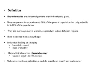
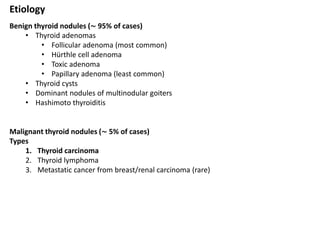
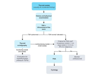
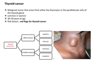
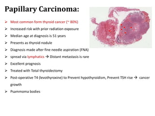
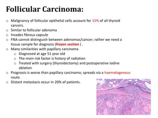
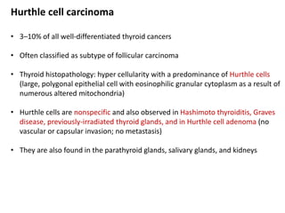
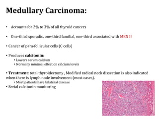
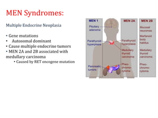
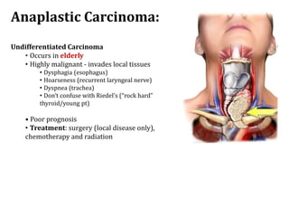
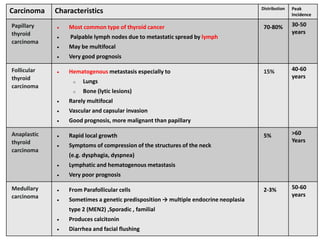
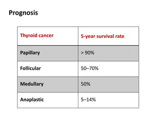
Thyroid Nodules and Cancer
By Lara Masri
4th year medical student
Sources : Step up, Amboss
Outlines
 Thyroid nodules
 Definition
 Etiology
 Diagnosis
 Thyroid cancers
 Papillary
 Follicular
 Medullary
 Anaplastic
• Definition
• Thyroid nodules are abnormal growths within the thyroid gland.
• They are present in approximately 50% of the general population but only palpable
in 5–10% of the population.
• They are more common in women, especially in iodine-deficient regions
• Their incidence increases with age
• Incidental finding on imaging
• Carotid ultrasound
• Neck or chest CT
• Major clinical concern: thyroid cancer
• Cause of about 4 to 10% nodules
• To be detectable on palpation, a nodule must be at least 1 cm in diameter
Etiology
Benign thyroid nodules (∼ 95% of cases)
• Thyroid adenomas
• Follicular adenoma (most common)
• Hürthle cell adenoma
• Toxic adenoma
• Papillary adenoma (least common)
• Thyroid cysts
• Dominant nodules of multinodular goiters
• Hashimoto thyroiditis
Malignant thyroid nodules (∼ 5% of cases)
Types
1. Thyroid carcinoma
2. Thyroid lymphoma
3. Metastatic cancer from breast/renal carcinoma (rare)
Red flags for thyroid cancer
o Patient characteristics
o Male sex
o Age: < 14 years or > 70 years
o History of radiation to the head or neck
o Family history of:
o MEN2 syndrome
o Differentiated thyroid cancer (i.E., Papillary, follicular,
or medullary thyroid cancer)
o Gardner syndrome
o Symptoms
o Rapid growth of thyroid nodule
o Recent onset of persistent hoarseness, dysphagia, or
dyspnea
o Palpatory findings
o Firm or hard nodule
o Fixed nodule
o Cervical lymphadenopathy
o solid nodule on thyroid ultrasound or a cold nodule on thyroid
scintigraphy
Diagnosis
TSH : may be normal, elevated or low
• Elevated TSH: associated with a higher risk of malignancy in thyroid nodules
• Low TSH: indication for thyroid scintigraphy
Thyroid ultrasound
• Differentiates a solid from a cystic nodule; most cancers are solid.
• Can identify nodules 1 to 3 mm in diameter.
• Cystic masses larger than 4 cm in diameter are not malignant.
• Cannot distinguish between benign and malignant thyroid nodules
suspicion for malignancy and require further evaluation with FNAC:
1. Solid
2. hypoechoic nodules with irregular margins
3. Microcalcifications
4. taller-than-wide shape
5. extrathyroidal growth
6. cervical lymphadenopathy
Thyroid scintigraphy
 Indication: thyroid nodules in a patient with low TSH
 Contraindications: pregnant and breastfeeding women
 Findings and interpretation
• Cold nodule (hypofunctioning nodule): Evaluate for indications for FNAC of thyroid
nodules with ultrasound.
• Hot nodule (hyperfunctioning nodule): rarely malignant, FNAC not recommended
• Solitary hot nodule: toxic adenoma
• Multiple hot nodules: toxic multinodular goiter
FNAC
• FNA has a sensitivity of 95% and a specificity of 95%.
• 5% false-negative results, so follow up with periodic FNA if thyroid nodularity
persists.
• FNA is reliable for all cancers (papillary, medullary, anaplastic) except
follicular.
• Patient should first be evaluated with ultrasound and TSH level.
• This is the only test that can reliably differentiate between benign and
malignant nodules.
• FNA findings:
1. Probable cancer (15%) : Most of these are really cancers. Surgery is
indicated.
2. Indeterminate (19%) : A thyroid scan should be performed, and if the lesion
is “cold” by the scan, surgical resection is indicated because about 20% of
these lesions are found to be malignant.
3. Benign (66%) : Most of these are benign. Observe for 1 year, then follow up
with an ultrasound.
4. Follicular neoplasm : Surgery is recommended because it is difficult to
distinguish between benign and malignant follicular cells on histology.
Thyroid cancer
 Malignant tumor that arises from either the thyrocytes or the parafollicular cells of
the thyroid gland.
 common in women
 30–50 years of age
 Risk factors : red flags for thyroid cancer
Thyroid
carcinoma
differentiated
papillary
follicular
Undifferentiated
medullary
anaplastic
Papillary Carcinoma:
 Most common form thyroid cancer (~ 80%)
 Increased risk with prior radiation exposure
 Median age at diagnosis is 51 years
 Presents as thyroid nodule
 Diagnosis made after fine needle aspiration (FNA)
 spread via lymphatics  Distant metastasis is rare
 Excellent prognosis
 Treated with Total thyroidectomy
 Post-operative T4 (levothyroxine) to Prevent hypothyroidism, Prevent TSH rise  cancer
growth
 Psammoma bodies
Follicular Carcinoma:
o Malignancy of follicular epithelial cells account for 15% of all thyroid
cancers.
o Similar to follicular adenoma
o Invades fibrous capsule
o FNA cannot distinguish between adenomas/cancer; rather we need a
tissue sample for diagnosis (frozen section ) .
o Many similarities with papillary carcinoma
o Diagnosed at age 51 year old
o The main risk factor is history of radiation
o Treated with surgery (thyroidectomy) and postoperative iodine
ablation
o Prognosis is worse than papillary carcinoma; spreads via a haematogenous
route.
o Distant metastasis occur in 20% of patients.
Hurthle cell carcinoma
• 3–10% of all well-differentiated thyroid cancers
• Often classified as subtype of follicular carcinoma
• Thyroid histopathology: hyper cellularity with a predominance of Hurthle cells
(large, polygonal epithelial cell with eosinophilic granular cytoplasm as a result of
numerous altered mitochondria)
• Hurthle cells are nonspecific and also observed in Hashimoto thyroiditis, Graves
disease, previously-irradiated thyroid glands, and in Hurthle cell adenoma (no
vascular or capsular invasion; no metastasis)
• They are also found in the parathyroid glands, salivary glands, and kidneys
Medullary Carcinoma:
• Accounts for 2% to 3% of all thyroid cancers
• One-third sporadic, one-third familial, one-third associated with MEN II
• Cancer of para-follicular cells (C cells)
• Produces calcitonin:
• Lowers serum calcium
• Normally minimal effect on calcium levels
• Treatment: total thyroidectomy , Modified radical neck dissection is also indicated
when there is lymph node involvement (most cases).
• Most patients have bilateral disease
• Serial calcitonin monitoring
MEN Syndromes:
Multiple Endocrine Neoplasia
• Gene mutations
• Autosomal dominant
• Cause multiple endocrine tumors
• MEN 2A and 2B associated with
medullary carcinoma
• Caused by RET oncogene mutation
Anaplastic Carcinoma:
Undifferentiated Carcinoma
• Occurs in elderly
• Highly malignant - invades local tissues
• Dysphagia (esophagus)
• Hoarseness (recurrent laryngeal nerve)
• Dyspnea (trachea)
• Don’t confuse with Riedel’s (“rock hard”
thyroid/young pt)
• Poor prognosis
• Treatment: surgery (local disease only),
chemotherapy and radiation
Carcinoma Characteristics Distribution Peak
Incidence
Papillary
thyroid
carcinoma
 Most common type of thyroid cancer
 Palpable lymph nodes due to metastatic spread by lymph
 May be multifocal
 Very good prognosis
70-80% 30-50
years
Follicular
thyroid
carcinoma
 Hematogenous metastasis especially to
o Lungs
o Bone (lytic lesions)
 Rarely multifocal
 Vascular and capsular invasion
 Good prognosis, more malignant than papillary
15% 40-60
years
Anaplastic
thyroid
carcinoma
 Rapid local growth
 Symptoms of compression of the structures of the neck
(e.g. dysphagia, dyspnea)
 Lymphatic and hematogenous metastasis
 Very poor prognosis
5% >60
Years
Medullary
carcinoma
 From Parafollicular cells
 Sometimes a genetic predisposition → multiple endocrine neoplasia
type 2 (MEN2) ,Sporadic , familial
 Produces calcitonin
 Diarrhea and facial flushing
2-3% 50-60
years
Prognosis
Thyroid cancer 5-year survival rate
Papillary > 90%
Follicular 50–70%
Medullary 50%
Anaplastic 5–14%

thyroid nodules and cancer.pptx

  • 1.
    Thyroid Nodules andCancer By Lara Masri 4th year medical student Sources : Step up, Amboss
  • 2.
    Outlines  Thyroid nodules Definition  Etiology  Diagnosis  Thyroid cancers  Papillary  Follicular  Medullary  Anaplastic
  • 3.
    • Definition • Thyroidnodules are abnormal growths within the thyroid gland. • They are present in approximately 50% of the general population but only palpable in 5–10% of the population. • They are more common in women, especially in iodine-deficient regions • Their incidence increases with age • Incidental finding on imaging • Carotid ultrasound • Neck or chest CT • Major clinical concern: thyroid cancer • Cause of about 4 to 10% nodules • To be detectable on palpation, a nodule must be at least 1 cm in diameter
  • 4.
    Etiology Benign thyroid nodules(∼ 95% of cases) • Thyroid adenomas • Follicular adenoma (most common) • Hürthle cell adenoma • Toxic adenoma • Papillary adenoma (least common) • Thyroid cysts • Dominant nodules of multinodular goiters • Hashimoto thyroiditis Malignant thyroid nodules (∼ 5% of cases) Types 1. Thyroid carcinoma 2. Thyroid lymphoma 3. Metastatic cancer from breast/renal carcinoma (rare)
  • 5.
    Red flags forthyroid cancer o Patient characteristics o Male sex o Age: < 14 years or > 70 years o History of radiation to the head or neck o Family history of: o MEN2 syndrome o Differentiated thyroid cancer (i.E., Papillary, follicular, or medullary thyroid cancer) o Gardner syndrome o Symptoms o Rapid growth of thyroid nodule o Recent onset of persistent hoarseness, dysphagia, or dyspnea o Palpatory findings o Firm or hard nodule o Fixed nodule o Cervical lymphadenopathy o solid nodule on thyroid ultrasound or a cold nodule on thyroid scintigraphy
  • 6.
    Diagnosis TSH : maybe normal, elevated or low • Elevated TSH: associated with a higher risk of malignancy in thyroid nodules • Low TSH: indication for thyroid scintigraphy Thyroid ultrasound • Differentiates a solid from a cystic nodule; most cancers are solid. • Can identify nodules 1 to 3 mm in diameter. • Cystic masses larger than 4 cm in diameter are not malignant. • Cannot distinguish between benign and malignant thyroid nodules suspicion for malignancy and require further evaluation with FNAC: 1. Solid 2. hypoechoic nodules with irregular margins 3. Microcalcifications 4. taller-than-wide shape 5. extrathyroidal growth 6. cervical lymphadenopathy
  • 7.
    Thyroid scintigraphy  Indication:thyroid nodules in a patient with low TSH  Contraindications: pregnant and breastfeeding women  Findings and interpretation • Cold nodule (hypofunctioning nodule): Evaluate for indications for FNAC of thyroid nodules with ultrasound. • Hot nodule (hyperfunctioning nodule): rarely malignant, FNAC not recommended • Solitary hot nodule: toxic adenoma • Multiple hot nodules: toxic multinodular goiter
  • 8.
    FNAC • FNA hasa sensitivity of 95% and a specificity of 95%. • 5% false-negative results, so follow up with periodic FNA if thyroid nodularity persists. • FNA is reliable for all cancers (papillary, medullary, anaplastic) except follicular. • Patient should first be evaluated with ultrasound and TSH level. • This is the only test that can reliably differentiate between benign and malignant nodules. • FNA findings: 1. Probable cancer (15%) : Most of these are really cancers. Surgery is indicated. 2. Indeterminate (19%) : A thyroid scan should be performed, and if the lesion is “cold” by the scan, surgical resection is indicated because about 20% of these lesions are found to be malignant. 3. Benign (66%) : Most of these are benign. Observe for 1 year, then follow up with an ultrasound. 4. Follicular neoplasm : Surgery is recommended because it is difficult to distinguish between benign and malignant follicular cells on histology.
  • 10.
    Thyroid cancer  Malignanttumor that arises from either the thyrocytes or the parafollicular cells of the thyroid gland.  common in women  30–50 years of age  Risk factors : red flags for thyroid cancer Thyroid carcinoma differentiated papillary follicular Undifferentiated medullary anaplastic
  • 11.
    Papillary Carcinoma:  Mostcommon form thyroid cancer (~ 80%)  Increased risk with prior radiation exposure  Median age at diagnosis is 51 years  Presents as thyroid nodule  Diagnosis made after fine needle aspiration (FNA)  spread via lymphatics  Distant metastasis is rare  Excellent prognosis  Treated with Total thyroidectomy  Post-operative T4 (levothyroxine) to Prevent hypothyroidism, Prevent TSH rise  cancer growth  Psammoma bodies
  • 12.
    Follicular Carcinoma: o Malignancyof follicular epithelial cells account for 15% of all thyroid cancers. o Similar to follicular adenoma o Invades fibrous capsule o FNA cannot distinguish between adenomas/cancer; rather we need a tissue sample for diagnosis (frozen section ) . o Many similarities with papillary carcinoma o Diagnosed at age 51 year old o The main risk factor is history of radiation o Treated with surgery (thyroidectomy) and postoperative iodine ablation o Prognosis is worse than papillary carcinoma; spreads via a haematogenous route. o Distant metastasis occur in 20% of patients.
  • 13.
    Hurthle cell carcinoma •3–10% of all well-differentiated thyroid cancers • Often classified as subtype of follicular carcinoma • Thyroid histopathology: hyper cellularity with a predominance of Hurthle cells (large, polygonal epithelial cell with eosinophilic granular cytoplasm as a result of numerous altered mitochondria) • Hurthle cells are nonspecific and also observed in Hashimoto thyroiditis, Graves disease, previously-irradiated thyroid glands, and in Hurthle cell adenoma (no vascular or capsular invasion; no metastasis) • They are also found in the parathyroid glands, salivary glands, and kidneys
  • 14.
    Medullary Carcinoma: • Accountsfor 2% to 3% of all thyroid cancers • One-third sporadic, one-third familial, one-third associated with MEN II • Cancer of para-follicular cells (C cells) • Produces calcitonin: • Lowers serum calcium • Normally minimal effect on calcium levels • Treatment: total thyroidectomy , Modified radical neck dissection is also indicated when there is lymph node involvement (most cases). • Most patients have bilateral disease • Serial calcitonin monitoring
  • 15.
    MEN Syndromes: Multiple EndocrineNeoplasia • Gene mutations • Autosomal dominant • Cause multiple endocrine tumors • MEN 2A and 2B associated with medullary carcinoma • Caused by RET oncogene mutation
  • 16.
    Anaplastic Carcinoma: Undifferentiated Carcinoma •Occurs in elderly • Highly malignant - invades local tissues • Dysphagia (esophagus) • Hoarseness (recurrent laryngeal nerve) • Dyspnea (trachea) • Don’t confuse with Riedel’s (“rock hard” thyroid/young pt) • Poor prognosis • Treatment: surgery (local disease only), chemotherapy and radiation
  • 17.
    Carcinoma Characteristics DistributionPeak Incidence Papillary thyroid carcinoma  Most common type of thyroid cancer  Palpable lymph nodes due to metastatic spread by lymph  May be multifocal  Very good prognosis 70-80% 30-50 years Follicular thyroid carcinoma  Hematogenous metastasis especially to o Lungs o Bone (lytic lesions)  Rarely multifocal  Vascular and capsular invasion  Good prognosis, more malignant than papillary 15% 40-60 years Anaplastic thyroid carcinoma  Rapid local growth  Symptoms of compression of the structures of the neck (e.g. dysphagia, dyspnea)  Lymphatic and hematogenous metastasis  Very poor prognosis 5% >60 Years Medullary carcinoma  From Parafollicular cells  Sometimes a genetic predisposition → multiple endocrine neoplasia type 2 (MEN2) ,Sporadic , familial  Produces calcitonin  Diarrhea and facial flushing 2-3% 50-60 years
  • 18.
    Prognosis Thyroid cancer 5-yearsurvival rate Papillary > 90% Follicular 50–70% Medullary 50% Anaplastic 5–14%