Polycystic Kidney Disease
Lara Masri, 4th year
Sources: BnB, Amboss, Step-Up
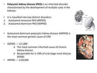
• Polycystic kidney disease (PKD) is an inherited disorder
characterized by the development of multiple cysts in the
kidneys.
• It is classified into two distinct disorders:
1. Autosomal recessive PKD (ARPKD)
2. Autosomal dominant PKD (ADPKD)
• Autosomal dominant polycystic kidney disease (ADPKD) is
the most common genetic cause of CKD
• ADPKD: ∼ 1/1,000
 The most common inherited cause of chronic
kidney disease
 Responsible for 5–10% of end-stage renal disease
(ESRD)
• ARPKD: ∼ 1/20,000
Etiology
• ADPKD
• Mutation in
1. PKD1 on chromosome 16 (85% of cases), the gene that encodes
polycystin-1
2. PKD2 on chromosome 4 (15% of cases), the gene that encodes
polycystin-2
3. Other genes identified in the etiology of ADPKD include GANAB, ALG9,
DNAJB11 and LRP5
• Positive family history
• ARPKD
• Mutation in PKHD1 gene on chromosome 6, the gene that encodes for
fibrocystin, a protein involved in the maintenance of primary cilia of the
renal collecting duct and biliary epithelial cells
• DZIP1L Mutation
Pathophysiology
ADPKD
• Two-hit hypothesis
1. Inherited mutation in one allele of PKD1 or PKD2 genes
2. Additional second somatic acquired mutation in tubule epithelial cells
• Although the disease is transmitted as an autosomal dominant trait, it is
recessive on a cellular level since both alleles must be mutated in order to cause
cyst formation. The timing, location, and frequency of the second mutation
determine the severity of the disease
• Abnormal cilia-mediated signaling pathways → formation and expansion of cysts
in the renal cortex and medulla → compression of renal vessels with activation of
the renin-angiotensin-aldosterone system (RAAS), ischemia, and destruction of
the kidney parenchyma
• Cyst formation occur at any point in the nephron.
ARPKD: inherited mutation in the PKHD1 gene → defective
fibrocystin → cystic dilation of collecting ducts and bile
ducts
Defect in Fibrocystin  Dysfunction of renal cilia
Clinical features
ADPKD
 Symptom onset
• Microscopic cysts present at birth  Too small to visualize with ultrasound 
Kidneys appear normal at birth
• Symptoms usually occur after 30 years of age, but the disease may also manifest
during childhood.
• Symptoms are less severe and tend to manifest later ( 15 years) in individuals with
the PKD2 mutation compared to those with the PKD1 mutation.
 Renal manifestations
1. Gross hematuria
2. Proteinuria
3. Flank or abdominal pain
4. Recurrent UTI
5. Nephrolithiasis
6. Kidneys might be palpable and enlarged on abdominal exam (they are usually
normal at birth)
7. Signs of chronic kidney disease (e.g., hypertension, fluid overload, uremia)
 Extrarenal manifestations
• Multiple benign asymptomatic hepatic cysts 70%
• Cysts in pancreas (10%) , spleen (5%) , ovary, and testicles.
• Cerebral berry aneurysm (8-12%)  most serious
 The risk is higher in patients with a family history positive of ADPKD.
 4 times higher risk than the general population
 May rupture and cause subarachnoid hemorrhage or ICH
• Cardiovascular
 Signs of Arterial hypertension (e.g., morning headaches) through increased renin
production
 Heart valve defects (particularly mitral valve prolapse or aortic regurgitation)
 Left ventricular hypertrophy
 Potential association with coronary artery aneurysm and aortic aneurysm.
• Colon diverticula (diverticulosis 80% )
• Abdominal or inguinal hernias
The pain can be caused by any of the following:
1. Enlargement of one or more cysts
2. Bleeding Hematuria
3. UTI (acute pyelonephritis, infected cysts, perinephric
abscess)
4. Nephrolithiasis and renal colic
5. Rarely, a coincidental hypernephroma
Clinical features
ARPKD
 Symptom onset: Symptoms most commonly manifest in infancy or childhood.
Renal manifestations
1. Protruding abdomen (nontender abdominal mass) due to bilateral renal
enlargement and/or hepatomegaly
2. Chronic renal failure: frequently hematuria, proteinuria, and oliguria
3. Severe in-utero renal impairment → oliguria in utero → maternal
oligohydramnios → Potter sequence ( by 24 weeks GA)
 Craniofacial abnormalities (retrognathia, low-set ears, flat nose) and clubbed
feet
 Pulmonary hypoplasia → respiratory insufficiency in neonates
 Extrarenal manifestations
• Hypertension
 Early manifestation
 Often not responsive to monotherapy
 Can result in complications if inadequately controlled (e.g., cardiac
hypertrophy, heart failure, cerebrovascular diseases, progressive renal
impairment)
• Liver involvement: congenital hepatic and portal fibrosis → progressive liver
failure and portal hypertension * always present *
• Respiratory distress 50%
Diagnosis
• US
ADPKD enlarged kidneys with multiple cysts bilaterally of varying sizes
ARPKD  Enlarged kidneys with multiple cysts bilaterally of equal size
• CT: Not the method of choice
• MR angiography: screening for berry aneurysms in patients with ADPKD is
recommended in the following scenarios:
1. Family history of aneurysm
2. Prior to extensive elective surgeries (e.g., transplantation)
3. Occupations in which unconsciousness represents a public safety risk (e.g., driver)
4. The patient wishes to be screened
• Genetic testing: DNA linkage analysis to identify ADPKD1 and ADPKD2
• Labs: to evaluate and monitor renal function  Blood cell count, Creatinine clearance,
Urinalysis
• Liver Biopsy
 US diagnostic criteria for ADPKD1 are as follows
• At least 2 cysts in 1 kidney or 1 cyst in each kidney in an at-risk patient
younger than 30 years
• At least 2 cysts in each kidney in an at-risk patient aged 30-59 years
• At least 4 cysts in each kidney for an at-risk patient aged 60 years or older
 US diagnostic criteria for ADPKD in patients with a family history but
unknown genotype are as follows
• Three or more (unilateral or bilateral) renal cysts in patients aged 15-39
years
• Two or more cysts in each kidney in patients aged 30-59 years
Pathology
ADPKD  Cystic dilatation of the kidney tubular system
ARPKD  Cystic dilatation of the Collecting ducts
Treatment
ACEIs/ ARBs  to prevent/treat hypertension as well as to slow proteinuria and ESRD
progression
Tolvaptan
• Indicated in patients with
1. Rapidly progressing ADPKD
2. Mild chronic kidney disease (GFR ≥ 25 mL/min/1.73 m2
3. Chronic kidney function estimated between 30–90%)
• Slows down the growth of kidney cysts in ADPKD patients
• Delays progression to ESRD
• Contraindicated with abnormal serum sodium
Early treatment of urinary tract infections
High fluid intake to prevent kidney stone formation and to possibly slow cyst progression
Avoid nephrotoxic substances (e.g., NSAIDs, sulfonamide antibiotics, aminoglycosides)
Avoid ADH (vasopressin may stimulate cyst growth)
In severe cases
o Hemodialysis or peritoneal dialysis
o Kidney transplantation is the only curative option
o Portosystemic shunting or liver transplantation may be required in ARPKD
patients with severe hepatic involvement.
o Treatment of complications (e.g., portal hypertension, subarachnoid
hemorrhage)
PCKD.pptx
PCKD.pptx
PCKD.pptx
PCKD.pptx

PCKD.pptx

  • 1.
    Polycystic Kidney Disease LaraMasri, 4th year Sources: BnB, Amboss, Step-Up
  • 2.
    • Polycystic kidneydisease (PKD) is an inherited disorder characterized by the development of multiple cysts in the kidneys. • It is classified into two distinct disorders: 1. Autosomal recessive PKD (ARPKD) 2. Autosomal dominant PKD (ADPKD) • Autosomal dominant polycystic kidney disease (ADPKD) is the most common genetic cause of CKD • ADPKD: ∼ 1/1,000  The most common inherited cause of chronic kidney disease  Responsible for 5–10% of end-stage renal disease (ESRD) • ARPKD: ∼ 1/20,000
  • 3.
    Etiology • ADPKD • Mutationin 1. PKD1 on chromosome 16 (85% of cases), the gene that encodes polycystin-1 2. PKD2 on chromosome 4 (15% of cases), the gene that encodes polycystin-2 3. Other genes identified in the etiology of ADPKD include GANAB, ALG9, DNAJB11 and LRP5 • Positive family history • ARPKD • Mutation in PKHD1 gene on chromosome 6, the gene that encodes for fibrocystin, a protein involved in the maintenance of primary cilia of the renal collecting duct and biliary epithelial cells • DZIP1L Mutation
  • 4.
    Pathophysiology ADPKD • Two-hit hypothesis 1.Inherited mutation in one allele of PKD1 or PKD2 genes 2. Additional second somatic acquired mutation in tubule epithelial cells • Although the disease is transmitted as an autosomal dominant trait, it is recessive on a cellular level since both alleles must be mutated in order to cause cyst formation. The timing, location, and frequency of the second mutation determine the severity of the disease • Abnormal cilia-mediated signaling pathways → formation and expansion of cysts in the renal cortex and medulla → compression of renal vessels with activation of the renin-angiotensin-aldosterone system (RAAS), ischemia, and destruction of the kidney parenchyma • Cyst formation occur at any point in the nephron.
  • 5.
    ARPKD: inherited mutationin the PKHD1 gene → defective fibrocystin → cystic dilation of collecting ducts and bile ducts Defect in Fibrocystin  Dysfunction of renal cilia
  • 6.
    Clinical features ADPKD  Symptomonset • Microscopic cysts present at birth  Too small to visualize with ultrasound  Kidneys appear normal at birth • Symptoms usually occur after 30 years of age, but the disease may also manifest during childhood. • Symptoms are less severe and tend to manifest later ( 15 years) in individuals with the PKD2 mutation compared to those with the PKD1 mutation.  Renal manifestations 1. Gross hematuria 2. Proteinuria 3. Flank or abdominal pain 4. Recurrent UTI 5. Nephrolithiasis 6. Kidneys might be palpable and enlarged on abdominal exam (they are usually normal at birth) 7. Signs of chronic kidney disease (e.g., hypertension, fluid overload, uremia)
  • 7.
     Extrarenal manifestations •Multiple benign asymptomatic hepatic cysts 70% • Cysts in pancreas (10%) , spleen (5%) , ovary, and testicles. • Cerebral berry aneurysm (8-12%)  most serious  The risk is higher in patients with a family history positive of ADPKD.  4 times higher risk than the general population  May rupture and cause subarachnoid hemorrhage or ICH • Cardiovascular  Signs of Arterial hypertension (e.g., morning headaches) through increased renin production  Heart valve defects (particularly mitral valve prolapse or aortic regurgitation)  Left ventricular hypertrophy  Potential association with coronary artery aneurysm and aortic aneurysm. • Colon diverticula (diverticulosis 80% ) • Abdominal or inguinal hernias
  • 8.
    The pain canbe caused by any of the following: 1. Enlargement of one or more cysts 2. Bleeding Hematuria 3. UTI (acute pyelonephritis, infected cysts, perinephric abscess) 4. Nephrolithiasis and renal colic 5. Rarely, a coincidental hypernephroma
  • 9.
    Clinical features ARPKD  Symptomonset: Symptoms most commonly manifest in infancy or childhood. Renal manifestations 1. Protruding abdomen (nontender abdominal mass) due to bilateral renal enlargement and/or hepatomegaly 2. Chronic renal failure: frequently hematuria, proteinuria, and oliguria 3. Severe in-utero renal impairment → oliguria in utero → maternal oligohydramnios → Potter sequence ( by 24 weeks GA)  Craniofacial abnormalities (retrognathia, low-set ears, flat nose) and clubbed feet  Pulmonary hypoplasia → respiratory insufficiency in neonates
  • 11.
     Extrarenal manifestations •Hypertension  Early manifestation  Often not responsive to monotherapy  Can result in complications if inadequately controlled (e.g., cardiac hypertrophy, heart failure, cerebrovascular diseases, progressive renal impairment) • Liver involvement: congenital hepatic and portal fibrosis → progressive liver failure and portal hypertension * always present * • Respiratory distress 50%
  • 12.
    Diagnosis • US ADPKD enlargedkidneys with multiple cysts bilaterally of varying sizes ARPKD  Enlarged kidneys with multiple cysts bilaterally of equal size • CT: Not the method of choice • MR angiography: screening for berry aneurysms in patients with ADPKD is recommended in the following scenarios: 1. Family history of aneurysm 2. Prior to extensive elective surgeries (e.g., transplantation) 3. Occupations in which unconsciousness represents a public safety risk (e.g., driver) 4. The patient wishes to be screened • Genetic testing: DNA linkage analysis to identify ADPKD1 and ADPKD2 • Labs: to evaluate and monitor renal function  Blood cell count, Creatinine clearance, Urinalysis • Liver Biopsy
  • 13.
     US diagnosticcriteria for ADPKD1 are as follows • At least 2 cysts in 1 kidney or 1 cyst in each kidney in an at-risk patient younger than 30 years • At least 2 cysts in each kidney in an at-risk patient aged 30-59 years • At least 4 cysts in each kidney for an at-risk patient aged 60 years or older  US diagnostic criteria for ADPKD in patients with a family history but unknown genotype are as follows • Three or more (unilateral or bilateral) renal cysts in patients aged 15-39 years • Two or more cysts in each kidney in patients aged 30-59 years
  • 15.
    Pathology ADPKD  Cysticdilatation of the kidney tubular system ARPKD  Cystic dilatation of the Collecting ducts
  • 16.
    Treatment ACEIs/ ARBs to prevent/treat hypertension as well as to slow proteinuria and ESRD progression Tolvaptan • Indicated in patients with 1. Rapidly progressing ADPKD 2. Mild chronic kidney disease (GFR ≥ 25 mL/min/1.73 m2 3. Chronic kidney function estimated between 30–90%) • Slows down the growth of kidney cysts in ADPKD patients • Delays progression to ESRD • Contraindicated with abnormal serum sodium Early treatment of urinary tract infections High fluid intake to prevent kidney stone formation and to possibly slow cyst progression Avoid nephrotoxic substances (e.g., NSAIDs, sulfonamide antibiotics, aminoglycosides) Avoid ADH (vasopressin may stimulate cyst growth)
  • 17.
    In severe cases oHemodialysis or peritoneal dialysis o Kidney transplantation is the only curative option o Portosystemic shunting or liver transplantation may be required in ARPKD patients with severe hepatic involvement. o Treatment of complications (e.g., portal hypertension, subarachnoid hemorrhage)