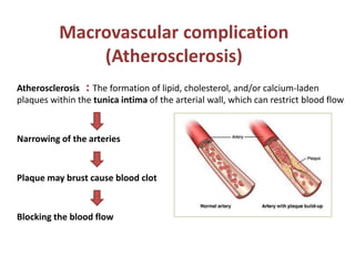
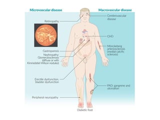
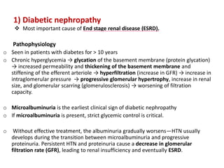
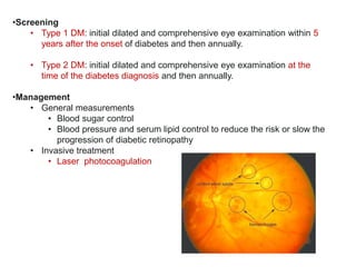
Chronic complication of
DM
By Lara Masri
4th year medical student
Types of chronic complications of DM
1) Macrovascular complications
2) Microvascular complications
Macrovascular complication
(Atherosclerosis)
Atherosclerosis : The formation of lipid, cholesterol, and/or calcium-laden
plaques within the tunica intima of the arterial wall, which can restrict blood flow
Narrowing of the arteries
Plaque may brust cause blood clot
Blocking the blood flow
Prevalence: more common in patients with type 2 diabetes.
Risk factors: The major determinants are metabolic risk factors, which include
obesity, dyslipidemia, and arterial hypertension.
Hyperglycemia may be less related to the development of macrovascular disease.
Manifestations
• Coronary heart disease
 RISK 2-4 times greater than non diabetics.
 Most common cause of death in diabetic patients.
 Silent myocardial infarctions are common.
• Cerebrovascular disease (Strokes)
• Peripheral artery disease (up to 60%)
• Monckeberg arteriosclerosis
• Gangrene
• Monckeberg arteriosclerosis
 A form of arteriosclerosis characterized by calcification of the
media and internal elastic lamina that do not cause arterial
stenosis.
 Associated with diabetes mellitus and/or progressive kidney
disease.
 Usually affects arteries in the extremities.
 PAD diagnostic tools are unreliable in patients with
Mönckeberg's arteriosclerosis.
 X-rays show long, pipestem arteries (calcifications)
The interventions
 Target LDL < 100 mgdl
 Target HTN < 13080
1.Diabetic Nephropathy
2.Diabetic Retinopathy
3.Diabetic Neuropathy
4.Diabetic Foot
1) Diabetic nephropathy
 Most important cause of End stage renal disease (ESRD).
Pathophysiology
o Seen in patients with diabetes for > 10 years
o Chronic hyperglycemia → glycation of the basement membrane (protein glycation)
→ increased permeability and thickening of the basement membrane and
stiffening of the efferent arteriole → hyperfiltration (increase in GFR) → increase in
intraglomerular pressure → progressive glomerular hypertrophy, increase in renal
size, and glomerular scarring (glomerulosclerosis) → worsening of filtration
capacity.
o Microalbuminuria is the earliest clinical sign of diabetic nephropathy
o If microalbuminuria is present, strict glycemic control is critical.
o Without effective treatment, the albuminuria gradually worsens—HTN usually
develops during the transition between microalbuminuria and progressive
proteinuria. Persistent HTN and proteinuria cause a decrease in glomerular
filtration rate (GFR), leading to renal insufficiency and eventually ESRD.
 Initiate ACE inhibitors or ARB immediately to
decrease the rate of progression of nephropathy.
 Microalbuminuria is the screening test!
It usually takes 1 to 5 years for microalbuminuria
to advance to full-blown proteinuria. However,
with proper treatment time can be prolonged.
Pathology: Three major histological changes can be seen on light microscopy.
1. Diffuse glomerular sclerosis
2. nodular glomerulosclerosis (Kimmelstiel-Wilson nodules)
3. Glomerular basement membrane thickening
Clinical features
 Often asymptomatic  patients may complain of foamy urine
 Progressive diabetic kidney disease with signs of renal failure and risk of uremia (e.g.,
uremic polyneuropathy)
 Arterial hypertension
Urine analysis - proteinuria
 Initially moderately increased albuminuria (microalbuminuria) ,
 Eventually significantly increased albuminuria (macroproteinuria)  nephrotic
syndrome may develop.
2) Diabetic retinopathy
• Prevalence is 75% after 20 years of DM
• The most common cause of visual impairment and blindness in patients aged 25–
74 years.
• Clinical features
– Asymptomatic until very late stages of disease
– Visual impairment
– Progression to blindness
• Ocular problems in diabetic patients include cataracts, retinopathy, and glaucoma.
• (Non-proliferative) Retinopathy
 Funduscopic examination shows hemorrhages, exudates, microaneurysms, and venous
dilatation.
 Usually asymptomatic unless retinal edema or ischemia involves the central macula.
 HTN and fluid retention exacerbate this condition.
• Proliferative Retinopathy
 Neovascularization and scarring formation.
 Two serious complications are vitreous hemorrhage and retinal detachment.
 Can lead to blindness. Laser photocoagulation is the treatment.
Overview of diabetic retinopathies
Nonproliferative retinopathy Proliferative retinopathy (PDR)
Mechanism
 Retinal vessel microangiopathy →
blood leaks → retinal
hemorrhages → retinal infiltration
with lipids and fluid → macular
edema
 Retinal vessel microangiopathy →
chronic retinal hypoxia →
abnormal proliferation of blood
vessels → traction on retina → retinal
detachment
Clinical
features
 Intraretinal microvascular abnorma
lities (IRMA)
o Microaneurysms;
o Caliber changes in venous
vessels;
o Intraretinal hemorrhage
o Hard exudates
o Retinal edema
o Cotton-wool spots
 Findings of nonproliferative
retinopathy are usually present.
 Preretinal neovascularization is the
hallmark
of PDR , fibrovascular proliferation , vi
treous hemorrhage, traction retinal
detachment , rubeosis iridis →
secondary glaucoma.
Visual loss
 Most commonly due to macular
edema
 Usually due to vitreous
hemorrhage, retinal detachment,
or neovascular glaucoma.
•Screening
• Type 1 DM: initial dilated and comprehensive eye examination within 5
years after the onset of diabetes and then annually.
• Type 2 DM: initial dilated and comprehensive eye examination at the
time of the diabetes diagnosis and then annually.
•Management
• General measurements
• Blood sugar control
• Blood pressure and serum lipid control to reduce the risk or slow the
progression of diabetic retinopathy
• Invasive treatment
• Laser photocoagulation
3) Diabetic neuropathy
 Pathophysiology: Chronic hyperglycemia causes glycation of axon
proteins.
 Peripheral neuropathy (distal symmetric neuropathy)
 Typically begins in feet, later involves hands (longest nerves affected first).
Numbness and paresthesia are common.
 Loss of sensation leads to: ulcer with subsequent ischemia of pressure
point areas; Charcot joints.
 Painful diabetic neuropathy—hypersensitivity to light touch; severe
“burning” pain (dysesthesia) , especially at night, that can be difficult to
tolerate.
secondary to nerve infarction:
• Types
– Mononeuropathy
• Cranial mononeuropathy
• Peripheral mononeuropathy
• Mononeuropathy multiplex: asymmetric neuropathy,
affecting the multiple peripheral and cranial nerves
• Diabetic truncal neuropathy
– Diabetic lumbosacral plexopathy
• A form of lumbosacral plexus injury caused by diabetes
mellitus
• Microvasculitis causes damage to the plexus formed by
the spinal roots T12-S4, which results in a variety of
symptoms, including pain in the lower back and legs and
progressive weakness of the proximal legs.
Autonomic neuropathy
• Impotence in men (most common presentation)
• Decreased sweating , dry and further increase the risk for ulceration
• Neurogenic bladder—retention, incontinence
• Gastroparesis—chronic nausea and vomiting,
early satiety
• Constipation and diarrhea (alternating)
• Postural hypotension
Screening
• Tuning fork: decreased vibration sense
(https://www.youtube.com/watch?v=X3kW26L_7dA)
• Monofilament test: decreased pressure sense
• Pinprick (pain assessment) or temperature assessment: decreased sensation
(https://www.youtube.com/watch?v=yERSYlRpnh4)
4) Diabetic foot
o A combination of artery disease (ischemia) & neuropathy.
o The patient does not feel pain.
o In addition, neuropathy may mask symptoms of Peripheral Vascular
Diseases (claudication/rest pain). Also, calcific medial arterial
disease is common.
o Increased susceptibility to infection due to impaired WBC function,
reduced blood supply, and Wound healing.
o Patients are at increased risk of: cellulitis, candidiasis, pneumonia,
osteomyelitis, and polymicrobial foot ulcers.
• Malum perforans
• hammer toe
• hallux valgus
Specific Treatment of Chronic Diabetic Complications
1. Macrovascular disease: reduction of risk factors (e.g., BP
reduction, lipid-lowering agents, smoking cessation, exercise), a daily
aspirin and strict glycemic control.
2. Nephropathy: ACE inhibitors
3. Retinopathy: laser photocoagulation.
4. Neuropathy: NSAIDs, tricyclic antidepressants, and gabapentin.
For gastroparesis, a promotility agent such as metoclopramide, in
addition to exercise and a low-fat diet.
5. Diabetic foot: Amputation is a last resort.

Chronic-complication-of-DM.pptx

  • 1.
    Chronic complication of DM ByLara Masri 4th year medical student
  • 2.
    Types of chroniccomplications of DM 1) Macrovascular complications 2) Microvascular complications
  • 3.
    Macrovascular complication (Atherosclerosis) Atherosclerosis :The formation of lipid, cholesterol, and/or calcium-laden plaques within the tunica intima of the arterial wall, which can restrict blood flow Narrowing of the arteries Plaque may brust cause blood clot Blocking the blood flow
  • 4.
    Prevalence: more commonin patients with type 2 diabetes. Risk factors: The major determinants are metabolic risk factors, which include obesity, dyslipidemia, and arterial hypertension. Hyperglycemia may be less related to the development of macrovascular disease. Manifestations • Coronary heart disease  RISK 2-4 times greater than non diabetics.  Most common cause of death in diabetic patients.  Silent myocardial infarctions are common. • Cerebrovascular disease (Strokes) • Peripheral artery disease (up to 60%) • Monckeberg arteriosclerosis • Gangrene
  • 5.
    • Monckeberg arteriosclerosis A form of arteriosclerosis characterized by calcification of the media and internal elastic lamina that do not cause arterial stenosis.  Associated with diabetes mellitus and/or progressive kidney disease.  Usually affects arteries in the extremities.  PAD diagnostic tools are unreliable in patients with Mönckeberg's arteriosclerosis.  X-rays show long, pipestem arteries (calcifications)
  • 6.
    The interventions  TargetLDL < 100 mgdl  Target HTN < 13080
  • 7.
  • 9.
    1) Diabetic nephropathy Most important cause of End stage renal disease (ESRD). Pathophysiology o Seen in patients with diabetes for > 10 years o Chronic hyperglycemia → glycation of the basement membrane (protein glycation) → increased permeability and thickening of the basement membrane and stiffening of the efferent arteriole → hyperfiltration (increase in GFR) → increase in intraglomerular pressure → progressive glomerular hypertrophy, increase in renal size, and glomerular scarring (glomerulosclerosis) → worsening of filtration capacity. o Microalbuminuria is the earliest clinical sign of diabetic nephropathy o If microalbuminuria is present, strict glycemic control is critical. o Without effective treatment, the albuminuria gradually worsens—HTN usually develops during the transition between microalbuminuria and progressive proteinuria. Persistent HTN and proteinuria cause a decrease in glomerular filtration rate (GFR), leading to renal insufficiency and eventually ESRD.
  • 11.
     Initiate ACEinhibitors or ARB immediately to decrease the rate of progression of nephropathy.  Microalbuminuria is the screening test! It usually takes 1 to 5 years for microalbuminuria to advance to full-blown proteinuria. However, with proper treatment time can be prolonged.
  • 12.
    Pathology: Three majorhistological changes can be seen on light microscopy. 1. Diffuse glomerular sclerosis 2. nodular glomerulosclerosis (Kimmelstiel-Wilson nodules) 3. Glomerular basement membrane thickening Clinical features  Often asymptomatic  patients may complain of foamy urine  Progressive diabetic kidney disease with signs of renal failure and risk of uremia (e.g., uremic polyneuropathy)  Arterial hypertension Urine analysis - proteinuria  Initially moderately increased albuminuria (microalbuminuria) ,  Eventually significantly increased albuminuria (macroproteinuria)  nephrotic syndrome may develop.
  • 13.
    2) Diabetic retinopathy •Prevalence is 75% after 20 years of DM • The most common cause of visual impairment and blindness in patients aged 25– 74 years. • Clinical features – Asymptomatic until very late stages of disease – Visual impairment – Progression to blindness • Ocular problems in diabetic patients include cataracts, retinopathy, and glaucoma. • (Non-proliferative) Retinopathy  Funduscopic examination shows hemorrhages, exudates, microaneurysms, and venous dilatation.  Usually asymptomatic unless retinal edema or ischemia involves the central macula.  HTN and fluid retention exacerbate this condition. • Proliferative Retinopathy  Neovascularization and scarring formation.  Two serious complications are vitreous hemorrhage and retinal detachment.  Can lead to blindness. Laser photocoagulation is the treatment.
  • 14.
    Overview of diabeticretinopathies Nonproliferative retinopathy Proliferative retinopathy (PDR) Mechanism  Retinal vessel microangiopathy → blood leaks → retinal hemorrhages → retinal infiltration with lipids and fluid → macular edema  Retinal vessel microangiopathy → chronic retinal hypoxia → abnormal proliferation of blood vessels → traction on retina → retinal detachment Clinical features  Intraretinal microvascular abnorma lities (IRMA) o Microaneurysms; o Caliber changes in venous vessels; o Intraretinal hemorrhage o Hard exudates o Retinal edema o Cotton-wool spots  Findings of nonproliferative retinopathy are usually present.  Preretinal neovascularization is the hallmark of PDR , fibrovascular proliferation , vi treous hemorrhage, traction retinal detachment , rubeosis iridis → secondary glaucoma. Visual loss  Most commonly due to macular edema  Usually due to vitreous hemorrhage, retinal detachment, or neovascular glaucoma.
  • 15.
    •Screening • Type 1DM: initial dilated and comprehensive eye examination within 5 years after the onset of diabetes and then annually. • Type 2 DM: initial dilated and comprehensive eye examination at the time of the diabetes diagnosis and then annually. •Management • General measurements • Blood sugar control • Blood pressure and serum lipid control to reduce the risk or slow the progression of diabetic retinopathy • Invasive treatment • Laser photocoagulation
  • 16.
    3) Diabetic neuropathy Pathophysiology: Chronic hyperglycemia causes glycation of axon proteins.  Peripheral neuropathy (distal symmetric neuropathy)  Typically begins in feet, later involves hands (longest nerves affected first). Numbness and paresthesia are common.  Loss of sensation leads to: ulcer with subsequent ischemia of pressure point areas; Charcot joints.  Painful diabetic neuropathy—hypersensitivity to light touch; severe “burning” pain (dysesthesia) , especially at night, that can be difficult to tolerate.
  • 17.
    secondary to nerveinfarction: • Types – Mononeuropathy • Cranial mononeuropathy • Peripheral mononeuropathy • Mononeuropathy multiplex: asymmetric neuropathy, affecting the multiple peripheral and cranial nerves • Diabetic truncal neuropathy – Diabetic lumbosacral plexopathy • A form of lumbosacral plexus injury caused by diabetes mellitus • Microvasculitis causes damage to the plexus formed by the spinal roots T12-S4, which results in a variety of symptoms, including pain in the lower back and legs and progressive weakness of the proximal legs.
  • 18.
    Autonomic neuropathy • Impotencein men (most common presentation) • Decreased sweating , dry and further increase the risk for ulceration • Neurogenic bladder—retention, incontinence • Gastroparesis—chronic nausea and vomiting, early satiety • Constipation and diarrhea (alternating) • Postural hypotension
  • 19.
    Screening • Tuning fork:decreased vibration sense (https://www.youtube.com/watch?v=X3kW26L_7dA) • Monofilament test: decreased pressure sense • Pinprick (pain assessment) or temperature assessment: decreased sensation (https://www.youtube.com/watch?v=yERSYlRpnh4)
  • 20.
    4) Diabetic foot oA combination of artery disease (ischemia) & neuropathy. o The patient does not feel pain. o In addition, neuropathy may mask symptoms of Peripheral Vascular Diseases (claudication/rest pain). Also, calcific medial arterial disease is common. o Increased susceptibility to infection due to impaired WBC function, reduced blood supply, and Wound healing. o Patients are at increased risk of: cellulitis, candidiasis, pneumonia, osteomyelitis, and polymicrobial foot ulcers.
  • 21.
    • Malum perforans •hammer toe • hallux valgus
  • 22.
    Specific Treatment ofChronic Diabetic Complications 1. Macrovascular disease: reduction of risk factors (e.g., BP reduction, lipid-lowering agents, smoking cessation, exercise), a daily aspirin and strict glycemic control. 2. Nephropathy: ACE inhibitors 3. Retinopathy: laser photocoagulation. 4. Neuropathy: NSAIDs, tricyclic antidepressants, and gabapentin. For gastroparesis, a promotility agent such as metoclopramide, in addition to exercise and a low-fat diet. 5. Diabetic foot: Amputation is a last resort.