Lung Cancer
By: Lara Masri
Reference: step up to medicine
Outline:
 Definition
Types
 Risk factors
 Clinical features
 Diagnosis
Treatment
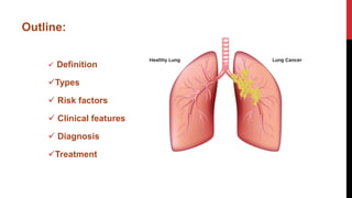
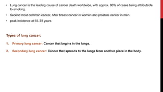
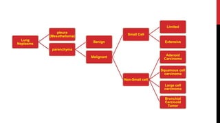
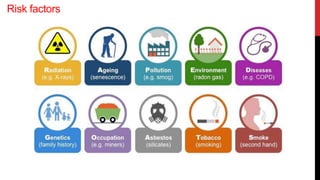
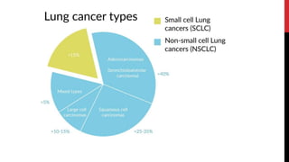
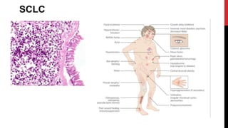
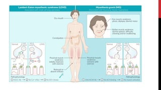
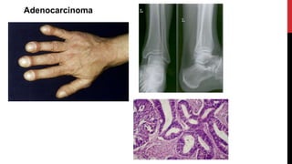
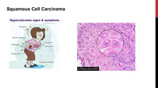
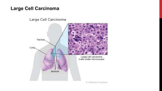
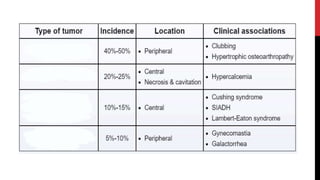
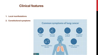
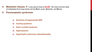
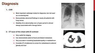
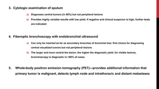
• Lung cancer is the leading cause of cancer death worldwide, with approx. 90% of cases being attributable
to smoking.
• Second most common cancer, After breast cancer in women and prostate cancer in men.
• peak incidence at 65–75 years
Types of lung cancer:
1. Primary lung cancer: Cancer that begins in the lungs.
2. Secondary lung cancer: Cancer that spreads to the lungs from another place in the body.
Lung
Neplasms
pleura
(Mesothelioma)
parenchyma
Benign
Malignant
Small Cell
Limited
Extensive
Non-Small cell
Adenoid
Carcinoma
Squamous cell
carcinoma
Large cell
carcinoma
Bronchial
Carcinoid
Tumor
Risk factors
SCLC
Adenocarcinoma
Squamous Cell Carcinoma
Large Cell Carcinoma
Clinical features
1. Local manifestations
2. Constitutional symptoms
3. Local invasion
A. Superior vena cava (SVC) syndrome  5%
B. Phrenic nerve palsy  1%
C. Recurrent laryngeal nerve palsy (3% of patients)
D. Horner syndrome
E. Malignant pleural effusion —10% to 15%
F. Pancoast tumor
4. Metastatic disease  “Lung cancer loves to BLAB:” the most common sites
of metastasis from lung cancer are the Brain, Liver, Adrenals, and Bones.
5. Paraneoplastic syndromes
a) Syndrome of inappropriate ADH
b) Cushing syndrome
c) Eaton–Lambert syndrome
d) Hypercalcemia
e) Hypertrophic pulmonary osteoarthropathy
Diagnosis
1. CXR
a) Most important radiologic study for diagnosis, but not used
as a screening test
b) Demonstrates abnormal findings in nearly all patients with
lung cancer
c) Stability of an abnormality over a 2-year period is almost
always associated with a benign lesion
2. CT scan of the chest with IV contrast
a) Very useful for staging
b) Can demonstrate extent of local and distant metastasis
c) Very accurate in revealing lymphadenopathy in mediastinum
d) Consider CT of abdomen to screen for metastases to adrenal
glands and liver
3. Cytologic examination of sputum
a) Diagnoses central tumors (in 80%) but not peripheral lesions
b) Provides highly variable results with low yield; if negative and clinical suspicion is high, further tests
are indicated
4. Fiberoptic bronchoscopy with endobronchial ultrasound
a) Can only be inserted as far as secondary branches of bronchial tree; first choice for diagnosing
central visualized tumors but not peripheral lesions
b) The larger and more central the lesion, the higher the diagnostic yield; for visible lesions,
bronchoscopy is diagnostic in >90% of cases
5. Whole-body positron emission tomography (PET)—provides additional information that
primary tumor is malignant, detects lymph node and intrathoracic and distant metastases
6. Transthoracic needle biopsy (under fluoroscopic or CT guidance)
a) Needle biopsy of suspicious pulmonary masses is highly accurate, and is useful for diagnosing
peripheral lesions as well
b) Needle biopsy is invasive and must be used only in selected patients. This is a better biopsy
method for peripheral lesions, whereas central, peribronchial lesions should be biopsied using
bronchoscopy
7. Mediastinoscopy
a) Allows direct visualization of the superior mediastinum
b) Identifies patients with advanced disease who would not benefit from surgical resection
Treatment
1. NSCLC
1. Surgery is the best option for limited disease.
1. A definitive pathologic diagnosis must be made prior to surgery.
2. Patients with metastatic disease outside the chest are not candidates for surgery.
3. Recurrence may occur even after complete resection.
2. Radiation therapy is an important adjunct to surgery.
3. Chemotherapy is of uncertain benefit. Some studies show a modest increase in
survival especially with novel immunotherapy agents. More trials are underway.
2. SCLC
a) For limited disease, combination of chemotherapy and radiation therapy is used
initially.
b) For extensive disease, chemotherapy is used alone as initial treatment. If patient
responds to initial chemotherapy treatment, prophylactic whole-brain irradiation
decreases incidence of brain metastases and prolongs survival.
c) Surgery has a limited role because these tumors are usually nonresectable.
Surgery is not possible in these cases:
• Bilateral disease or lymph nodes involved on opposite side.
• Malignant pleural effusion.
• Heart, carina, aorta, or vena cava is involved

lung cancer.pptx

  • 1.
    Lung Cancer By: LaraMasri Reference: step up to medicine
  • 2.
    Outline:  Definition Types  Riskfactors  Clinical features  Diagnosis Treatment
  • 3.
    • Lung canceris the leading cause of cancer death worldwide, with approx. 90% of cases being attributable to smoking. • Second most common cancer, After breast cancer in women and prostate cancer in men. • peak incidence at 65–75 years Types of lung cancer: 1. Primary lung cancer: Cancer that begins in the lungs. 2. Secondary lung cancer: Cancer that spreads to the lungs from another place in the body.
  • 5.
  • 6.
  • 8.
  • 10.
  • 11.
  • 12.
  • 14.
    Clinical features 1. Localmanifestations 2. Constitutional symptoms
  • 15.
    3. Local invasion A.Superior vena cava (SVC) syndrome  5%
  • 17.
    B. Phrenic nervepalsy  1% C. Recurrent laryngeal nerve palsy (3% of patients)
  • 18.
    D. Horner syndrome E.Malignant pleural effusion —10% to 15%
  • 19.
  • 20.
    4. Metastatic disease “Lung cancer loves to BLAB:” the most common sites of metastasis from lung cancer are the Brain, Liver, Adrenals, and Bones. 5. Paraneoplastic syndromes a) Syndrome of inappropriate ADH b) Cushing syndrome c) Eaton–Lambert syndrome d) Hypercalcemia e) Hypertrophic pulmonary osteoarthropathy
  • 21.
    Diagnosis 1. CXR a) Mostimportant radiologic study for diagnosis, but not used as a screening test b) Demonstrates abnormal findings in nearly all patients with lung cancer c) Stability of an abnormality over a 2-year period is almost always associated with a benign lesion 2. CT scan of the chest with IV contrast a) Very useful for staging b) Can demonstrate extent of local and distant metastasis c) Very accurate in revealing lymphadenopathy in mediastinum d) Consider CT of abdomen to screen for metastases to adrenal glands and liver
  • 22.
    3. Cytologic examinationof sputum a) Diagnoses central tumors (in 80%) but not peripheral lesions b) Provides highly variable results with low yield; if negative and clinical suspicion is high, further tests are indicated 4. Fiberoptic bronchoscopy with endobronchial ultrasound a) Can only be inserted as far as secondary branches of bronchial tree; first choice for diagnosing central visualized tumors but not peripheral lesions b) The larger and more central the lesion, the higher the diagnostic yield; for visible lesions, bronchoscopy is diagnostic in >90% of cases 5. Whole-body positron emission tomography (PET)—provides additional information that primary tumor is malignant, detects lymph node and intrathoracic and distant metastases
  • 23.
    6. Transthoracic needlebiopsy (under fluoroscopic or CT guidance) a) Needle biopsy of suspicious pulmonary masses is highly accurate, and is useful for diagnosing peripheral lesions as well b) Needle biopsy is invasive and must be used only in selected patients. This is a better biopsy method for peripheral lesions, whereas central, peribronchial lesions should be biopsied using bronchoscopy 7. Mediastinoscopy a) Allows direct visualization of the superior mediastinum b) Identifies patients with advanced disease who would not benefit from surgical resection
  • 24.
    Treatment 1. NSCLC 1. Surgeryis the best option for limited disease. 1. A definitive pathologic diagnosis must be made prior to surgery. 2. Patients with metastatic disease outside the chest are not candidates for surgery. 3. Recurrence may occur even after complete resection. 2. Radiation therapy is an important adjunct to surgery. 3. Chemotherapy is of uncertain benefit. Some studies show a modest increase in survival especially with novel immunotherapy agents. More trials are underway.
  • 25.
    2. SCLC a) Forlimited disease, combination of chemotherapy and radiation therapy is used initially. b) For extensive disease, chemotherapy is used alone as initial treatment. If patient responds to initial chemotherapy treatment, prophylactic whole-brain irradiation decreases incidence of brain metastases and prolongs survival. c) Surgery has a limited role because these tumors are usually nonresectable.
  • 26.
    Surgery is notpossible in these cases: • Bilateral disease or lymph nodes involved on opposite side. • Malignant pleural effusion. • Heart, carina, aorta, or vena cava is involved