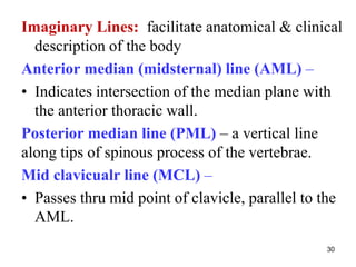
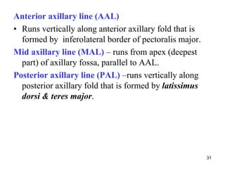
The document provides information on the anatomy of the thoracic wall and diaphragm. It discusses the following key points:
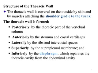
1. The thoracic wall is formed by bones including the ribs, sternum, and thoracic vertebrae. It is covered by muscles and fascia externally and pleura internally.
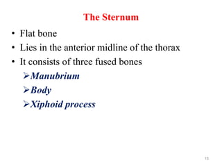
2. The 12 pairs of ribs are connected to the sternum by costal cartilages. There are three types of ribs.
3. The diaphragm is a double-domed muscle that separates the thoracic and abdominal cavities. It has openings for structures to pass through.
4. During inspiration, contraction of the diaphragm and external inter