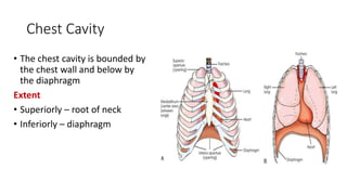
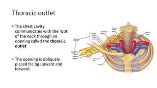
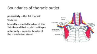
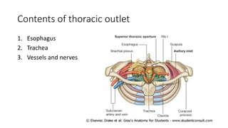
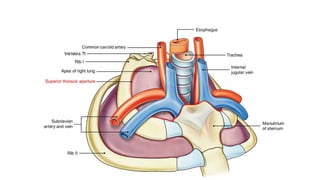
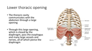
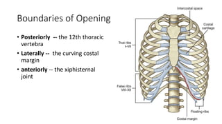
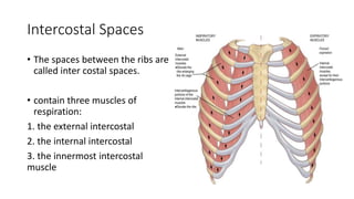
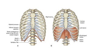
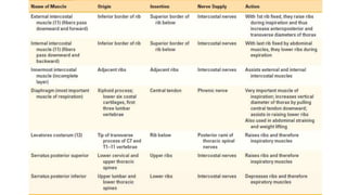
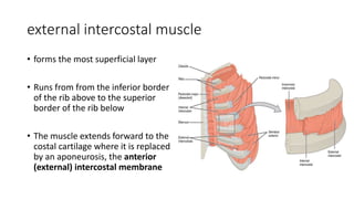
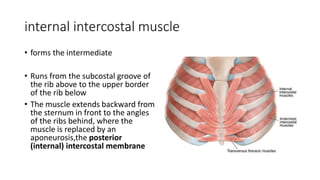
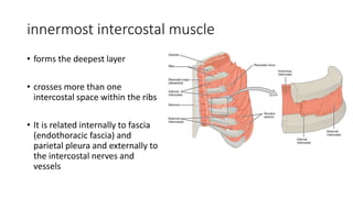
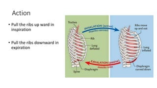
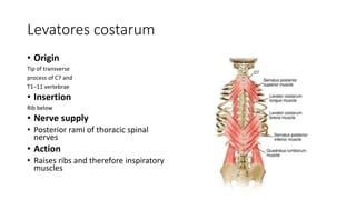
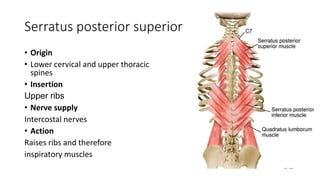
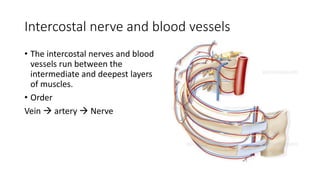
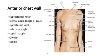
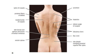
THORACIC CAVITY
By DR. Aliza Moin
Studying objectives
• To learn chest cavity
• Thoracic outlet
• Lower thoracic opening
• Intercostal space and muscles
• Anterior and posterior chest walls
Chest Cavity
• The chest cavity is bounded by
the chest wall and below by
the diaphragm
Extent
• Superiorly – root of neck
• Inferiorly – diaphragm
Openings of the Thorax
• Thorax has two openings
1. Thoracic inlet
2. Thoracic outlet
Thoracic outlet
• The chest cavity
communicates with the root
of the neck through an
opening called the thoracic
outlet
• The opening is obliquely
placed facing upward and
forward
Boundaries of thoracic outlet
posteriorly -- the 1st thoracic
Vertebra
laterally -- medial borders of the
1st ribs and their costal cartilages
anteriorly -- superior border of
the manubrium sterni
Contents of thoracic outlet
1. Esophagus
2. Trachea
3. Vessels and nerves
Lower thoracic opening
• The thoracic cavity
communicates with the
abdomen through a large
opening
• Through this large opening,
which is closed by the
diaphragm, pass the esophagus
and many large vessels and
nerves, all of which pierce the
diaphragm
Boundaries of Opening
• Posteriorly -- the 12th thoracic
vertebra
• Laterally -- the curving costal
margin
• anteriorly -- the xiphisternal
joint
Intercostal spaces and muscles
Intercostal Spaces
• The spaces between the ribs are
called inter costal spaces.
• contain three muscles of
respiration:
1. the external intercostal
2. the internal intercostal
3. the innermost intercostal
muscle
endothoracic fascia
• The innermost intercostal
• muscle is lined internally by the
endothoracic fascia, which
• is lined internally by the parietal
pleura
external intercostal muscle
• forms the most superficial layer
• Runs from from the inferior border
of the rib above to the superior
border of the rib below
• The muscle extends forward to the
costal cartilage where it is replaced
by an aponeurosis, the anterior
(external) intercostal membrane
internal intercostal muscle
• forms the intermediate
• Runs from the subcostal groove of
the rib above to the upper border
of the rib below
• The muscle extends backward from
the sternum in front to the angles
of the ribs behind, where the
muscle is replaced by an
aponeurosis,the posterior
(internal) intercostal membrane
innermost intercostal muscle
• forms the deepest layer
• crosses more than one
intercostal space within the ribs
• It is related internally to fascia
(endothoracic fascia) and
parietal pleura and externally to
the intercostal nerves and
vessels
Action
• Pull the ribs up ward in
inspiration
• Pull the ribs downward in
expiration
Levatores costarum
• Origin
Tip of transverse
process of C7 and
T1–11 vertebrae
• Insertion
Rib below
• Nerve supply
• Posterior rami of thoracic spinal
nerves
• Action
• Raises ribs and therefore inspiratory
muscles
Serratus posterior superior
• Origin
• Lower cervical and upper thoracic
spines
• Insertion
Upper ribs
• Nerve supply
Intercostal nerves
• Action
Raises ribs and therefore
inspiratory muscles
Serratus posterior inferior
• Origin
• Upper lumbar and lower thoracic
spines
• Insertion
Lower ribs
• Nerve supply
Intercostal nerves
• Action
• Depresses ribs and therefore
expiratory muscles
Intercostal nerve and blood vessels
• The intercostal nerves and blood
vessels run between the
intermediate and deepest layers
of muscles.
• Order
Vein  artery  Nerve
Chest walls
Anterior chest wall
• suprasternal notch
• sternal angle (angle of Louis)
• xiphisternal joint
• subcostal angle
• costal margin
• Clivicle
• Nipple
Posterior chest wall
• spinous processes
• The first spinous process to be
felt is that of the seventh
cervical vertebrae
• Scapula
1. Superior angle
2. Spine
3. Inferior angle
Thorax cavity.pptx

Thorax cavity.pptx

  • 1.
  • 2.
    Studying objectives • Tolearn chest cavity • Thoracic outlet • Lower thoracic opening • Intercostal space and muscles • Anterior and posterior chest walls
  • 3.
    Chest Cavity • Thechest cavity is bounded by the chest wall and below by the diaphragm Extent • Superiorly – root of neck • Inferiorly – diaphragm
  • 4.
    Openings of theThorax • Thorax has two openings 1. Thoracic inlet 2. Thoracic outlet
  • 5.
    Thoracic outlet • Thechest cavity communicates with the root of the neck through an opening called the thoracic outlet • The opening is obliquely placed facing upward and forward
  • 6.
    Boundaries of thoracicoutlet posteriorly -- the 1st thoracic Vertebra laterally -- medial borders of the 1st ribs and their costal cartilages anteriorly -- superior border of the manubrium sterni
  • 7.
    Contents of thoracicoutlet 1. Esophagus 2. Trachea 3. Vessels and nerves
  • 9.
    Lower thoracic opening •The thoracic cavity communicates with the abdomen through a large opening • Through this large opening, which is closed by the diaphragm, pass the esophagus and many large vessels and nerves, all of which pierce the diaphragm
  • 10.
    Boundaries of Opening •Posteriorly -- the 12th thoracic vertebra • Laterally -- the curving costal margin • anteriorly -- the xiphisternal joint
  • 11.
  • 12.
    Intercostal Spaces • Thespaces between the ribs are called inter costal spaces. • contain three muscles of respiration: 1. the external intercostal 2. the internal intercostal 3. the innermost intercostal muscle
  • 14.
    endothoracic fascia • Theinnermost intercostal • muscle is lined internally by the endothoracic fascia, which • is lined internally by the parietal pleura
  • 16.
    external intercostal muscle •forms the most superficial layer • Runs from from the inferior border of the rib above to the superior border of the rib below • The muscle extends forward to the costal cartilage where it is replaced by an aponeurosis, the anterior (external) intercostal membrane
  • 17.
    internal intercostal muscle •forms the intermediate • Runs from the subcostal groove of the rib above to the upper border of the rib below • The muscle extends backward from the sternum in front to the angles of the ribs behind, where the muscle is replaced by an aponeurosis,the posterior (internal) intercostal membrane
  • 18.
    innermost intercostal muscle •forms the deepest layer • crosses more than one intercostal space within the ribs • It is related internally to fascia (endothoracic fascia) and parietal pleura and externally to the intercostal nerves and vessels
  • 19.
    Action • Pull theribs up ward in inspiration • Pull the ribs downward in expiration
  • 20.
    Levatores costarum • Origin Tipof transverse process of C7 and T1–11 vertebrae • Insertion Rib below • Nerve supply • Posterior rami of thoracic spinal nerves • Action • Raises ribs and therefore inspiratory muscles
  • 21.
    Serratus posterior superior •Origin • Lower cervical and upper thoracic spines • Insertion Upper ribs • Nerve supply Intercostal nerves • Action Raises ribs and therefore inspiratory muscles
  • 22.
    Serratus posterior inferior •Origin • Upper lumbar and lower thoracic spines • Insertion Lower ribs • Nerve supply Intercostal nerves • Action • Depresses ribs and therefore expiratory muscles
  • 23.
    Intercostal nerve andblood vessels • The intercostal nerves and blood vessels run between the intermediate and deepest layers of muscles. • Order Vein  artery  Nerve
  • 24.
  • 25.
    Anterior chest wall •suprasternal notch • sternal angle (angle of Louis) • xiphisternal joint • subcostal angle • costal margin • Clivicle • Nipple
  • 26.
    Posterior chest wall •spinous processes • The first spinous process to be felt is that of the seventh cervical vertebrae • Scapula 1. Superior angle 2. Spine 3. Inferior angle