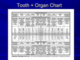
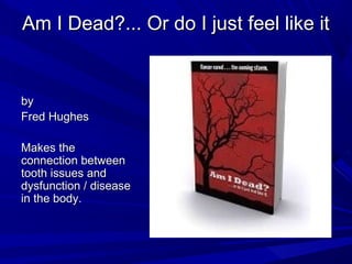
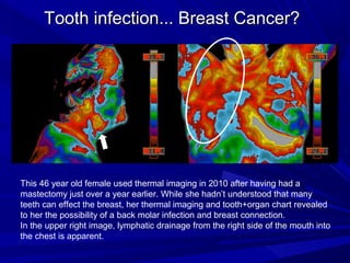
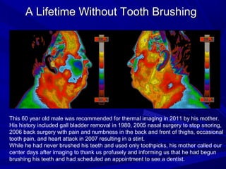
Dental Thermal Imaging
       Examples


               From
The Thermogram Center, Inc. of Colorado
     www.ThermogramCenter.com ● 303-664-1139
                    Est. 2002
Tooth + Organ Chart
Am I Dead?... Or do I just feel like it


by
Fred Hughes

Makes the
connection between
tooth issues and
dysfunction / disease
in the body.
* The Thermogram Center (TTC) is not a
   treatment or diagnosing center. The
 following images were acquired at The
         Thermogram Center and
 analyzed/interpreted by its interpretive
    service provider. Any treatment or
    diagnoses obtained thereafter was
  through the client’s health provider(s).
TTC’s Thermal Imaging
TTC’s camera maps 76,000+ temperature readings to
0.06°C sensitivity and 1.3mrad spatial resolution, with
images in B+W, reversed B+W, and color
Each image has a built-in scale in Celsius
– The higher the color, the higher the temperature
– The lower the color, the lower the temperature
Thermal patterns of the left should be somewhat
symmetrical to the right
– Distal body parts are colder, i.e. noses, ear lobes
– Proximal body parts are hotter, i.e. eye sockets, inner ear;
  scalenes and SCM can be “tight” and hotter
Temperature differences that exceed 1ºC can be
significant
TTC’s Thermal Imaging

Teeth, glands, and organs should be operating
at relatively the same temperature and should
NOT stand out on images.
– When intense excess heat exhibits on a jaw with
  apparent lymphatic inflammation, it can result from
  tooth infection, among other causes.
– When a little excess heat exhibits on a jaw, it can
  result from stress, irritation or inflammation from TMJ
  muscle tightness, clenching or grinding, bite
  misalignment/occlusion, cracked/chipped tooth, deep
  gum pocket, among other causes.
About Sinus Drainage

Prior to 2010’s Colorado Summer wildfires,
about half of all clients exhibited sinus/lymphatic
drainage, typically draining bilaterally below the
nostrils and down the canine regions under the
chin and into the neck.
Post wildfires... 99% of all clients exhibit such
signs.
The majority of clients have no awareness of
such drainage, which can be caused by
pollutants, food/chemical sensitivities, allergies,
infections including mold, fungus, yeast,
parasites, microbes, viruses, bacteria, etc.
About Tooth Findings
Over the past 11 years, over 7,750 imaging
sessions have been provided to men, women
and children:
– Thermal findings suggesting the possibility of tooth
  infection has exceeded findings suggesting highest
  risk for breast cancer by a substantial amount,
  perhaps 50:1.
– Each Face Series is made up of 7 views (each in 3
  color schemes): anterior neck with head tilted back,
  anterior and bilateral face, and open-mouth anterior
  and bilateral face.
– When thermal findings present possible areas of
  concern for teeth, clients are recommended to seek
  further evaluation with an appropriate dental
  professional.
Typical “Normal” includes Lymphatic Sinus Drainage
+ TMJ Tension
Sinus Infection
         Regarding the top and bottom images at left:
         This 51 year old female had no symptoms in
         the teeth, mouth, cheek, sinuses, or head.
         Imaging revealed temperatures 3.95ºC warmer
         on the left cheek than on the right cheek.
         This thermal pattern was consistent with
         infection/inflammation, and prompted the client
         to consult her ENT Doctor who prescribed oral
         antibiotics.

         Regarding the top and bottom images at right:
         One month later, the images on the right
         resulted, consistent with resolution.
         Note the change in cheek patterns, going from
         very asymmetrical to relatively symmetrical in
         just one month’s time.
Severe Sinus Infection


                    This 56 year old female
                    had complaints of chronic
                    lower back pain and
                    asthma.

                    While she had no idea
                    she may have severe
                    sinus and/or tooth
                    infections, her thermal
                    imaging strongly suggests
                    it.
Cracked Tooth... Early Infection

               This 45 year old female had a complaint
               with an upper right front tooth. Her dental
               hygienist indicated that all was well.
               During an imaging appointment in 2008,
               thermal findings consistent with mild oral
               infection were revealed.

               Still symptomatic and with her thermal
               imaging report, she requested that her
               dentist conduct a thorough exam.
               The dentist found that the tooth was
               cracked and that infection had developed
               under the tooth that required care and a
               crown.

               She later reported that the tooth feels
               much better.
Root Canal... Infection


This female had no concerns regarding her oral health. Note the significant
heat/thermal finding at "B", which is consistent with an active lymph node, among
other things. In the event of an underlying tooth infection, she sought dentistry to
determine the cause of this finding. Dental xray of tooth #18 indicated abnormality at
"C", consistent with infection. This tooth had previously undergone root canal and was
crowned. Months of treatment to improve the health of the tooth proved unsuccessful,
and the tooth was extracted. After extraction, a contributing factor/likely cause of
infection was observed: a long/vertical fracture/crack at "D". Note the soft tissue
infection between the roots on the opposite side of the tooth at "E".
Root Canal.... Infection...Heart Attack?
                                         This 72 year old male had experienced
                                         innumerable root canals and decades of
                                         dental work which was completed in 1998.

                                         When thermally imaged in 2009, he was
                                         free of symptoms or concerns for his teeth
                                         and therefore surprised when his thermal
                                         imaging revealed signs consistent with
                                         dental issues and infection.


Afterwards, he obtained a second opinion from a dentist very experienced with
diagnosing/treating infections. Not only were three tooth infections diagnosed,
but this male also learned that tooth #18 could have contributed to his heart
attack two years earlier.
Wisdom Teeth Extraction... Cavitation...
             Lymphatic Drainage


This 54 year old female reported depleting energy levels and concern for developing
auto-immune disorders if the source of her chronic fatigue could not be resolved.
Her thermal imaging revealed signs consistent with potential dental issues and
infection with lymphatic involvement/drainage into the chest. Note the excess heat
and thermal patterns along the left chest. While her follow-up tests were normal for
the breast, her dentist diagnosed 2 cavitations in her right jaws - in wisdom teeth
sockets where the teeth had been extracted twenty years earlier.
Tooth infection... Breast Cancer?


This 46 year old female used thermal imaging in 2010 after having had a
mastectomy just over a year earlier. While she hadn’t understood that many
teeth can effect the breast, her thermal imaging and tooth+organ chart revealed
to her the possibility of a back molar infection and breast connection.
In the upper right image, lymphatic drainage from the right side of the mouth into
the chest is apparent.
A Lifetime Without Tooth Brushing


This 60 year old male was recommended for thermal imaging in 2011 by his mother.
His history included gall bladder removal in 1980, 2005 nasal surgery to stop snoring,
2006 back surgery with pain and numbness in the back and front of thighs, occasional
tooth pain, and heart attack in 2007 resulting in a stint.
While he had never brushed his teeth and used only toothpicks, his mother called our
center days after imaging to thank us profusely and informing us that he had begun
brushing his teeth and had scheduled an appointment to see a dentist.

Thermal Imaging Examples of Dental Issues

  • 1.
    Dental Thermal Imaging Examples From The Thermogram Center, Inc. of Colorado www.ThermogramCenter.com ● 303-664-1139 Est. 2002
  • 2.
  • 3.
    Am I Dead?...Or do I just feel like it by Fred Hughes Makes the connection between tooth issues and dysfunction / disease in the body.
  • 4.
    * The ThermogramCenter (TTC) is not a treatment or diagnosing center. The following images were acquired at The Thermogram Center and analyzed/interpreted by its interpretive service provider. Any treatment or diagnoses obtained thereafter was through the client’s health provider(s).
  • 5.
    TTC’s Thermal Imaging TTC’scamera maps 76,000+ temperature readings to 0.06°C sensitivity and 1.3mrad spatial resolution, with images in B+W, reversed B+W, and color Each image has a built-in scale in Celsius – The higher the color, the higher the temperature – The lower the color, the lower the temperature Thermal patterns of the left should be somewhat symmetrical to the right – Distal body parts are colder, i.e. noses, ear lobes – Proximal body parts are hotter, i.e. eye sockets, inner ear; scalenes and SCM can be “tight” and hotter Temperature differences that exceed 1ºC can be significant
  • 6.
    TTC’s Thermal Imaging Teeth,glands, and organs should be operating at relatively the same temperature and should NOT stand out on images. – When intense excess heat exhibits on a jaw with apparent lymphatic inflammation, it can result from tooth infection, among other causes. – When a little excess heat exhibits on a jaw, it can result from stress, irritation or inflammation from TMJ muscle tightness, clenching or grinding, bite misalignment/occlusion, cracked/chipped tooth, deep gum pocket, among other causes.
  • 7.
    About Sinus Drainage Priorto 2010’s Colorado Summer wildfires, about half of all clients exhibited sinus/lymphatic drainage, typically draining bilaterally below the nostrils and down the canine regions under the chin and into the neck. Post wildfires... 99% of all clients exhibit such signs. The majority of clients have no awareness of such drainage, which can be caused by pollutants, food/chemical sensitivities, allergies, infections including mold, fungus, yeast, parasites, microbes, viruses, bacteria, etc.
  • 8.
    About Tooth Findings Overthe past 11 years, over 7,750 imaging sessions have been provided to men, women and children: – Thermal findings suggesting the possibility of tooth infection has exceeded findings suggesting highest risk for breast cancer by a substantial amount, perhaps 50:1. – Each Face Series is made up of 7 views (each in 3 color schemes): anterior neck with head tilted back, anterior and bilateral face, and open-mouth anterior and bilateral face. – When thermal findings present possible areas of concern for teeth, clients are recommended to seek further evaluation with an appropriate dental professional.
  • 9.
    Typical “Normal” includesLymphatic Sinus Drainage
  • 10.
  • 11.
    Sinus Infection Regarding the top and bottom images at left: This 51 year old female had no symptoms in the teeth, mouth, cheek, sinuses, or head. Imaging revealed temperatures 3.95ºC warmer on the left cheek than on the right cheek. This thermal pattern was consistent with infection/inflammation, and prompted the client to consult her ENT Doctor who prescribed oral antibiotics. Regarding the top and bottom images at right: One month later, the images on the right resulted, consistent with resolution. Note the change in cheek patterns, going from very asymmetrical to relatively symmetrical in just one month’s time.
  • 12.
    Severe Sinus Infection This 56 year old female had complaints of chronic lower back pain and asthma. While she had no idea she may have severe sinus and/or tooth infections, her thermal imaging strongly suggests it.
  • 13.
    Cracked Tooth... EarlyInfection This 45 year old female had a complaint with an upper right front tooth. Her dental hygienist indicated that all was well. During an imaging appointment in 2008, thermal findings consistent with mild oral infection were revealed. Still symptomatic and with her thermal imaging report, she requested that her dentist conduct a thorough exam. The dentist found that the tooth was cracked and that infection had developed under the tooth that required care and a crown. She later reported that the tooth feels much better.
  • 14.
    Root Canal... Infection Thisfemale had no concerns regarding her oral health. Note the significant heat/thermal finding at "B", which is consistent with an active lymph node, among other things. In the event of an underlying tooth infection, she sought dentistry to determine the cause of this finding. Dental xray of tooth #18 indicated abnormality at "C", consistent with infection. This tooth had previously undergone root canal and was crowned. Months of treatment to improve the health of the tooth proved unsuccessful, and the tooth was extracted. After extraction, a contributing factor/likely cause of infection was observed: a long/vertical fracture/crack at "D". Note the soft tissue infection between the roots on the opposite side of the tooth at "E".
  • 15.
    Root Canal.... Infection...HeartAttack? This 72 year old male had experienced innumerable root canals and decades of dental work which was completed in 1998. When thermally imaged in 2009, he was free of symptoms or concerns for his teeth and therefore surprised when his thermal imaging revealed signs consistent with dental issues and infection. Afterwards, he obtained a second opinion from a dentist very experienced with diagnosing/treating infections. Not only were three tooth infections diagnosed, but this male also learned that tooth #18 could have contributed to his heart attack two years earlier.
  • 16.
    Wisdom Teeth Extraction...Cavitation... Lymphatic Drainage This 54 year old female reported depleting energy levels and concern for developing auto-immune disorders if the source of her chronic fatigue could not be resolved. Her thermal imaging revealed signs consistent with potential dental issues and infection with lymphatic involvement/drainage into the chest. Note the excess heat and thermal patterns along the left chest. While her follow-up tests were normal for the breast, her dentist diagnosed 2 cavitations in her right jaws - in wisdom teeth sockets where the teeth had been extracted twenty years earlier.
  • 17.
    Tooth infection... BreastCancer? This 46 year old female used thermal imaging in 2010 after having had a mastectomy just over a year earlier. While she hadn’t understood that many teeth can effect the breast, her thermal imaging and tooth+organ chart revealed to her the possibility of a back molar infection and breast connection. In the upper right image, lymphatic drainage from the right side of the mouth into the chest is apparent.
  • 18.
    A Lifetime WithoutTooth Brushing This 60 year old male was recommended for thermal imaging in 2011 by his mother. His history included gall bladder removal in 1980, 2005 nasal surgery to stop snoring, 2006 back surgery with pain and numbness in the back and front of thighs, occasional tooth pain, and heart attack in 2007 resulting in a stint. While he had never brushed his teeth and used only toothpicks, his mother called our center days after imaging to thank us profusely and informing us that he had begun brushing his teeth and had scheduled an appointment to see a dentist.