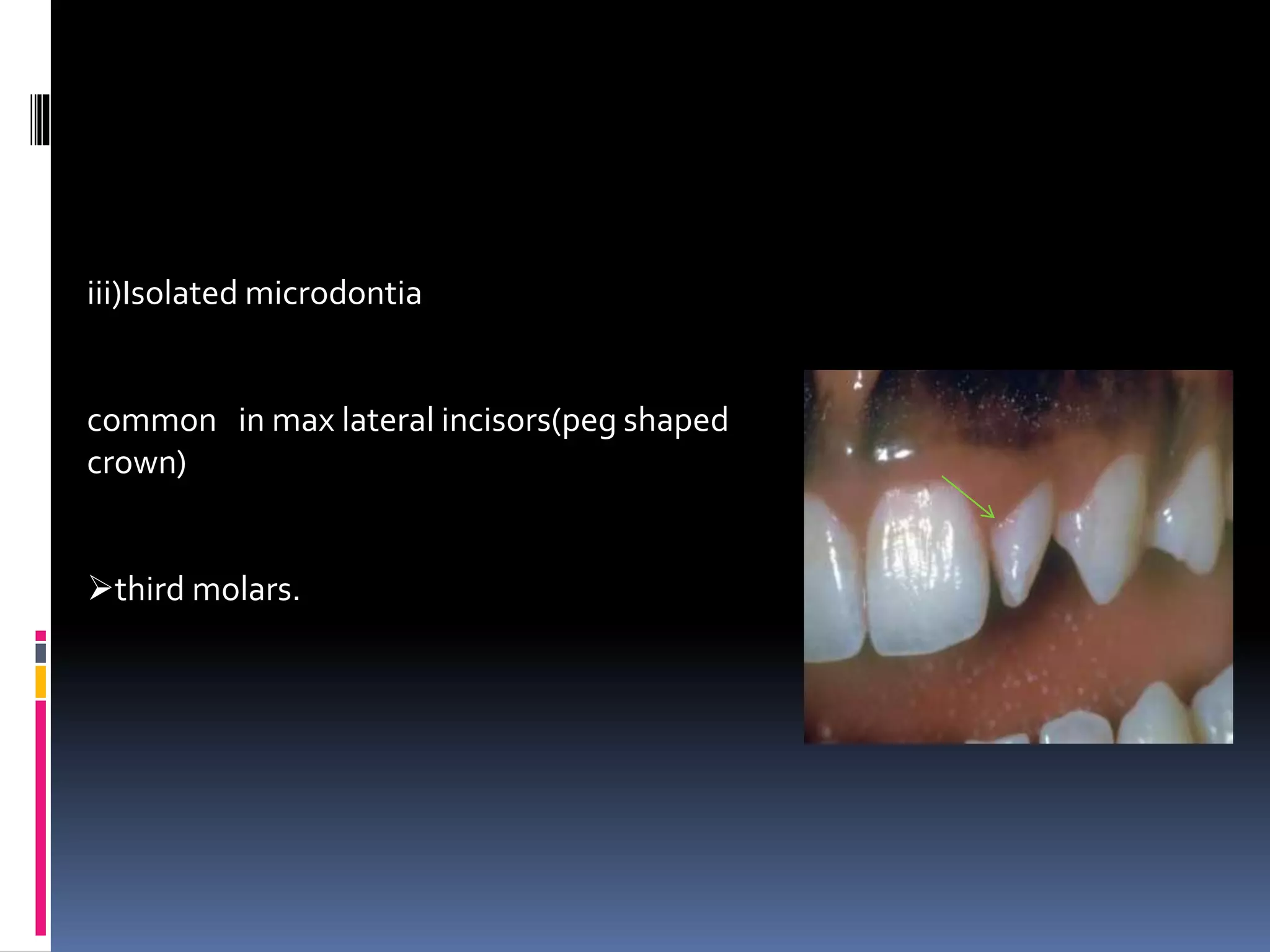
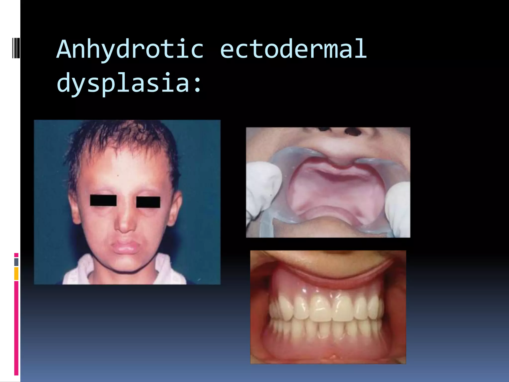
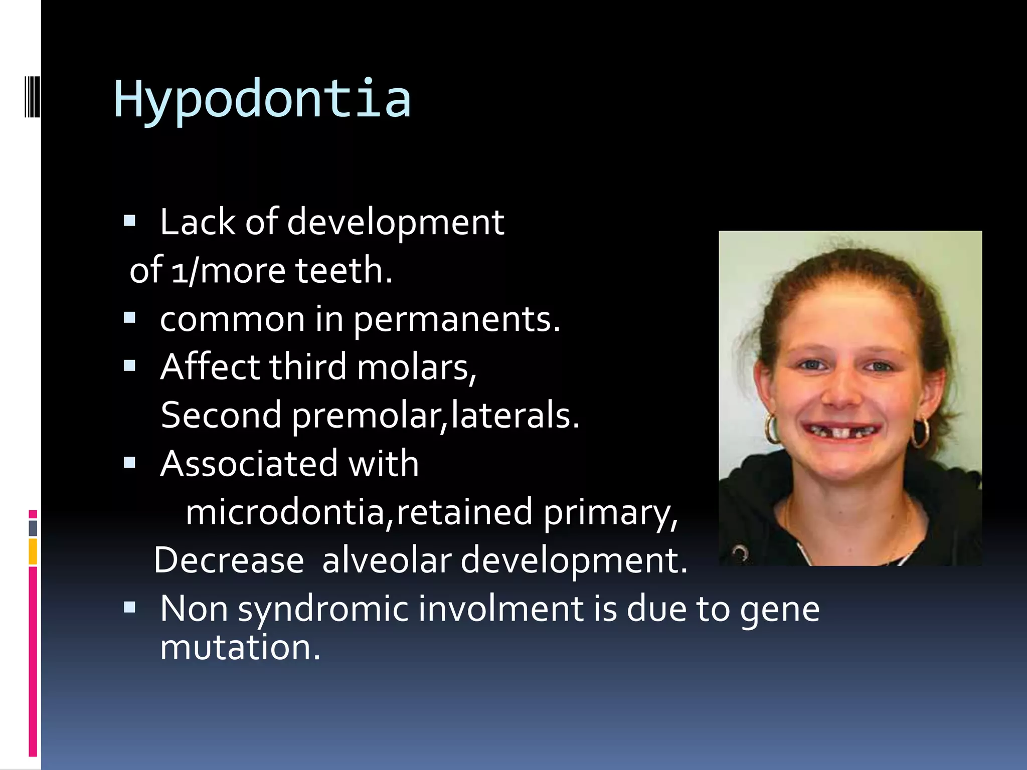
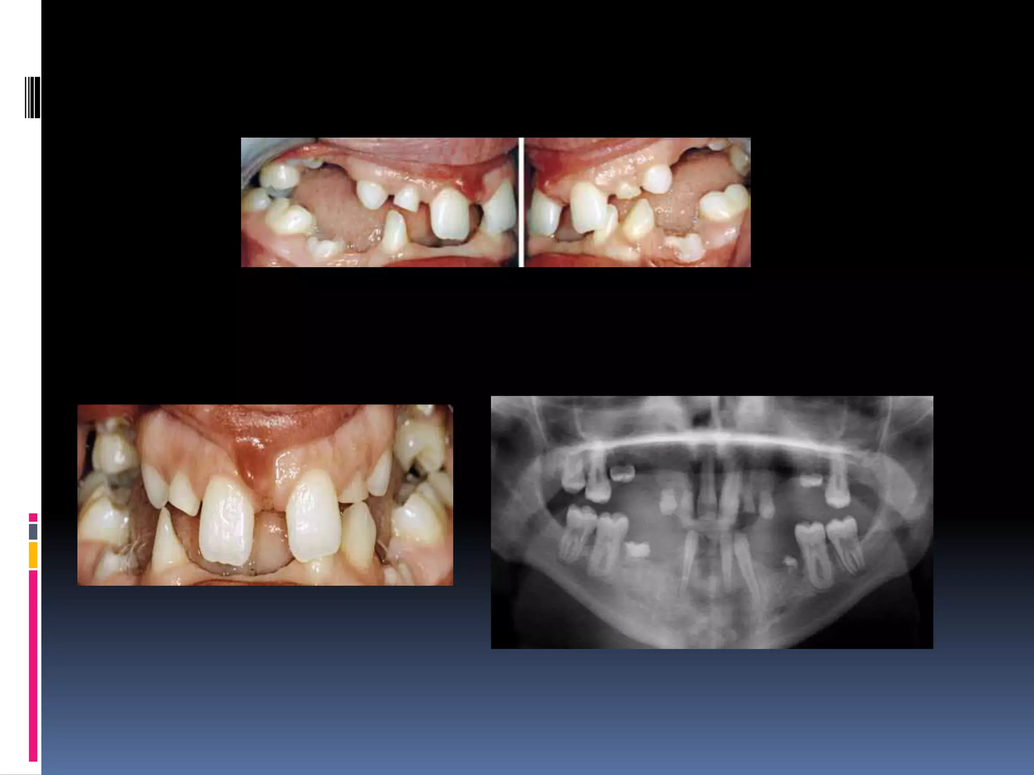
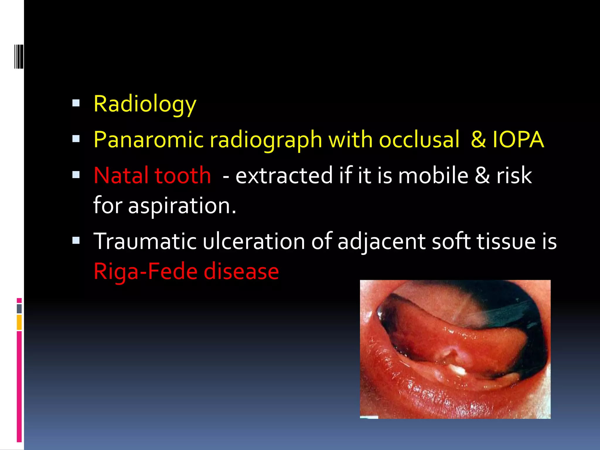
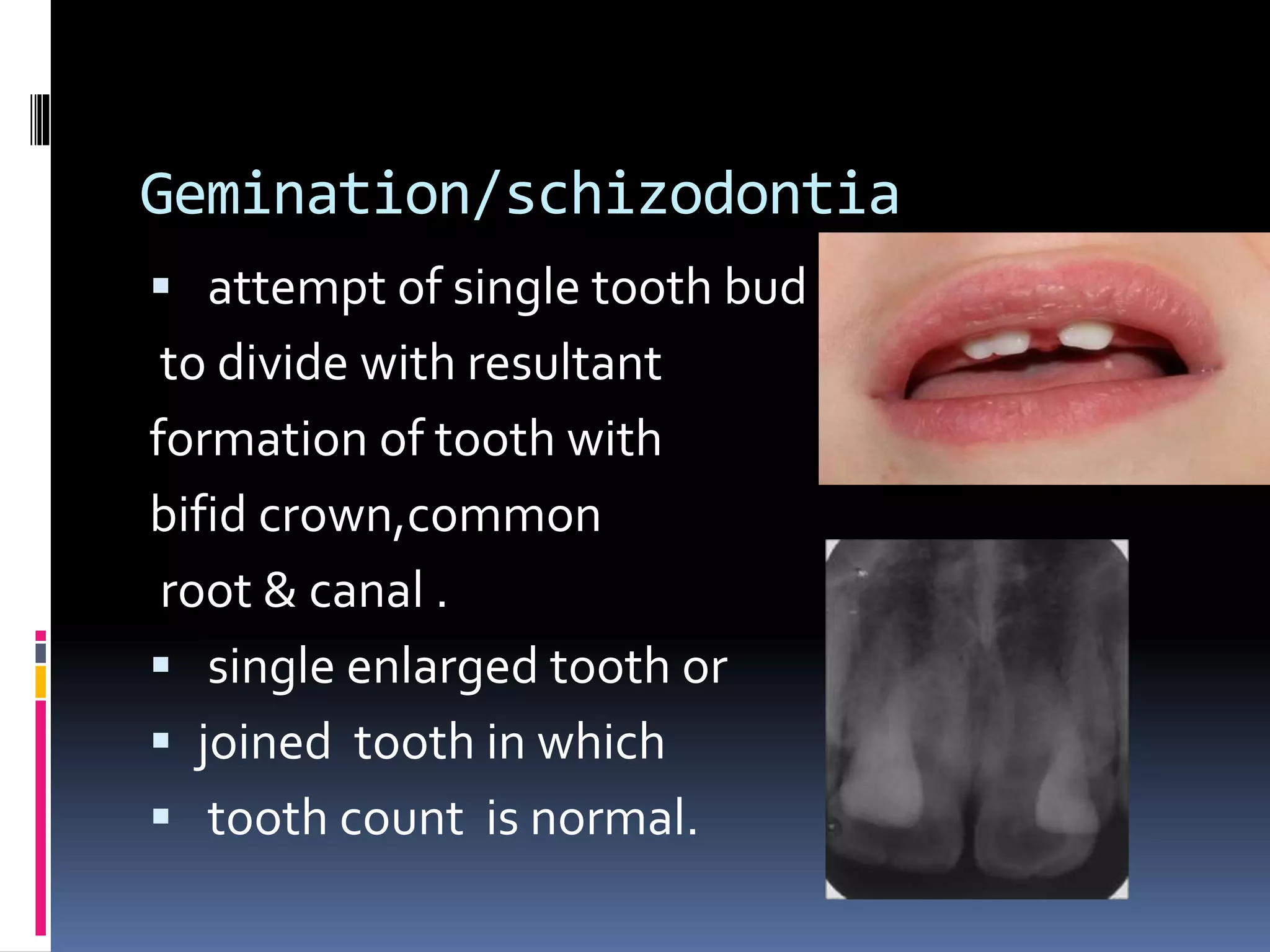
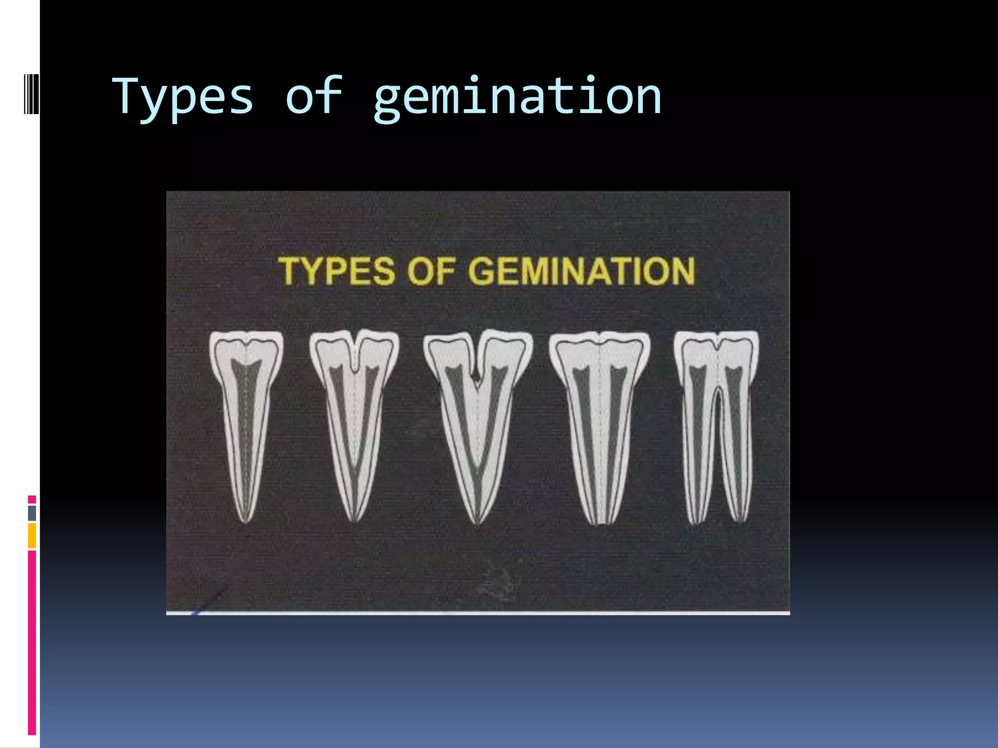
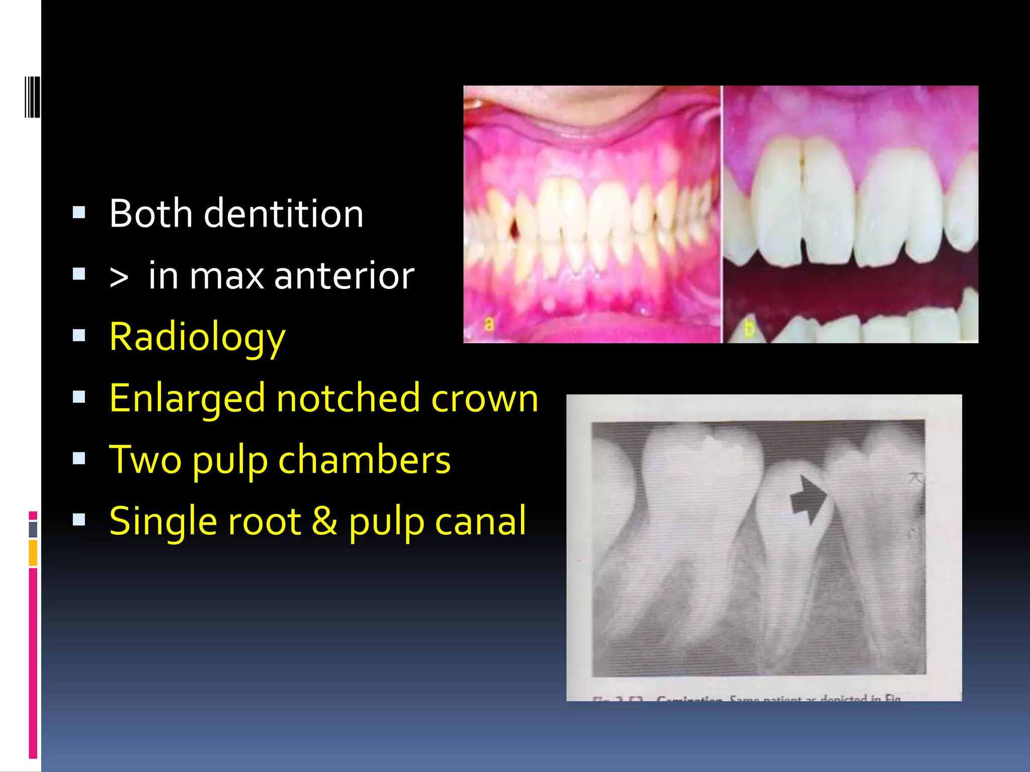
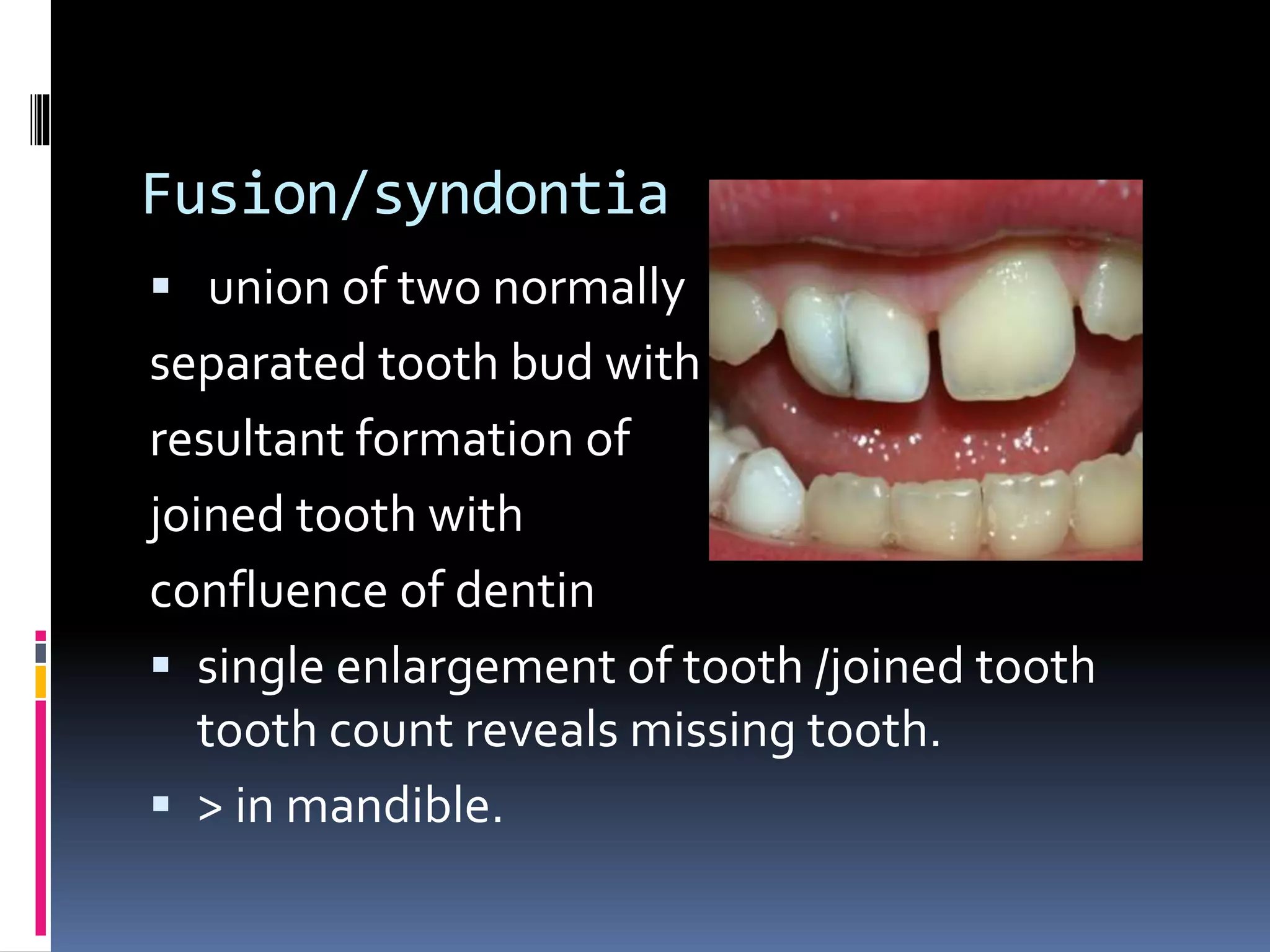
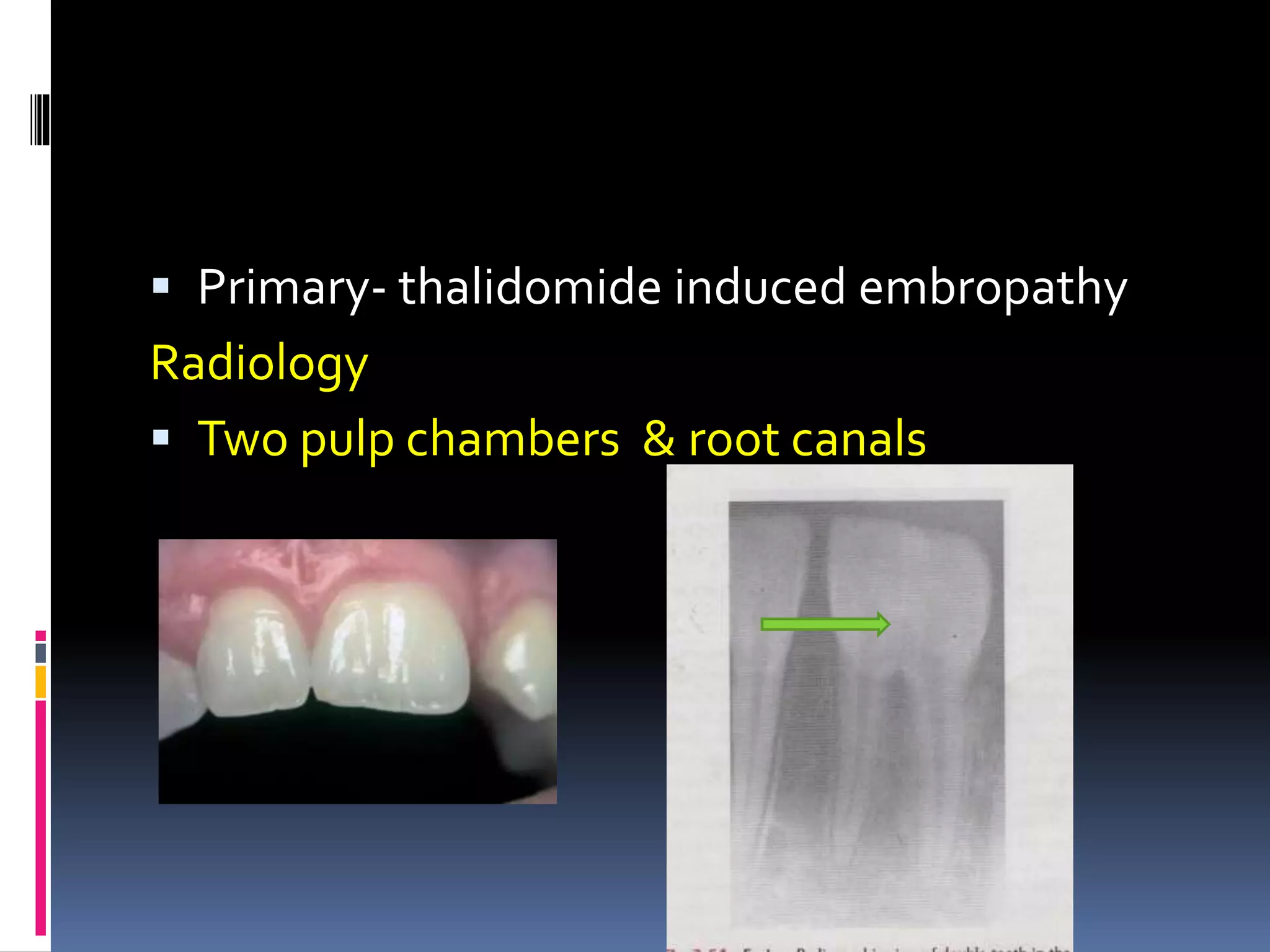
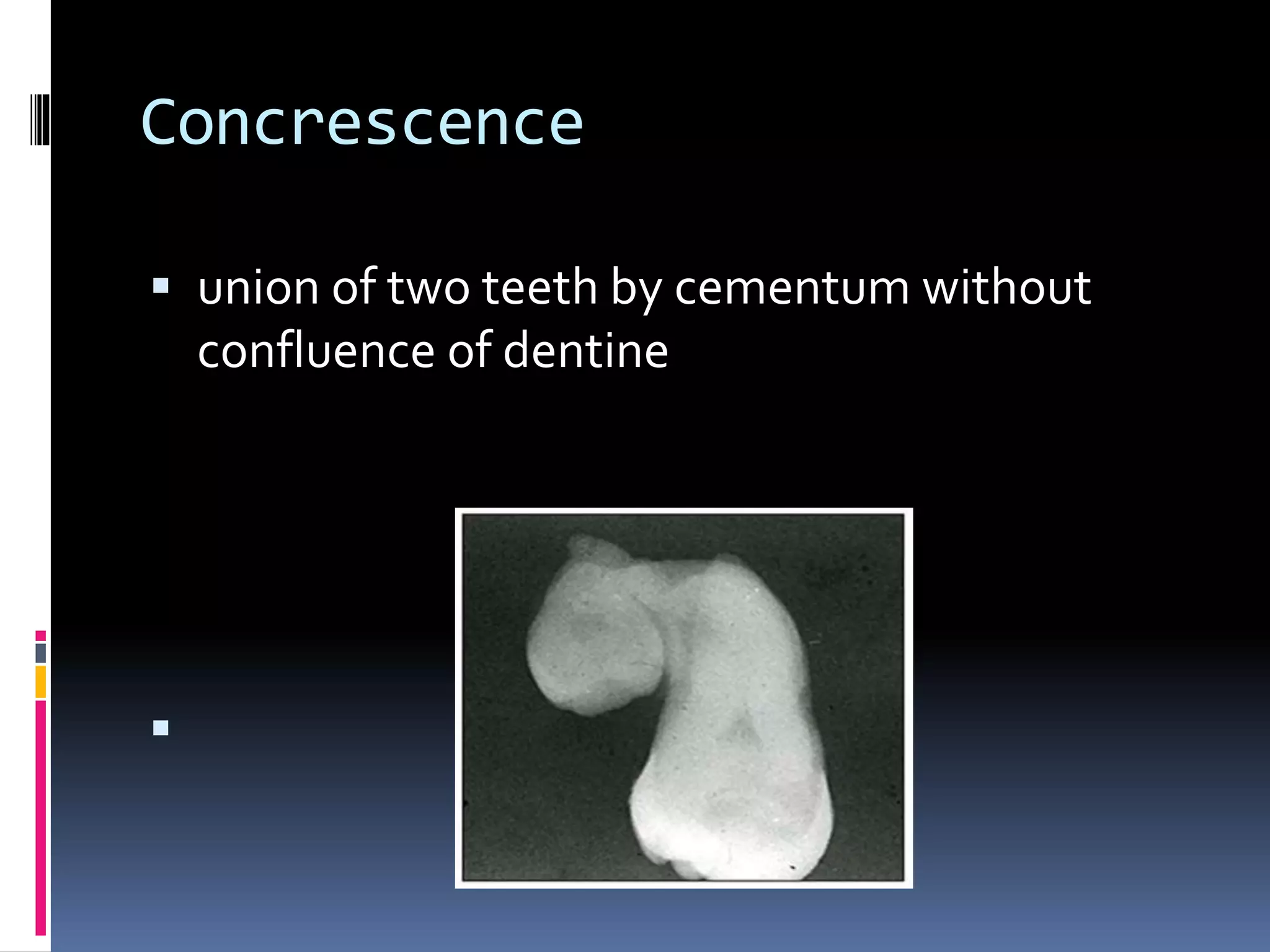
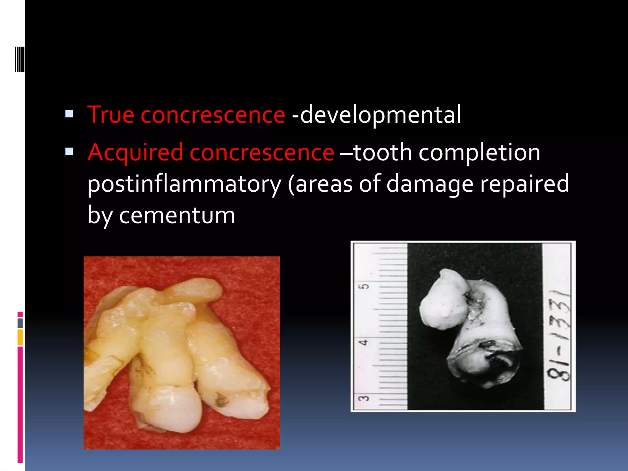
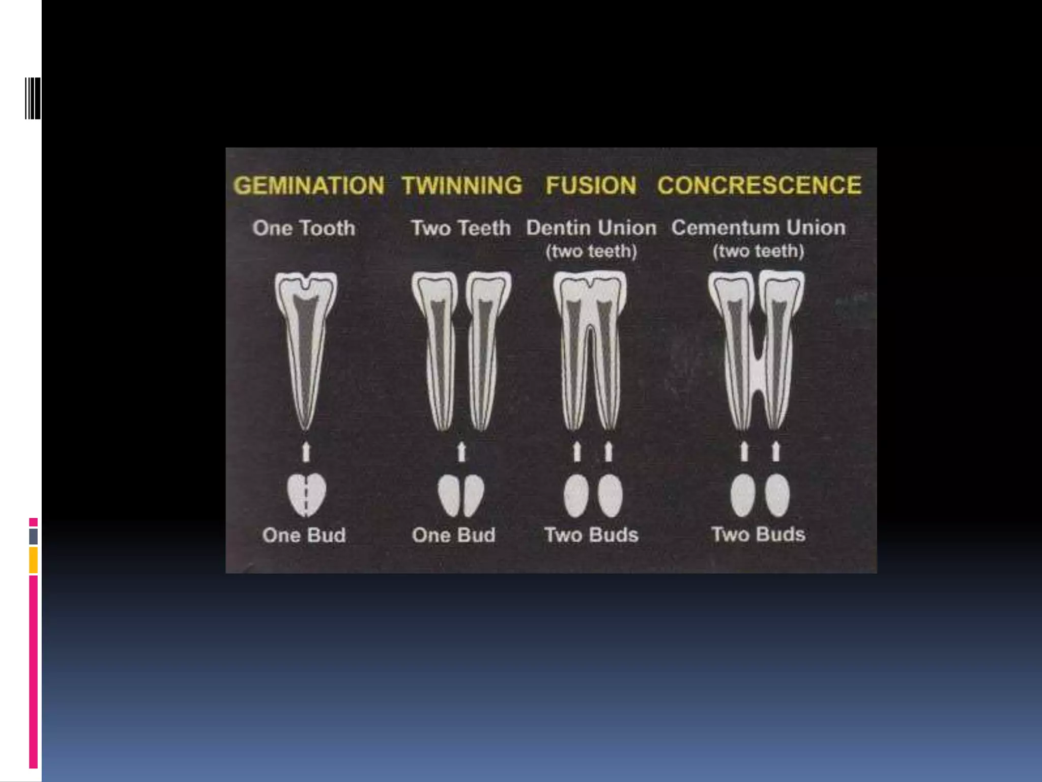
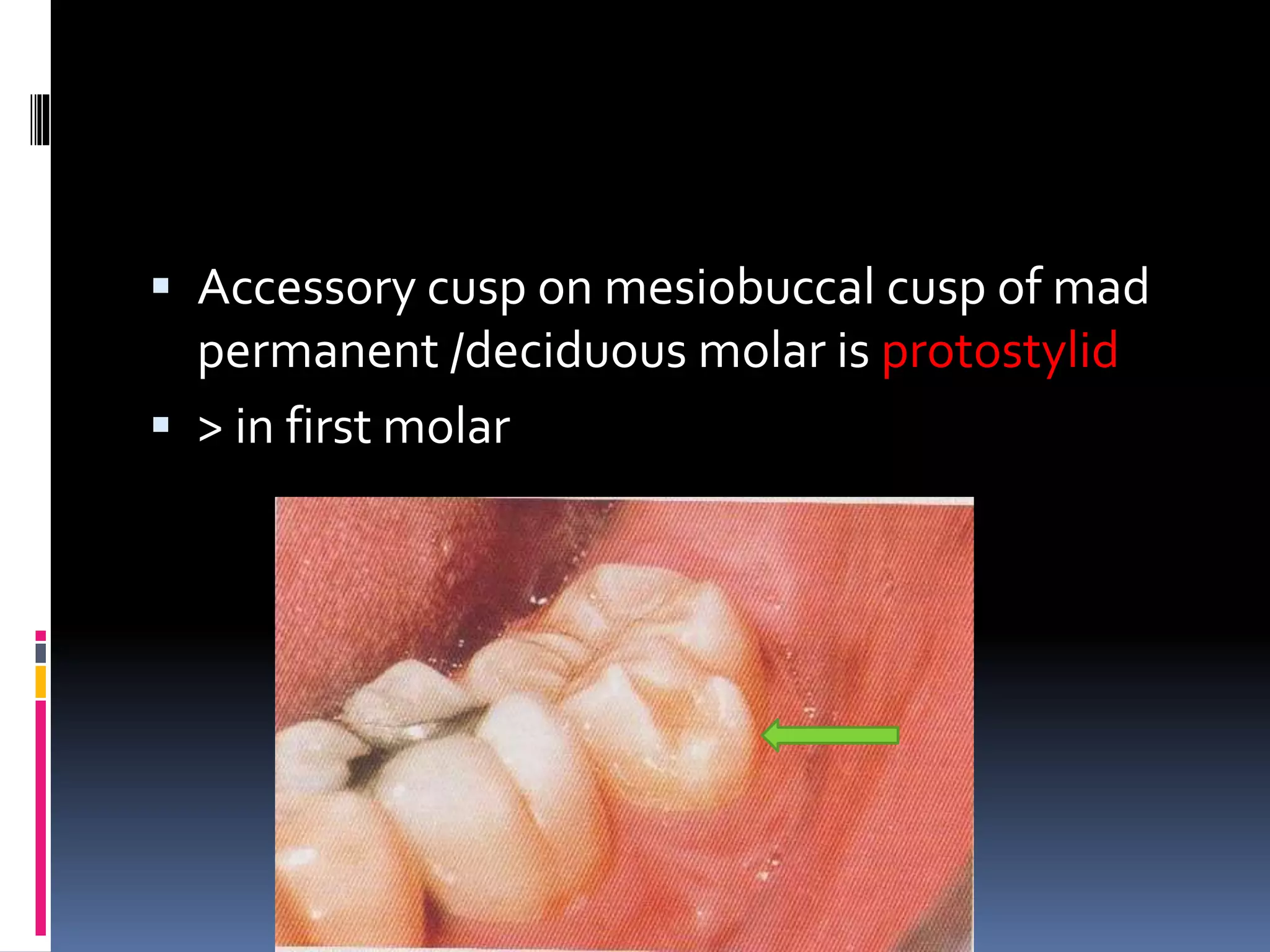
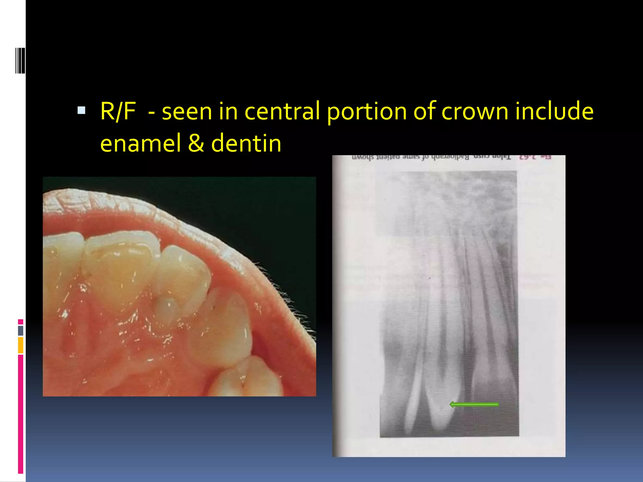
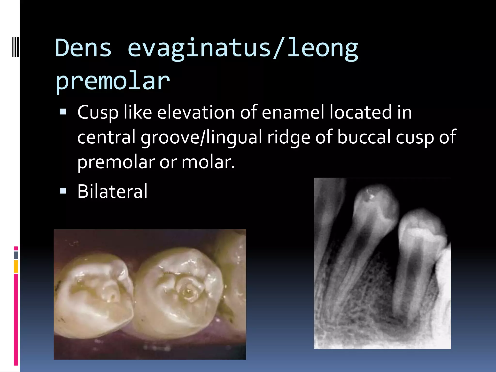
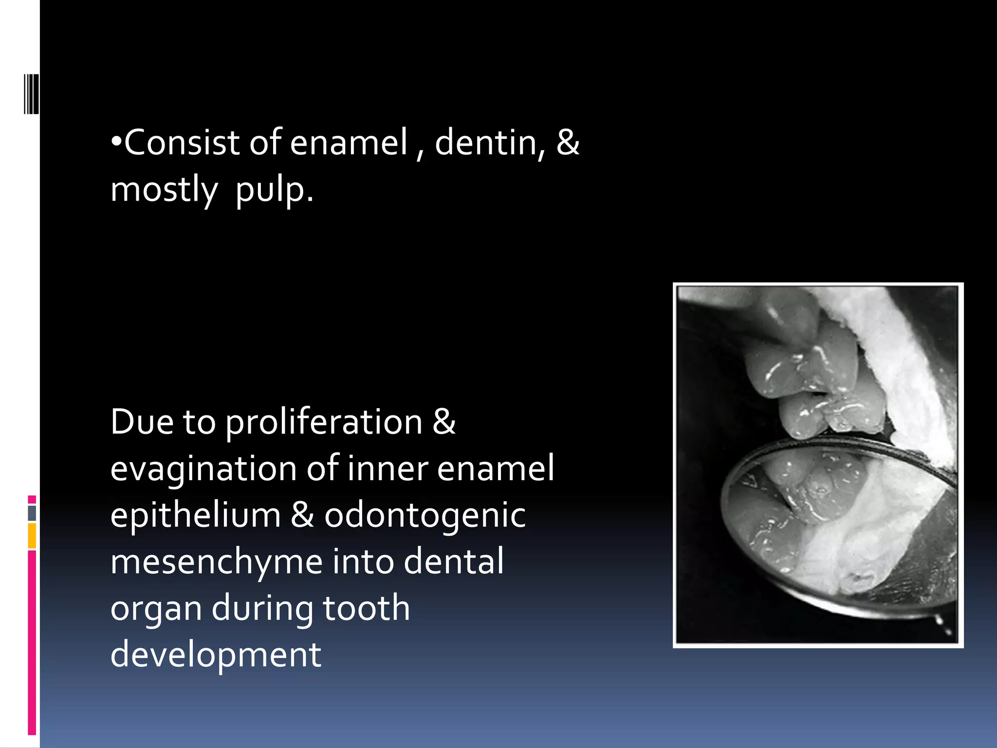
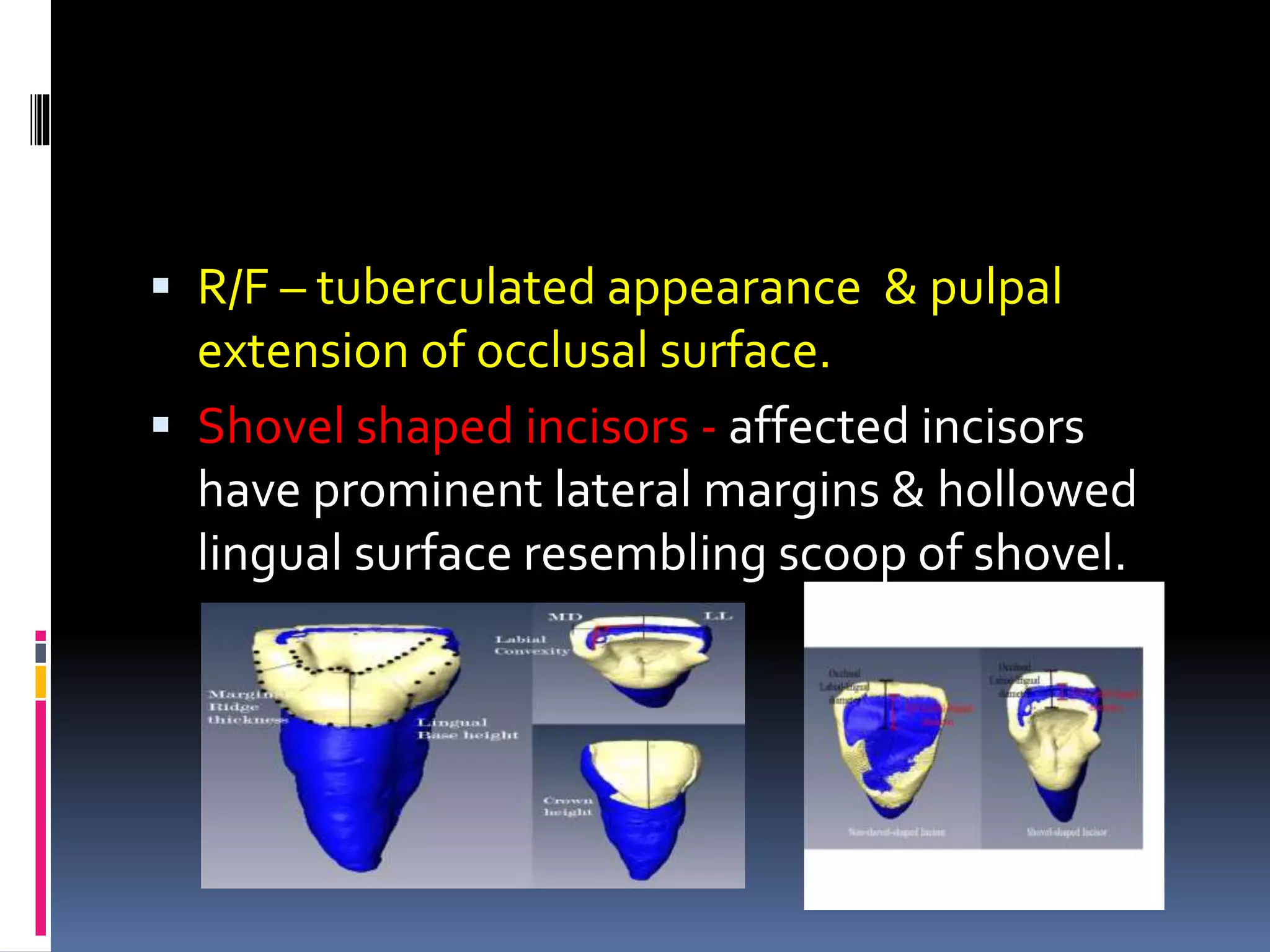
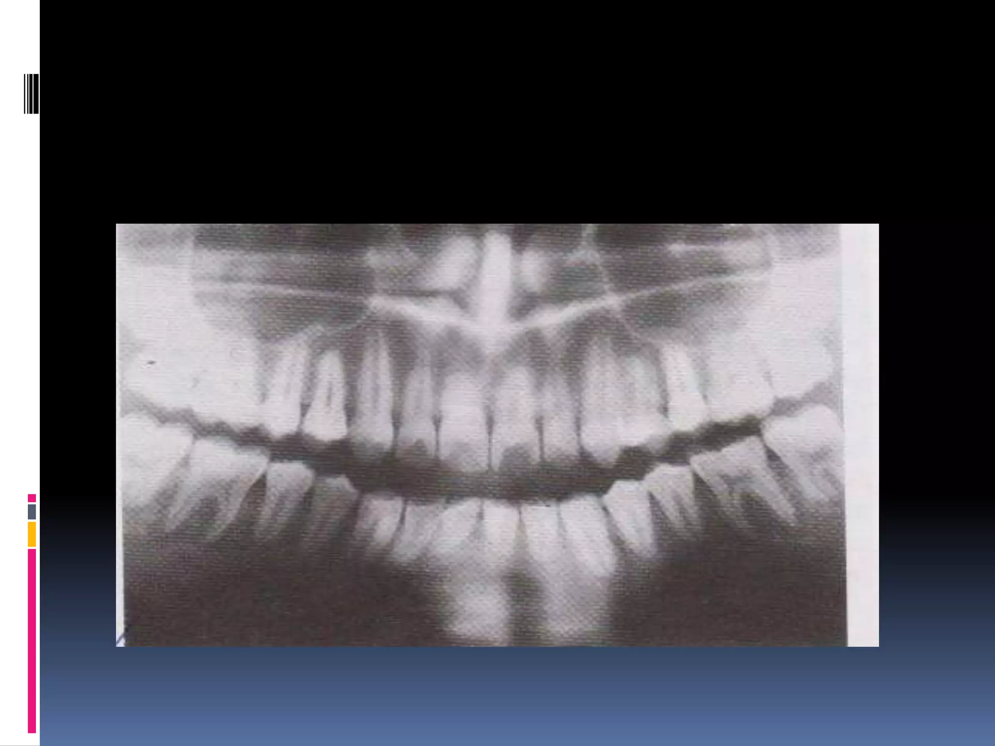
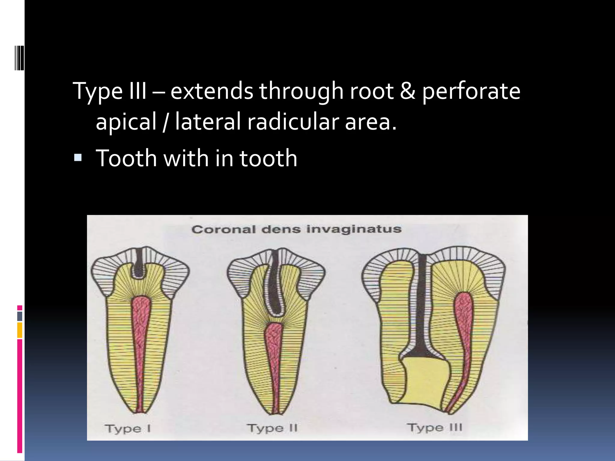
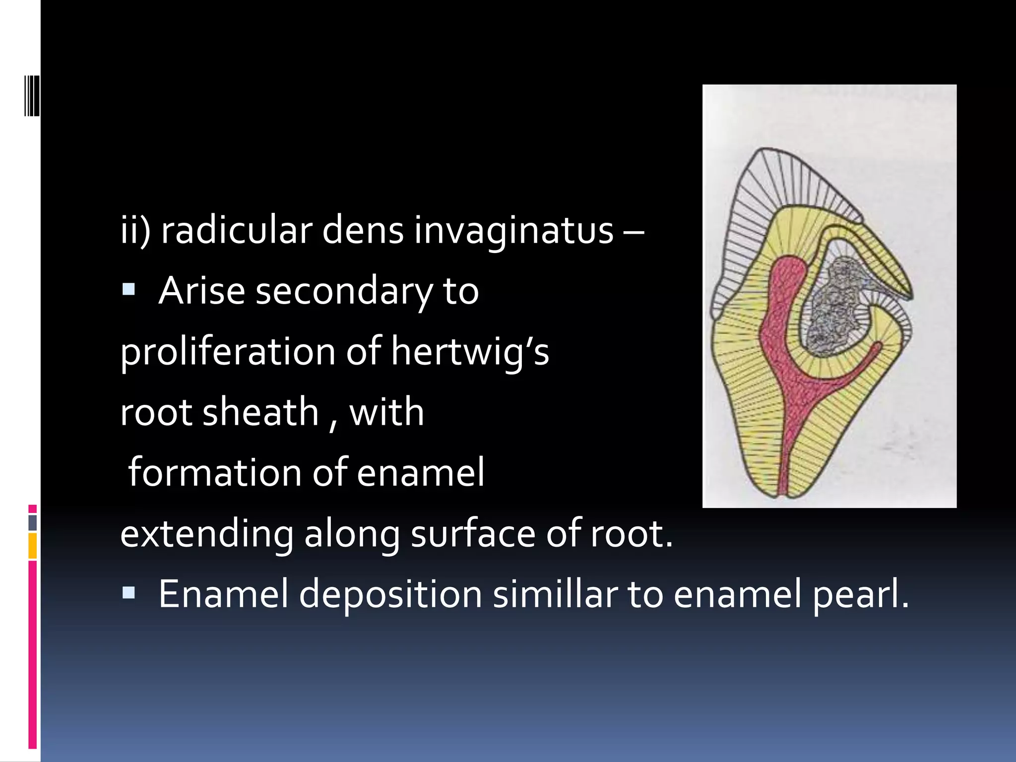
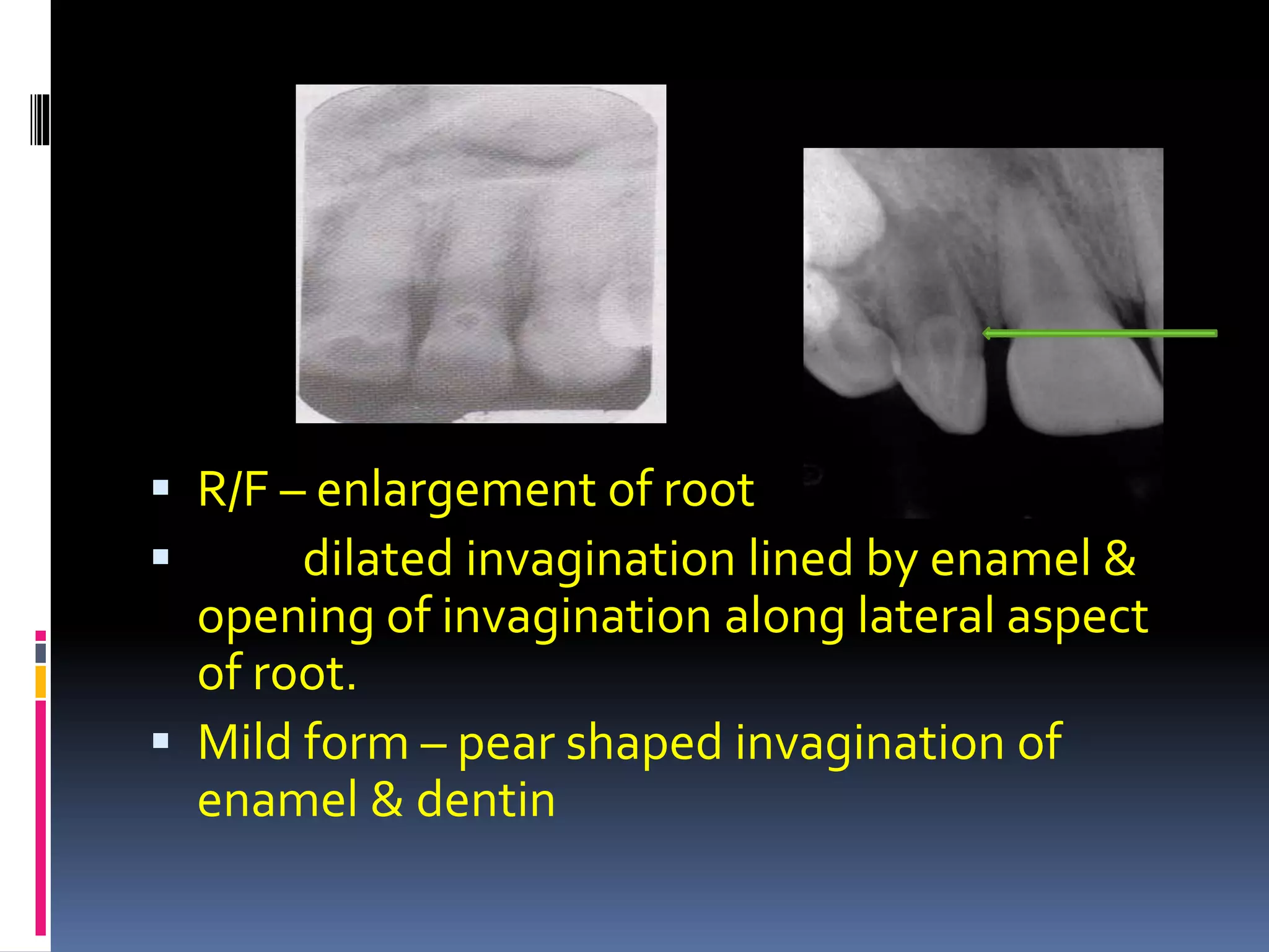
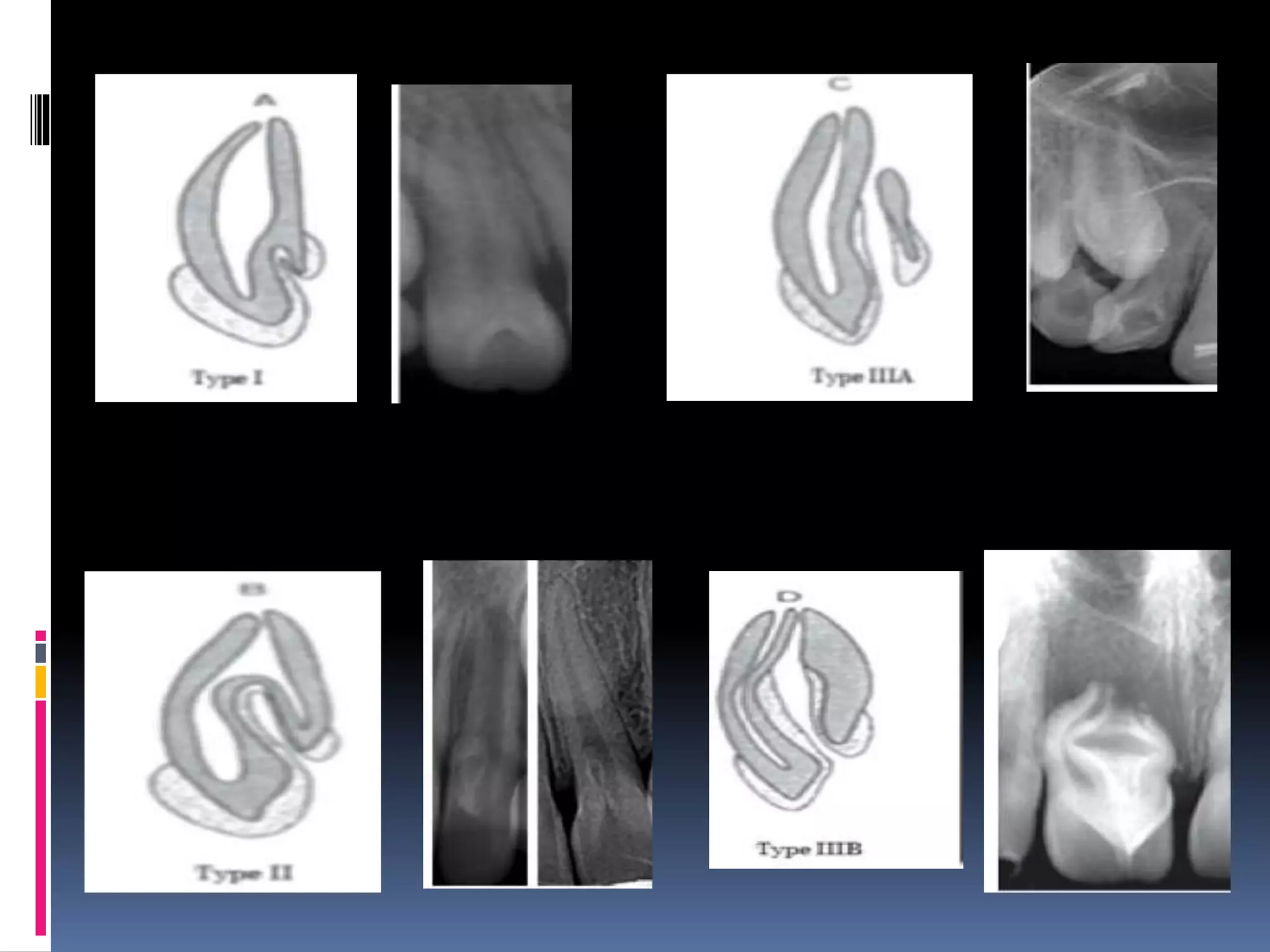
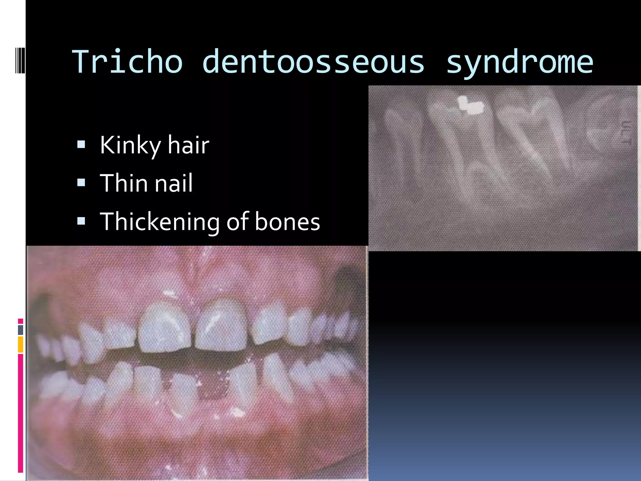
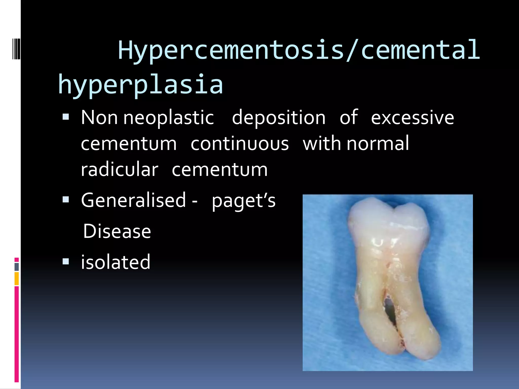
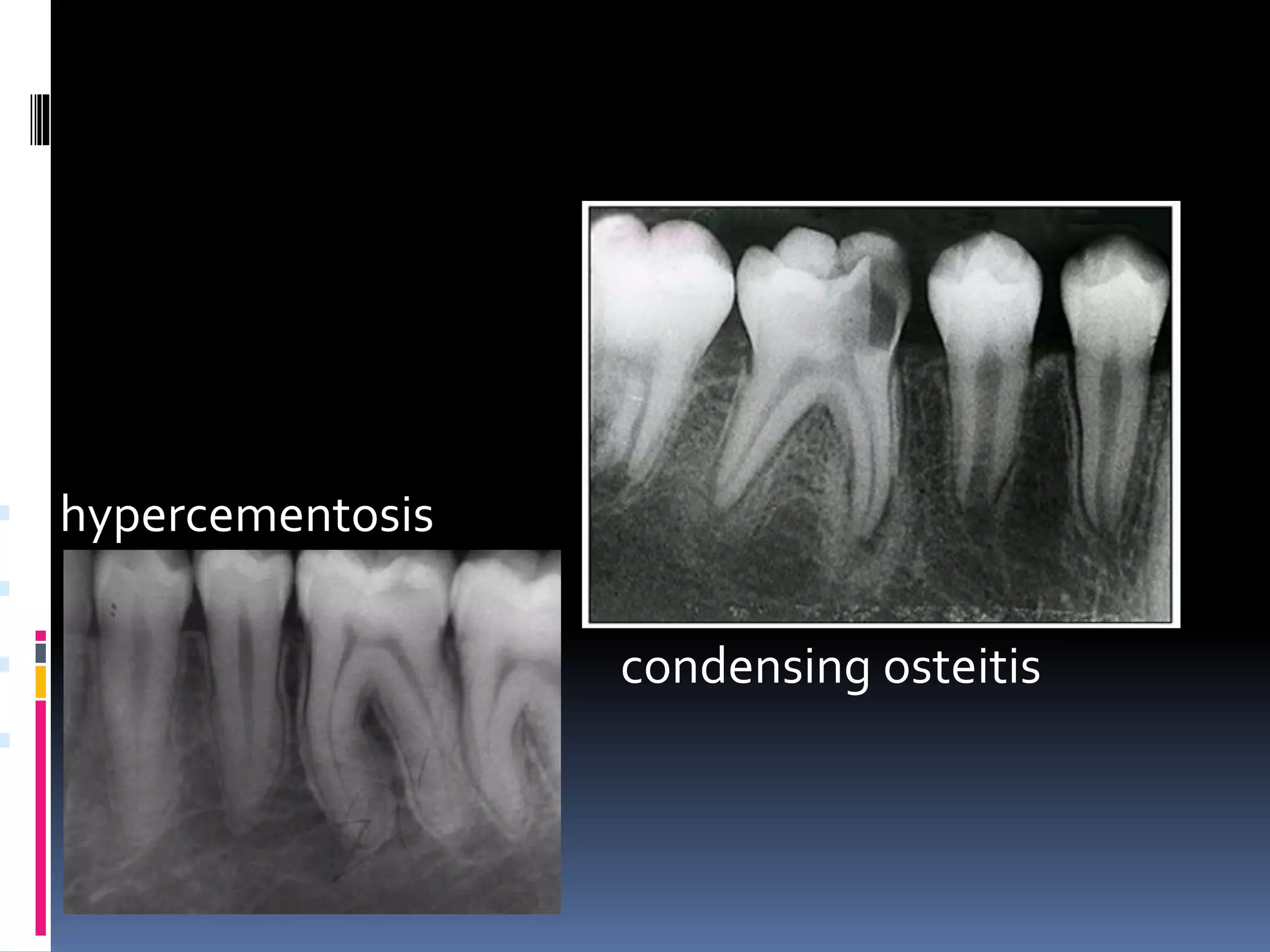
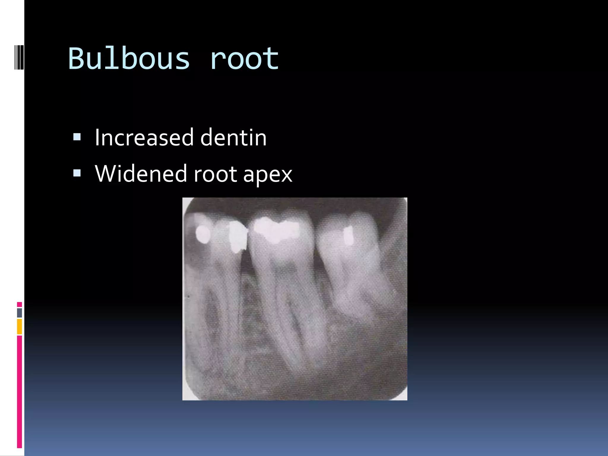
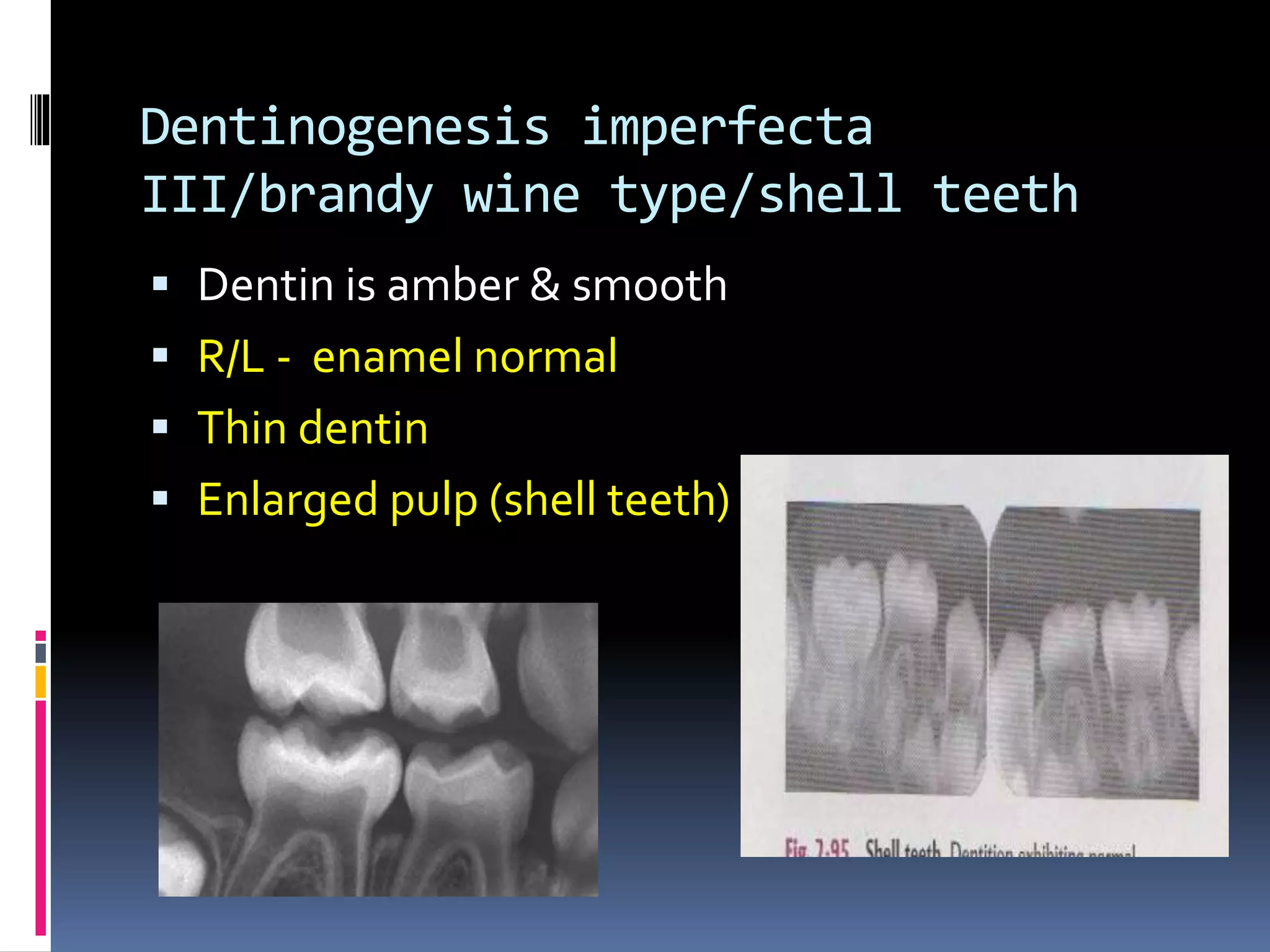
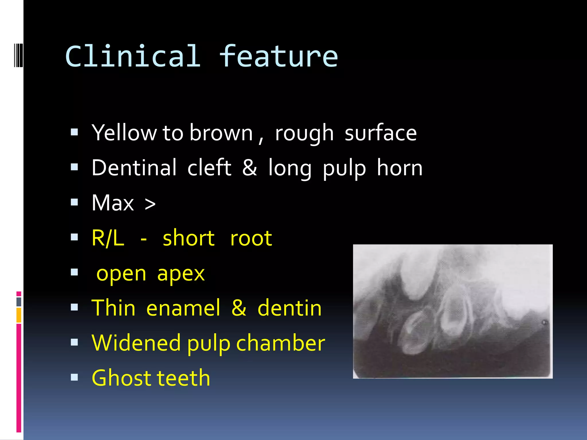
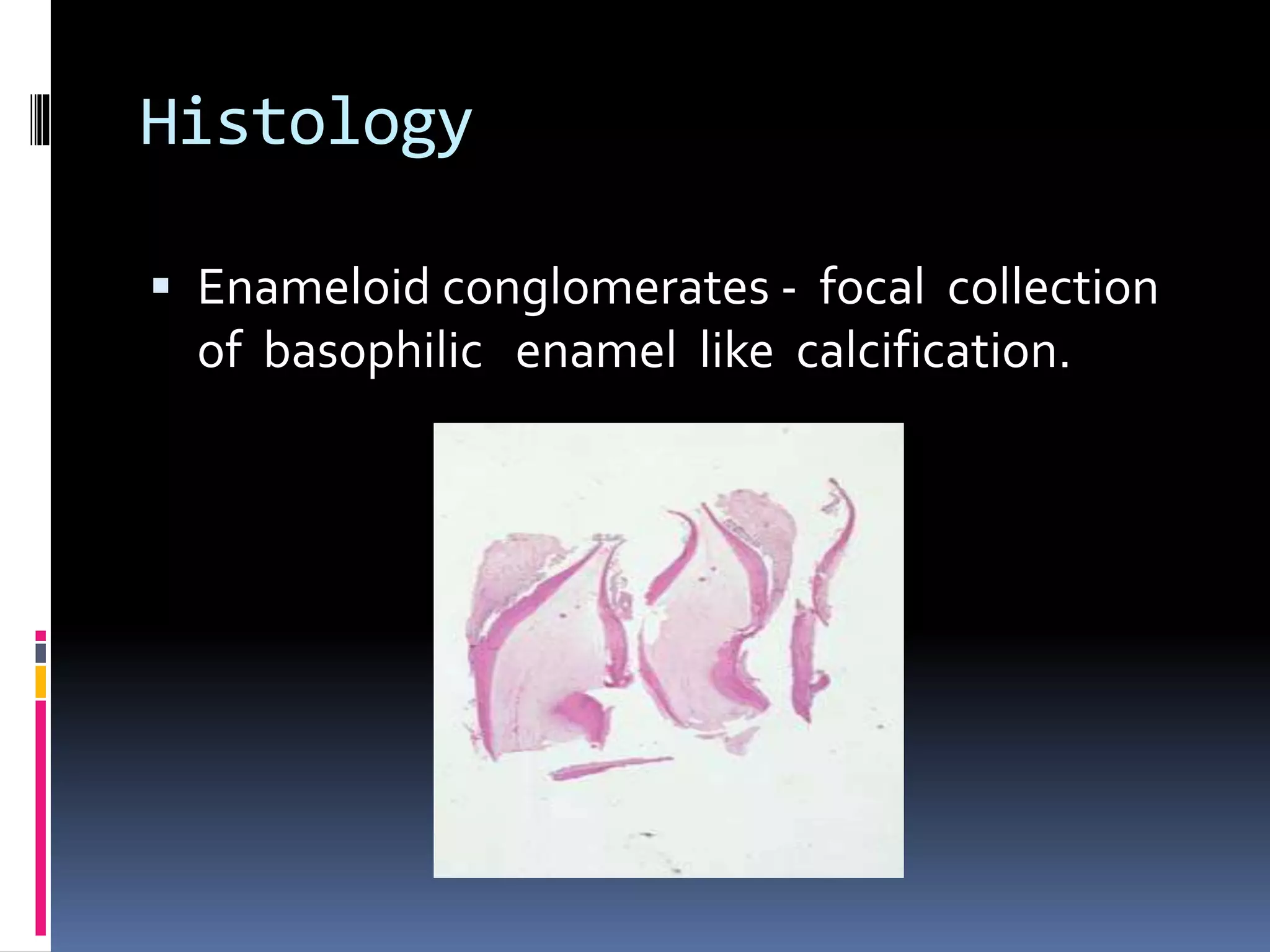
DEVELOPMENTAL DISTURBANCES
OF TEETH
Presented by
Melbia shiny
First MDS
contents
 Classification
 Clinical features
 Radiological features
 Treatment
Development of tooth
amelogenesis
 Formation of enamel matrix
 Mineralisation of matrix
 Maturation of enamel
dentinogenesis
 Cusp tip
 Predentin
 Dentin till tooth eruption
 Hydroxyapatite crystals
 Crystal deposition radially from centre
(spherulite)
classification
1)In number
 Hypodontia
 Anodontia
 Hyperdontia
2)In size
microdontia
macrodontia
3)in shape
 Gemination
 Fusion
 Concrescence
 Accessory cusp
 Dens invaginatus
 Ectopic enamel
 Taurodontism
 Hypercementosis
 Acessory roots
 dilaceration
In size of teeth
1)Microdontia
– teeth physically smaller than usual.
i)True generalised microdontia:
 all teeth smaller than normal
 uncommon
 in downs syndrome , pituitary dwarfism
ii)Relative generalised microdontia:
 normal sized teeth in larger jaws.
 hereditary
iii)Isolated microdontia
common in max lateral incisors(peg shaped
crown)
third molars.
Macrodontia/megalodontia
 Teeth are physically larger than usual.
i)True generalised macrodontia/diffuse:
 rare
 all teeth larger than normal.
 in pituitary gigantism ,otodental
syndrome & pineal hyperplasia with
hyperinsulinism.
i
ii) Relative generalised macrodontia:
– normal sized teeth crowed in
small jaw.
- hereditary.
iii)Isolated macrodontia:
uncommon
in incisors, canine, second
premolar & third molar.
HEMIFACIAL HYPERPLASIA
Treatment and prognosis
 Done for aesthetic purpose
 Peg laterals- full size porcelain crowns.
In number of teeth
Anodontia
 Total lack of tooth development.
i)True anodontia/congenital absence of teeth:
 all teeth are missing.
 affect both dentition.
 rare
 in hereditary hypohidrotic
ectodermal dysplasia
Anhydrotic ectodermal
dysplasia:
Hypodontia
 Lack of development
of 1/more teeth.
 common in permanents.
 Affect third molars,
Second premolar,laterals.
 Associated with
microdontia,retained primary,
Decrease alveolar development.
 Non syndromic involment is due to gene
mutation.
 Affected gene correlates to missing teeth:
AXIN2 gene - second & third molar,second
premolar, lower incisor.
Associated with adenomatous polyp of colon
& colorectal carcinoma.
 PAX9 gene – second molar
 MSX1 gene – second premolar & third molar
 He-Zhao gene – third molar, second
premolar,max laterals.
Syndromes associated with
hypodontia
Book’s syndrome:(PHC
syndrome)
agenesis of premolars
hypohidrosis
canities
white hair
Riegers syndrome:
agenesis of incisors.
congenital glaucoma.
Incontinentia
pigmenti/bloch- sulzberger
syndrome:
peg/cone shaped
baldness
Crouzon disease:
 early closure of dysostosis
 acanthosis nigricans
 hypoplastic maxilla
Ellis van crevold syndrome:
enamel hypoplasia
middle upper lip fuse with max
gingival margin.
delayed tooth eruption
Apert syndrome
Mid face hypoplasia
Syndactyly
Shovel shaped incisor
Down syndrome
Enamel hypoplasia
Hypo & hyperdontia
macroglossia
Ehler danlos syndrome
 Pulp stones
 Enamel hypoplasia
 both
Oligodontia
 Lack of development of 6/more teeth.
Radiology
 Panoramic radiographs
Hyperdontia
increase number of tooth
 Supernumerary- additional teeth
 Associated with macrodontia.
 Develop from third tooth bud from dental
lamina.
 Splitting of permanent bud
 Local , conditioned hyperactivity of dental
lamina.
Classification of supernumerary
 According to morphology :
1)Rudimentary
abnormal shape & size.
 Conical
Peg shaped, common, as mesiodens
 Tuberculate
barrel shape anterior with> 1 cusp, less
frequent , associated with delayed incisor
eruption.
 Molariform
small molar/premolar like.
2)supplemental:
normal size & shape
common in max laterals.
3)Odontome
hamartomas ,included in odontogenic
neoplasm.
 According to location:
1)Mesiodens
 in max anterior incisors.
.
2)Distomolars
 distal to third molar
3)Paramolar
lingual /buccal to molar.
 Supernumerary in soft palate, max sinus
,nasal cavity, orbit, brain ,sphenomaxillary
fissure.
Syndromes associated with
hyperdontia
cleidocranial dysplasia
Gardner syndrome
Hallermann streiff syndrome
 Dicephalia
 Parrot/beaked nose
 Mandibular hypoplasia
 Dwarfism
 Blue sclera
 hypertrichosis
 Dental transposition- normal teeth erupt into
inappropriate position
Commonly involved max. canines , first
premolars
 Natal teeth – accessory teeth present shortly
after birth
 Most common mad. Incisors
 Neonatal teeth
Teeth arising with in first 30 days of life is
 Arise from bud of accessory dental lamina.
Treatment & prognosis
Hypodontia is associated with
 Abnormal spacing of teeth
 Delayed tooth formation
 Delayed deciduous tooth exfoliation
 Late permanent tooth eruption
 Altered dimension of gnathic regions
Single missing tooth – no treatment
 Multiple teeth – prosthetic replacement with
RPD, FPD, osseointegrated implant & resin
bonded bridge.
 Children – FPD not indicated due to risk of
pulp exposure & implant not recommended
till completion of skeletal growth.
 Orthodontic treatment eliminate need for
restorative treatment follow up radiographs
since associated with external root
resorption.
 Radiology
 Panaromic radiograph with occlusal & IOPA
 Natal tooth - extracted if it is mobile & risk
for aspiration.
 Traumatic ulceration of adjacent soft tissue is
Riga-Fede disease
In shape of teeth
Gemination/schizodontia
 attempt of single tooth bud
to divide with resultant
formation of tooth with
bifid crown,common
root & canal .
 single enlarged tooth or
 joined tooth in which
 tooth count is normal.
Types of gemination
 Both dentition
 > in max anterior
 Radiology
 Enlarged notched crown
 Two pulp chambers
 Single root & pulp canal
Fusion/syndontia
 union of two normally
separated tooth bud with
resultant formation of
joined tooth with
confluence of dentin
 single enlargement of tooth /joined tooth
tooth count reveals missing tooth.
 > in mandible.
 Primary- thalidomide induced embropathy
Radiology
 Two pulp chambers & root canals
Concrescence
 union of two teeth by cementum without
confluence of dentine

 True concrescence -developmental
 Acquired concrescence –tooth completion
postinflammatory (areas of damage repaired
by cementum
Treatment & prognosis
 Result in crowding , abnormal spacing &
delayed /ectopic eruption of permanent
 pronounced labial /lingual groove are caries
prone so place fissure sealant or composite
restoration.
 Surgical division & endodontic treatment &
full crown.
 Concrescence interferes with eruption –
surgical removal done.
 Extraction difficulties & surgical separation
done.
Accessory cusps
 Cusp of carabelli
 Talon cusp
 Dens evaginatus
Cusp of carabelli
 Accessory cusp on palatal surface of mesio
lingual cusp of max molar.
 In both dentition
 Accessory cusp on mesiobuccal cusp of mad
permanent /deciduous molar is protostylid
 > in first molar
Talon cusp
 Is a well delineated additional cusp that is
located on anterior tooth extending from CEJ
to incisal edge
 > in permanent max incisors
 Resemble eagle’s talon
 Presence of deep developmental groove
 .
 R/F - seen in central portion of crown include
enamel & dentin
Syndroms associated
 Rubinstein taybi syndrome
 Mohr syndrome
 Ellis van creveld
 Incontinentia pigmenti achromias
 Sturge weber angiomatosis
Dens evaginatus/leong
premolar
 Cusp like elevation of enamel located in
central groove/lingual ridge of buccal cusp of
premolar or molar.
 Bilateral
•Consist of enamel , dentin, &
mostly pulp.
Due to proliferation &
evagination of inner enamel
epithelium & odontogenic
mesenchyme into dental
organ during tooth
development
 R/F – tuberculated appearance & pulpal
extension of occlusal surface.
 Shovel shaped incisors - affected incisors
have prominent lateral margins & hollowed
lingual surface resembling scoop of shovel.
Shovel shaped incisor
syndrome
 Interproximal caries
 Lingual pit caries
 Periapical & pulpal lesion
 Shortened & tapered roots
Treatment & prognosis
 Cusp of carabelli – no treatment.
 Deep grooves are sealed to prevent caries.
 Talon cusp – no therapy (mad teeth).
 In max – interfere with occlusion hence removal
 Periodic grinding for tertiary dentin deposition &
pulpal recession , apply fluoride varnish
(desensitizing).
 After removal , dentin covered with Ca(OH)2 &
enamel is etched & composite resin is placed.
 Dens evaginatus - occlusal interference
eliminated with removal of minimum dentin
& treatment with stannous fluoride.
 Shovel shaped incisors – restoration of deep
fissures to prevent caries.
Dens invaginatus/dens
indente
A deep surface invagination of crown/root
lined by enamel.
Due to invagination of surface of tooth crown
before calcification occurred.
 > max laterals
 i)coronal dens invaginatus:
 Type I – invagination confined to crown.
 Type II – extends below CEJ but not
communicating with pulp.
Type III – extends through root & perforate
apical / lateral radicular area.
 Tooth with in tooth
ii) radicular dens invaginatus –
 Arise secondary to
proliferation of hertwig’s
root sheath , with
formation of enamel
extending along surface of root.
 Enamel deposition simillar to enamel pearl.
 R/F – enlargement of root
 dilated invagination lined by enamel &
opening of invagination along lateral aspect
of root.
 Mild form – pear shaped invagination of
enamel & dentin
Treatment & prognosis
 Type I – restoration of invagination to prevent
caries.
 If caries – endodontic treatment
 Type III – temporary placement of Ca(OH)2
with endodontic treatment
 Radicular - openings are closed before pulpal
necrosis.
Palato gingival groove
 Associated with periodontal defect
Ectopic enamel
 Presence of enamel in unusual locations.
i)Enamel pearl /enameloma:
Hemispheric structures consist entirly of
enamel/ contain dentin & pulp.
 Due to prolonged contact b/t hertwig’s root
sheath & developing dentin inducing enamel
formation.
 1-4 epithelial pearls.
> in root furcation /near CEJ.
 R/F – well defined radiopaque nodules along
root surface.(CEJ)
enamel pearl pulp stone
iii)cervical enamel extension :
 Dipping of enamel from CEJ towards
bifurcation of molar teeth.
 Base of triangle is continuous with coronal
enamel.
Buccal bifurcation cyst
Treatment & prognosis
 Enamel pearl – area of weak point of
periodontal attachment .
 Oral hygiene maintained to prevent loss of
periodontal support.
 Flattening / removal of enamel & furcation
plasty .
Taurodontism
 Enlagement of body & pulp chamber of
multirooted tooth , with apical displacement
of pulpal floor & bifurcation of root.
 Bull like tooth.
 Unilateral/bilateral.
 rectangular teeth with pulp chambers
increased apico occlusal height & bifucation
close to apex
etiology
 Failure of hertwig’s epithelial sheath to
invaginate
 Mutation resulting from odontoblastic
deficiency during dentinogenesis of root.
Atavistic feature
Classification
 According to degree of apical displacement of
pulpal floor:
i)Hypotaurodontism – mild
ii)Mesotaurodontism - moderate
iii)Hypertaurodontism - severe
Types
Hypertaurodont
Radiological features
 Rectangular shape
 Large pulp chambers with > apico occlusal
height.
 Lack of pulpal constriction ot cervical region.
 Short roots
 Bifurcation few mm above root apex.
Syndrome associated
with taurodontism
Tricho dentoosseous syndrome
 Kinky hair
 Thin nail
 Thickening of bones
 Klinfelter syndrome
 Mohr syndrome
 Down’s syndrome
Treatment & prognosis
 No specific therapy
 Significant periodontal destruction before
furcation involvement occurs.
Roots
 i) dilaceration
 Abnormal angulation /bend in root.
 Mostly idiopathic , but can occur due to injury
, due to cyst /tumour.
 Mostly in mad third molar, then max secon
premolar, then mad second molar.
 Deciduous involvement - inappropriate
resorption & delayed eruption of permanent
Treatment & prognosis
 Extraction of deciduous teeth
 Endodontic treatment done carefully to avoid
root perforation.
 Splinting of dilacerated tooth done to
overcome stress related problems , when
used as abutment for prosthetic problems
Supernumerary root
 Development of increase number of root in
tooth.
 Both dentition.
 > in molars than cuspid & premolar
 Radix entomolaris
 R/F – some are seen , others superimposed
radix
entomolaris
Hypercementosis/cemental
hyperplasia
 Non neoplastic deposition of excessive
cementum continuous with normal
radicular cementum
 Generalised - paget’s
Disease
 isolated
radiology
thickening/
blunting of root
root outline is enlarged &
delineated by PDL
space & lamina
dura
 hypercementosis

 condensing osteitis

Bulbous root
 Increased dentin
 Widened root apex
Treatment & prognosis
 Detection of accessory root – decrease failure
of endodontic treatment
 Significance in exodontia.
Screw driver
incisors/hutchinson’s teeth
Mulberry molars/fournier
molar
Structure of teeth
a)Amelogenesis imperfecta/hereditary enamel
dysplasia:
Developmental alteration in structure
of enamel in absence of systemic disorders
Classification according toWit kop (based on
phenotype & pedigree.
 Type I -HYPOPLASTIC
 Generalised pitted
 Localised pitted
 Localised pitted
 Diffuse smooth
 Diffuse smooth
 Diffuse rough
 Enamel agenesis
 Type II – HYPOMATURATION
 Diffuse pigmented
 Diffuse
 Snow capped
 Snow capped
 Type III- HYPOCALCIFIED
 Diffuse
 Diffuse
 Type IV A– HYPOMATURATION-
HYPOPLASTIC
 Type IV B –HYPOPLASTIC -
HYPOMATURATION
Mutated genes
1)AMELX gene
amelogenin(protein for enamel
formation).
2)ENAM gene
enamelin
3)MMP-20 gene
enamelysin(proteinase)
4)KLK4 gene
kallikrein -4 (proteinase)
5)DLX3 gene
code for proteins for craniofacial, tooth ,
hair , brain & neural development.
6)AMBN gene
ameloblastin
Hypoplastic amelogenesis
imperfecta
 Inadequate deposition of enamel matrix.
Generalised pattern:
 Pin point to pinhead sized pits
 Affect buccal surface
 Staining of pit
Localised pattern
 Both dentition
 Incisal,middle & buccal surface.
Autosomal dominant smooth pattern:
 Enamel is thin hard & glossy
 Open contact points
 Opaque white to translucent brown.
Rough pattern:
 Thin , hard ,rough surfaced enamel.
 Colour – white to yellowish white
 Anterior open bite
X linked smooth pattern:
 Males - diffuse,thin, smooth, & shiny
 Females – vertical furrows
 Open bite
 Brown – yellow brown.
 .
Enamel agenesis
 Total lack of enamel formation
 Yellow brown hue
 Open contact points
Hypomaturation
 defect in maturation of enamel crystals
 Normal shape
 Mottled, opaque, white brown yellow
discoloratioN
Pigmented pattern:
 Agar brown & mottled enamel
 Enamel fracture from dentin.
X linked pattern :
 Lyonization seen
 Focal areas of brown discolouration
 Males – opaque with translucent mottling
 Females – vertical bands of white opaque
enamel.
 Snow capped pattern:
 White opaque enamel on incisal/occlusal one
third of crown.
 Both dentition.
 Anterior & posterior distribution.
Snow capped
Hypocalcified
 Enamel matrix is laid down appropriately but
no mineralization.
 Enamel is soft & easily lost.
 Yellow – brown / orange but exhibit stained
brown- black.
 Coronal enamel is removed with cervical
portion calcified
Amelogenesis imperfecta with
taurodontia
 hypomaturation/hypoplastic :
 Both dentition
 Enamel – mottled yellow- white to yellow-
brown.
 Buccal side – pits
 R/F – enamel & dentine same density
 - large pulp chambers.
 Hypoplastic hypomaturation:
 Enamel – thin
 R/F - enamel & dentin same density.
 - large pulp chambers.
 In tricho -dento- osseous syndrome
CLINICAL FEATURES
 Hypoplastic - pitted surface
 Hypocalcified - soft enamel removed with
 Prophylaxis scaler
 Hypomaturation - can be pierced by
explorer, lost by chipping
radiology
 Hypoplastic - tapered crown
 lack of contact
 Hypocalcified - no contrast b/t enamel &
dentin
 Enamel moth eaten appearance
 Hypomaturation - enamel chips & abrades
 H/F – decalcification – enamel is lost.
 Ground section of non- decalcified specimens
Treatment & prognosis
 Placement of full crowns
 Severe case – full dentures (over dentures)
Turner’s tooth
Hypoplasia due to
antineoplastic therapy
Environmental enamel
hypoplasia
Dentinogenesis Imperfecta
 Hereditary developmental disturbance of
dentin in absence of systemic disorder.
 Mutation of DSPP(dentin
sialophosphoprotein) gene
Shield’s classification
a)Dentinogenesis imperfecta I /capdepont
teeth/shield’s type II/DI with out osteogenesis
imperfecta.
b)Dentinogenesis imperfecta II/ shield’s type
I/DI with osteogenesis imperfecta.
c)Dentinogenesis imperfecta III/brandy wine
type/shell teeth
DI with out osteogenesis
imperfecta/opalescent dentin
 Blue brown discoloration
DI with osteogenesis
imperfecta
 Genes COL I A1, COL I A 2 code for type I
collagen.
radiology
.bulbous crown
.thin roots
.obliteration
of root canal & pulp
chamber.
.cervical constriction
.enamel hypoplasia.
Dentinogenesis imperfecta
III/brandy wine type/shell teeth
 Dentin is amber & smooth
 R/L - enamel normal
 Thin dentin
 Enlarged pulp (shell teeth)
Histology
 Dentin near enamel is normal, rest abnormal
 Atypical odontoblast lining pulp surface are
entrapped in defective dentin
 Enamel is normal
Treatment & prognosis
 Crown not recommended ( cervical
fracture) , so over lay dentures & teeth
covered with Fluorid releasing GIC
Dentin dysplasia
 Type I - radicular dentin dysplasia/ rootless
teeth
 Type II - coronal dentin dysplasia
Rootless tooth
 Short root - less root dentin
 Tooth mobility (premature exfoliation)
 Predispose to fracture
O’carroll’s classification
of Dentin dysplasia
Type I a
typeI d
Coronal dentin dysplasia
blue–amber-brown
R/L – thistle tube/
flamed shape
Pulpal dysplasia
 Normal clinically
 Both dentition thistle tube shaped / flamed
shaped
 Multiple pulp stones
Histology
 Type I coronal enamel & dentin normal
 Whorls of dentin & atypical osteodentin
(stream flowing around boulders)
 Type II numerous interglobular dentin near
pulp
Pulp stones
Treatment & prognosis
 Type I - short root s (early loss from
periodontitis)
 Pulp channels close to DEJ ( restoratio n
& endodontic treatment)
 Type II - permanent - endodontic
treatment
Regional
odontodysplasia/ghost teeth
 Localised , non hereditary developmental
abnormality of teeth with extensive
adverse effect on formation of enamel
dentin & pulp
Clinical feature
 Yellow to brown , rough surface
 Dentinal cleft & long pulp horn
 Max >
 R/L - short root
 open apex
 Thin enamel & dentin
 Widened pulp chamber
 Ghost teeth
Histology
 Enameloid conglomerates - focal collection
of basophilic enamel like calcification.
Treatment & prognosis
 Retension of altered teeth to pressure
surrounding alveolar ridge
 Unerupted teeth - left till skeletal growth
completes
 Erupted teeth - etched retained
restoration/stainless steel crowns
 Osseointegrated implant after pubertal
growth.
references
 Text book of oral pathology shafer, hine levy
 Colour atlas of common oral diseases
langlais miller
 Human embryology inderber singh
 Diagnostic imaging of jaws – langlais
langland
 Oral 7 maxillofacial pathology neville damm
 Orbans oral histology & embrology
Thank you

Developmental disturbances of teeth

  • 1.
  • 2.
    contents  Classification  Clinicalfeatures  Radiological features  Treatment
  • 3.
  • 5.
    amelogenesis  Formation ofenamel matrix  Mineralisation of matrix  Maturation of enamel
  • 6.
    dentinogenesis  Cusp tip Predentin  Dentin till tooth eruption  Hydroxyapatite crystals  Crystal deposition radially from centre (spherulite)
  • 7.
    classification 1)In number  Hypodontia Anodontia  Hyperdontia 2)In size microdontia macrodontia
  • 8.
    3)in shape  Gemination Fusion  Concrescence  Accessory cusp  Dens invaginatus  Ectopic enamel  Taurodontism  Hypercementosis  Acessory roots  dilaceration
  • 9.
    In size ofteeth 1)Microdontia – teeth physically smaller than usual. i)True generalised microdontia:  all teeth smaller than normal  uncommon  in downs syndrome , pituitary dwarfism ii)Relative generalised microdontia:  normal sized teeth in larger jaws.  hereditary
  • 10.
    iii)Isolated microdontia common inmax lateral incisors(peg shaped crown) third molars.
  • 11.
    Macrodontia/megalodontia  Teeth arephysically larger than usual. i)True generalised macrodontia/diffuse:  rare  all teeth larger than normal.  in pituitary gigantism ,otodental syndrome & pineal hyperplasia with hyperinsulinism. i
  • 13.
    ii) Relative generalisedmacrodontia: – normal sized teeth crowed in small jaw. - hereditary. iii)Isolated macrodontia: uncommon in incisors, canine, second premolar & third molar.
  • 14.
  • 15.
    Treatment and prognosis Done for aesthetic purpose  Peg laterals- full size porcelain crowns.
  • 16.
  • 17.
    Anodontia  Total lackof tooth development. i)True anodontia/congenital absence of teeth:  all teeth are missing.  affect both dentition.  rare  in hereditary hypohidrotic ectodermal dysplasia
  • 18.
  • 19.
    Hypodontia  Lack ofdevelopment of 1/more teeth.  common in permanents.  Affect third molars, Second premolar,laterals.  Associated with microdontia,retained primary, Decrease alveolar development.  Non syndromic involment is due to gene mutation.
  • 21.
     Affected genecorrelates to missing teeth: AXIN2 gene - second & third molar,second premolar, lower incisor. Associated with adenomatous polyp of colon & colorectal carcinoma.  PAX9 gene – second molar  MSX1 gene – second premolar & third molar  He-Zhao gene – third molar, second premolar,max laterals.
  • 22.
  • 23.
    Book’s syndrome:(PHC syndrome) agenesis ofpremolars hypohidrosis canities white hair
  • 24.
    Riegers syndrome: agenesis ofincisors. congenital glaucoma.
  • 25.
  • 26.
    Crouzon disease:  earlyclosure of dysostosis  acanthosis nigricans  hypoplastic maxilla
  • 27.
    Ellis van crevoldsyndrome: enamel hypoplasia middle upper lip fuse with max gingival margin. delayed tooth eruption
  • 28.
    Apert syndrome Mid facehypoplasia Syndactyly Shovel shaped incisor
  • 29.
    Down syndrome Enamel hypoplasia Hypo& hyperdontia macroglossia
  • 30.
    Ehler danlos syndrome Pulp stones  Enamel hypoplasia  both
  • 31.
    Oligodontia  Lack ofdevelopment of 6/more teeth. Radiology  Panoramic radiographs
  • 32.
    Hyperdontia increase number oftooth  Supernumerary- additional teeth  Associated with macrodontia.  Develop from third tooth bud from dental lamina.  Splitting of permanent bud  Local , conditioned hyperactivity of dental lamina.
  • 33.
    Classification of supernumerary According to morphology : 1)Rudimentary abnormal shape & size.  Conical Peg shaped, common, as mesiodens  Tuberculate barrel shape anterior with> 1 cusp, less frequent , associated with delayed incisor eruption.  Molariform small molar/premolar like.
  • 35.
    2)supplemental: normal size &shape common in max laterals. 3)Odontome hamartomas ,included in odontogenic neoplasm.
  • 36.
     According tolocation: 1)Mesiodens  in max anterior incisors. .
  • 37.
  • 38.
    3)Paramolar lingual /buccal tomolar.  Supernumerary in soft palate, max sinus ,nasal cavity, orbit, brain ,sphenomaxillary fissure.
  • 39.
  • 40.
  • 41.
  • 42.
    Hallermann streiff syndrome Dicephalia  Parrot/beaked nose  Mandibular hypoplasia  Dwarfism  Blue sclera  hypertrichosis
  • 43.
     Dental transposition-normal teeth erupt into inappropriate position Commonly involved max. canines , first premolars
  • 44.
     Natal teeth– accessory teeth present shortly after birth  Most common mad. Incisors
  • 45.
     Neonatal teeth Teetharising with in first 30 days of life is  Arise from bud of accessory dental lamina.
  • 46.
    Treatment & prognosis Hypodontiais associated with  Abnormal spacing of teeth  Delayed tooth formation  Delayed deciduous tooth exfoliation  Late permanent tooth eruption  Altered dimension of gnathic regions Single missing tooth – no treatment
  • 47.
     Multiple teeth– prosthetic replacement with RPD, FPD, osseointegrated implant & resin bonded bridge.  Children – FPD not indicated due to risk of pulp exposure & implant not recommended till completion of skeletal growth.  Orthodontic treatment eliminate need for restorative treatment follow up radiographs since associated with external root resorption.
  • 48.
     Radiology  Panaromicradiograph with occlusal & IOPA  Natal tooth - extracted if it is mobile & risk for aspiration.  Traumatic ulceration of adjacent soft tissue is Riga-Fede disease
  • 49.
  • 50.
    Gemination/schizodontia  attempt ofsingle tooth bud to divide with resultant formation of tooth with bifid crown,common root & canal .  single enlarged tooth or  joined tooth in which  tooth count is normal.
  • 51.
  • 52.
     Both dentition > in max anterior  Radiology  Enlarged notched crown  Two pulp chambers  Single root & pulp canal
  • 53.
    Fusion/syndontia  union oftwo normally separated tooth bud with resultant formation of joined tooth with confluence of dentin  single enlargement of tooth /joined tooth tooth count reveals missing tooth.  > in mandible.
  • 54.
     Primary- thalidomideinduced embropathy Radiology  Two pulp chambers & root canals
  • 55.
    Concrescence  union oftwo teeth by cementum without confluence of dentine 
  • 56.
     True concrescence-developmental  Acquired concrescence –tooth completion postinflammatory (areas of damage repaired by cementum
  • 58.
    Treatment & prognosis Result in crowding , abnormal spacing & delayed /ectopic eruption of permanent  pronounced labial /lingual groove are caries prone so place fissure sealant or composite restoration.  Surgical division & endodontic treatment & full crown.
  • 59.
     Concrescence interfereswith eruption – surgical removal done.  Extraction difficulties & surgical separation done.
  • 60.
    Accessory cusps  Cuspof carabelli  Talon cusp  Dens evaginatus
  • 61.
    Cusp of carabelli Accessory cusp on palatal surface of mesio lingual cusp of max molar.  In both dentition
  • 62.
     Accessory cuspon mesiobuccal cusp of mad permanent /deciduous molar is protostylid  > in first molar
  • 63.
    Talon cusp  Isa well delineated additional cusp that is located on anterior tooth extending from CEJ to incisal edge  > in permanent max incisors  Resemble eagle’s talon  Presence of deep developmental groove  .
  • 64.
     R/F -seen in central portion of crown include enamel & dentin
  • 65.
    Syndroms associated  Rubinsteintaybi syndrome  Mohr syndrome  Ellis van creveld  Incontinentia pigmenti achromias  Sturge weber angiomatosis
  • 66.
    Dens evaginatus/leong premolar  Cusplike elevation of enamel located in central groove/lingual ridge of buccal cusp of premolar or molar.  Bilateral
  • 67.
    •Consist of enamel, dentin, & mostly pulp. Due to proliferation & evagination of inner enamel epithelium & odontogenic mesenchyme into dental organ during tooth development
  • 68.
     R/F –tuberculated appearance & pulpal extension of occlusal surface.  Shovel shaped incisors - affected incisors have prominent lateral margins & hollowed lingual surface resembling scoop of shovel.
  • 69.
    Shovel shaped incisor syndrome Interproximal caries  Lingual pit caries  Periapical & pulpal lesion  Shortened & tapered roots
  • 71.
    Treatment & prognosis Cusp of carabelli – no treatment.  Deep grooves are sealed to prevent caries.  Talon cusp – no therapy (mad teeth).  In max – interfere with occlusion hence removal  Periodic grinding for tertiary dentin deposition & pulpal recession , apply fluoride varnish (desensitizing).  After removal , dentin covered with Ca(OH)2 & enamel is etched & composite resin is placed.
  • 72.
     Dens evaginatus- occlusal interference eliminated with removal of minimum dentin & treatment with stannous fluoride.  Shovel shaped incisors – restoration of deep fissures to prevent caries.
  • 73.
    Dens invaginatus/dens indente A deepsurface invagination of crown/root lined by enamel. Due to invagination of surface of tooth crown before calcification occurred.
  • 74.
     > maxlaterals  i)coronal dens invaginatus:  Type I – invagination confined to crown.  Type II – extends below CEJ but not communicating with pulp.
  • 75.
    Type III –extends through root & perforate apical / lateral radicular area.  Tooth with in tooth
  • 76.
    ii) radicular densinvaginatus –  Arise secondary to proliferation of hertwig’s root sheath , with formation of enamel extending along surface of root.  Enamel deposition simillar to enamel pearl.
  • 77.
     R/F –enlargement of root  dilated invagination lined by enamel & opening of invagination along lateral aspect of root.  Mild form – pear shaped invagination of enamel & dentin
  • 79.
    Treatment & prognosis Type I – restoration of invagination to prevent caries.  If caries – endodontic treatment  Type III – temporary placement of Ca(OH)2 with endodontic treatment  Radicular - openings are closed before pulpal necrosis.
  • 80.
    Palato gingival groove Associated with periodontal defect
  • 81.
    Ectopic enamel  Presenceof enamel in unusual locations. i)Enamel pearl /enameloma: Hemispheric structures consist entirly of enamel/ contain dentin & pulp.
  • 82.
     Due toprolonged contact b/t hertwig’s root sheath & developing dentin inducing enamel formation.  1-4 epithelial pearls. > in root furcation /near CEJ.  R/F – well defined radiopaque nodules along root surface.(CEJ)
  • 83.
  • 84.
    iii)cervical enamel extension:  Dipping of enamel from CEJ towards bifurcation of molar teeth.  Base of triangle is continuous with coronal enamel.
  • 85.
  • 86.
    Treatment & prognosis Enamel pearl – area of weak point of periodontal attachment .  Oral hygiene maintained to prevent loss of periodontal support.  Flattening / removal of enamel & furcation plasty .
  • 87.
    Taurodontism  Enlagement ofbody & pulp chamber of multirooted tooth , with apical displacement of pulpal floor & bifurcation of root.  Bull like tooth.  Unilateral/bilateral.  rectangular teeth with pulp chambers increased apico occlusal height & bifucation close to apex
  • 88.
    etiology  Failure ofhertwig’s epithelial sheath to invaginate  Mutation resulting from odontoblastic deficiency during dentinogenesis of root. Atavistic feature
  • 90.
    Classification  According todegree of apical displacement of pulpal floor: i)Hypotaurodontism – mild ii)Mesotaurodontism - moderate iii)Hypertaurodontism - severe
  • 91.
  • 92.
  • 93.
    Radiological features  Rectangularshape  Large pulp chambers with > apico occlusal height.  Lack of pulpal constriction ot cervical region.  Short roots  Bifurcation few mm above root apex.
  • 94.
  • 95.
    Tricho dentoosseous syndrome Kinky hair  Thin nail  Thickening of bones
  • 96.
     Klinfelter syndrome Mohr syndrome  Down’s syndrome
  • 97.
    Treatment & prognosis No specific therapy  Significant periodontal destruction before furcation involvement occurs.
  • 98.
    Roots  i) dilaceration Abnormal angulation /bend in root.  Mostly idiopathic , but can occur due to injury , due to cyst /tumour.  Mostly in mad third molar, then max secon premolar, then mad second molar.  Deciduous involvement - inappropriate resorption & delayed eruption of permanent
  • 100.
    Treatment & prognosis Extraction of deciduous teeth  Endodontic treatment done carefully to avoid root perforation.  Splinting of dilacerated tooth done to overcome stress related problems , when used as abutment for prosthetic problems
  • 101.
    Supernumerary root  Developmentof increase number of root in tooth.  Both dentition.  > in molars than cuspid & premolar  Radix entomolaris  R/F – some are seen , others superimposed
  • 102.
  • 103.
    Hypercementosis/cemental hyperplasia  Non neoplasticdeposition of excessive cementum continuous with normal radicular cementum  Generalised - paget’s Disease  isolated
  • 104.
    radiology thickening/ blunting of root rootoutline is enlarged & delineated by PDL space & lamina dura
  • 105.
  • 106.
    Bulbous root  Increaseddentin  Widened root apex
  • 107.
    Treatment & prognosis Detection of accessory root – decrease failure of endodontic treatment  Significance in exodontia.
  • 108.
  • 109.
  • 110.
    Structure of teeth a)Amelogenesisimperfecta/hereditary enamel dysplasia: Developmental alteration in structure of enamel in absence of systemic disorders Classification according toWit kop (based on phenotype & pedigree.
  • 111.
     Type I-HYPOPLASTIC  Generalised pitted  Localised pitted  Localised pitted  Diffuse smooth  Diffuse smooth  Diffuse rough  Enamel agenesis
  • 112.
     Type II– HYPOMATURATION  Diffuse pigmented  Diffuse  Snow capped  Snow capped
  • 113.
     Type III-HYPOCALCIFIED  Diffuse  Diffuse  Type IV A– HYPOMATURATION- HYPOPLASTIC  Type IV B –HYPOPLASTIC - HYPOMATURATION
  • 114.
    Mutated genes 1)AMELX gene amelogenin(proteinfor enamel formation). 2)ENAM gene enamelin 3)MMP-20 gene enamelysin(proteinase) 4)KLK4 gene kallikrein -4 (proteinase)
  • 115.
    5)DLX3 gene code forproteins for craniofacial, tooth , hair , brain & neural development. 6)AMBN gene ameloblastin
  • 116.
  • 117.
    Generalised pattern:  Pinpoint to pinhead sized pits  Affect buccal surface  Staining of pit
  • 118.
    Localised pattern  Bothdentition  Incisal,middle & buccal surface. Autosomal dominant smooth pattern:  Enamel is thin hard & glossy  Open contact points  Opaque white to translucent brown.
  • 120.
    Rough pattern:  Thin, hard ,rough surfaced enamel.  Colour – white to yellowish white  Anterior open bite
  • 122.
    X linked smoothpattern:  Males - diffuse,thin, smooth, & shiny  Females – vertical furrows  Open bite  Brown – yellow brown.
  • 123.
     . Enamel agenesis Total lack of enamel formation  Yellow brown hue  Open contact points
  • 124.
    Hypomaturation  defect inmaturation of enamel crystals  Normal shape  Mottled, opaque, white brown yellow discoloratioN
  • 125.
    Pigmented pattern:  Agarbrown & mottled enamel  Enamel fracture from dentin. X linked pattern :  Lyonization seen  Focal areas of brown discolouration  Males – opaque with translucent mottling  Females – vertical bands of white opaque enamel.
  • 127.
     Snow cappedpattern:  White opaque enamel on incisal/occlusal one third of crown.  Both dentition.  Anterior & posterior distribution.
  • 128.
  • 129.
    Hypocalcified  Enamel matrixis laid down appropriately but no mineralization.  Enamel is soft & easily lost.  Yellow – brown / orange but exhibit stained brown- black.  Coronal enamel is removed with cervical portion calcified
  • 131.
    Amelogenesis imperfecta with taurodontia hypomaturation/hypoplastic :  Both dentition  Enamel – mottled yellow- white to yellow- brown.  Buccal side – pits  R/F – enamel & dentine same density  - large pulp chambers.
  • 132.
     Hypoplastic hypomaturation: Enamel – thin  R/F - enamel & dentin same density.  - large pulp chambers.  In tricho -dento- osseous syndrome
  • 133.
    CLINICAL FEATURES  Hypoplastic- pitted surface  Hypocalcified - soft enamel removed with  Prophylaxis scaler  Hypomaturation - can be pierced by explorer, lost by chipping
  • 134.
    radiology  Hypoplastic -tapered crown  lack of contact  Hypocalcified - no contrast b/t enamel & dentin  Enamel moth eaten appearance  Hypomaturation - enamel chips & abrades
  • 135.
     H/F –decalcification – enamel is lost.  Ground section of non- decalcified specimens
  • 136.
    Treatment & prognosis Placement of full crowns  Severe case – full dentures (over dentures)
  • 137.
  • 138.
  • 139.
  • 140.
    Dentinogenesis Imperfecta  Hereditarydevelopmental disturbance of dentin in absence of systemic disorder.  Mutation of DSPP(dentin sialophosphoprotein) gene
  • 141.
    Shield’s classification a)Dentinogenesis imperfectaI /capdepont teeth/shield’s type II/DI with out osteogenesis imperfecta. b)Dentinogenesis imperfecta II/ shield’s type I/DI with osteogenesis imperfecta. c)Dentinogenesis imperfecta III/brandy wine type/shell teeth
  • 142.
    DI with outosteogenesis imperfecta/opalescent dentin  Blue brown discoloration
  • 143.
    DI with osteogenesis imperfecta Genes COL I A1, COL I A 2 code for type I collagen.
  • 144.
    radiology .bulbous crown .thin roots .obliteration ofroot canal & pulp chamber. .cervical constriction .enamel hypoplasia.
  • 145.
    Dentinogenesis imperfecta III/brandy winetype/shell teeth  Dentin is amber & smooth  R/L - enamel normal  Thin dentin  Enlarged pulp (shell teeth)
  • 146.
    Histology  Dentin nearenamel is normal, rest abnormal  Atypical odontoblast lining pulp surface are entrapped in defective dentin  Enamel is normal
  • 147.
    Treatment & prognosis Crown not recommended ( cervical fracture) , so over lay dentures & teeth covered with Fluorid releasing GIC
  • 148.
    Dentin dysplasia  TypeI - radicular dentin dysplasia/ rootless teeth  Type II - coronal dentin dysplasia
  • 149.
    Rootless tooth  Shortroot - less root dentin  Tooth mobility (premature exfoliation)  Predispose to fracture
  • 150.
  • 151.
  • 152.
  • 153.
    Pulpal dysplasia  Normalclinically  Both dentition thistle tube shaped / flamed shaped  Multiple pulp stones
  • 154.
    Histology  Type Icoronal enamel & dentin normal  Whorls of dentin & atypical osteodentin (stream flowing around boulders)  Type II numerous interglobular dentin near pulp Pulp stones
  • 155.
    Treatment & prognosis Type I - short root s (early loss from periodontitis)  Pulp channels close to DEJ ( restoratio n & endodontic treatment)  Type II - permanent - endodontic treatment
  • 156.
    Regional odontodysplasia/ghost teeth  Localised, non hereditary developmental abnormality of teeth with extensive adverse effect on formation of enamel dentin & pulp
  • 157.
    Clinical feature  Yellowto brown , rough surface  Dentinal cleft & long pulp horn  Max >  R/L - short root  open apex  Thin enamel & dentin  Widened pulp chamber  Ghost teeth
  • 158.
    Histology  Enameloid conglomerates- focal collection of basophilic enamel like calcification.
  • 159.
    Treatment & prognosis Retension of altered teeth to pressure surrounding alveolar ridge  Unerupted teeth - left till skeletal growth completes  Erupted teeth - etched retained restoration/stainless steel crowns  Osseointegrated implant after pubertal growth.
  • 160.
    references  Text bookof oral pathology shafer, hine levy  Colour atlas of common oral diseases langlais miller  Human embryology inderber singh  Diagnostic imaging of jaws – langlais langland  Oral 7 maxillofacial pathology neville damm  Orbans oral histology & embrology
  • 161.