Downloaded 251 times

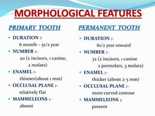
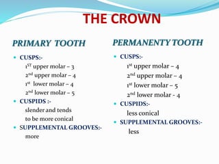
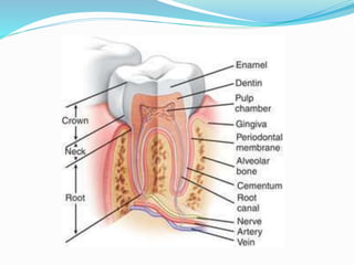
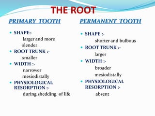
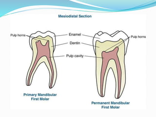
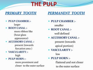
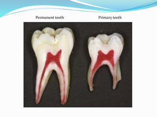
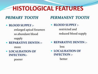
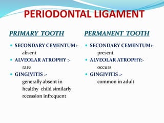


This document compares the anatomical, morphological, histological, and applied aspects of primary and permanent teeth. It outlines key differences between primary and permanent teeth, including their duration, number, enamel thickness, occlusal plane, cusps, roots, pulp chamber, dentin, and periodontal ligament structures. The morphological and histological differences between primary and permanent teeth have important applications in procedures like cavity preparation, extraction, endodontic treatment, and pulp therapy.