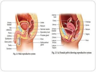
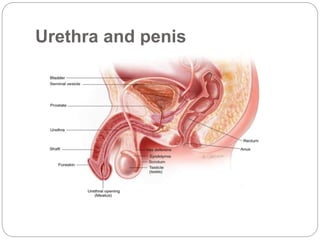
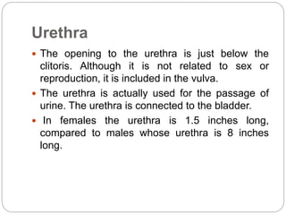
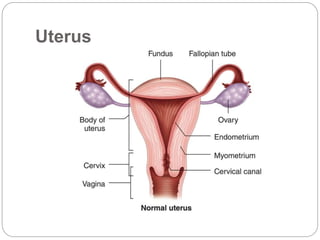
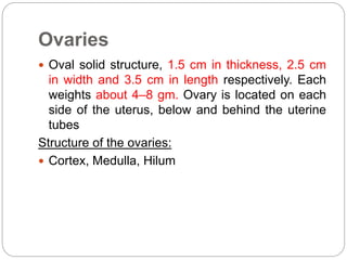
4. MALE REPRODUCTIVE
SYSTEM
SCROTUM
It is a pouch of deeply
pigmented skin, fibrous and
connective tissue and smooth
muscle.
It divided into two
compartments each of which
contain one testis, epididymis
and the testicular end of a
spermatic cord
It lies below the symphysis
pubis, in front of the upper
part of thigh and behind penis
6. Testes
Reproductive glands of the male
4.5 cm long, 2.5cm wide, 3cm thick
Suspended in the scrotum by the spermatic cords
3 layers - Tunica vaginalis, tunica albuginea, tunica
vasculosa
Each testes are 200 -300 lobules, each lobule are 1
to 4 convoluted loops composed of germinal epithelial
cells called seminiferous tubules.
Between the tubules are groups of
Interstitial cells- secret testosterone after
Puberty
7. Blood supply, lymph drainage and
nerve supply
Arterial supply : the testicular artery branches
from the abdominal aorta, just below the renal
arteries
Venous drainage : the testicular vein passes into
the abdominal cavity. The left vein opens in to the
left renal vein and the right into the inferior vena
cava
Lymph drainage: this through lymph nodes
around aorta
Nerve supply: this is provided by branches from
the 10th and 11th thoracic nerve
8. Spermatic cord
It suspend the testes in the scrotum
Each cord contains a testicular artery, testicular
veins, lymphatics, a deferent duct and testicular
nerves, which come together to form the cord
from their various origins in the abdomen
9. Seminal vesicle
These are two small fibromuscular pouches lined
with columnar epithelium, lying on the posterior
aspect of bladder
At the lower end each seminal vesicle opens into
a short duct, which joins with the corresponding
deferent duct to form an ejaculatory duct
10. Ejaculatory duct
Are two tubes about 2 cm long, each formed by
the union of the duct from seminal vesicle and
deferent duct.
They pass through the prostate gland and join the
prostate urethra, carrying seminal fluid and
spermatozoa to the urethra
11. Prostate gland
It lies in the pelvic cavity in front of the rectum
behind the symphysis pubis, surrounding the first
part of the urethra.
Consist of an outer fibrous covering, a layer of
smooth muscle and glandular substance
composed of columnar epithelial cells.
13. Urethra
It provides a common pathway for the flow of
urine and semen
It is about 19 to 20 cm long and consist of 3 part
The prostatic urethra originates at the urethral
orifice of the bladder and passes through the
prostate gland
The membranous urethra is the shortest and
narrowest part and extends from the prostate
gland to the bulb of the penis, after passing
through the perineal membrane
The penile urethra lies within the corpus
spongiosum of the penis and terminates at the
external urethral orifice in the glans penis
14. Cont…..
There are two urethral sphincters
Internal sphincter consists of smooth muscle
fibres at the neck of the bladder above the
prostate gland.
The external sphincter consists of skeletal muscle
fibres surrounding the membranous part
15. Penis
The penis has a root and a body.
The root lies in the perineum and the body
surrounds the urethra.
It is formed by three cylindrical masses of erectile
tissue and smooth muscle.
The erectile tissue is supported by fibrous tissue
and covered with skin and has a rich blood supply
Just above the glans the skin is folded upon itself
and forms a movable double layer, the foreskin or
prepuce
16. Female reproductive system
The reproductive organ in female are those which
are concerned with copulation, fertilization,
growth and development of the fetus and its
subsequent exit to the outer world.
18. Vulva
The external female genitalia is referred to as
vulva.
It consists of the labia majora and labia minora
(while these names translate as "large" and
"small" lips, often the "minora" can protrude
outside the "majora"), mons pubis, clitoris,
opening of the urethra (meatus), vaginal
vestibule, vestibular bulbs, vestibular glands.
19. Mons Veneris (mons pubis)
The mons veneris, Latin for "mound of Venus"
(Roman Goddess of love) is the soft mound at the
front of the vulva (fatty tissue covering the pubic
bone).
It is also referred to as the mons pubis.
The mons veneris protects the pubic bone and
vulva from the impact of sexual intercourse.
After puberty, it is covered with pubic hair, usually
in a triangular shape.
Heredity can play a role in the amount of pubic
hair an individual grows.
20. Labia Majora
The labia majora are the outer "lips" of the vulva.
They are pads of loose connective and adipose
tissue, as well as some smooth muscle.
The labia majora wrap around the vulva from the
mons pubis to the perineum.
There is also a longitudinal separation called the
pudendal cleft. These labia are usually covered with
pubic hair. The color of the outside skin of the labia
majora is usually close to the overall color of the
individual, although there may be some variation. The
inside skin is usually pink to light brown.
They contain numerous sweat and oil glands. It has
been suggested that the scent from these oils are
sexually arousing.
21. Labia Minora
Medial to the labia majora are the labia minora.
The labia minora are the inner lips of the vulva.
They are thin stretches of tissue within the labia
majora that fold and protect the vagina, urethra,
and clitoris.
There is no pubic hair on the labia minora, but
there are sebaceous glands. The two smaller lips
of the labia minora come together longitudinally to
form the prepuce, a fold that covers part of the
clitoris.
22. Clitoris
The clitoris, visible as the small white oval
between the top of the labia minora and the
clitoral hood, is a small body of spongy tissue that
functions solely for sexual pleasure.
It measuring about 1.5-2 cm and is consist of a
glans, a body and two crura.
The term clitoris comes from a Greek word
meaning key.
Erectile organ. It’s rich vascular, highly sensitive
to temperature, touch, and pressure sensation.
23. Vestibule
Is oval-shaped area formed between the labia
minora, clitoris, and fourchette. Vestibule contains
four openings are urethral opening, vaginal orifice
and hymen, opening of Bartholin’s duct, Skene’s
gland.
24. Urethra
The opening to the urethra is just below the
clitoris. Although it is not related to sex or
reproduction, it is included in the vulva.
The urethra is actually used for the passage of
urine. The urethra is connected to the bladder.
In females the urethra is 1.5 inches long,
compared to males whose urethra is 8 inches
long.
25. Hymen
The hymen is a thin fold of mucous membrane
that separates the lumen of the vagina from the
urethral sinus. Sometimes it may partially cover
the vaginal orifice. The hymen is usually
perforated during later fetal development.
26. Perineum
The perineum is the short stretch of skin starting
at the bottom of the vulva and extending to the
anus.
It is a diamond shaped area between the
symphysis pubis and the coccyx.
This area forms the floor of the pelvis and
contains the external sex organs and the anal
opening.
It can be further divided into the urogenital
triangle in front and the anal triangle in back.
28. Vagina
The vagina is a muscular, hollow tube that
extends from the vaginal opening to the cervix of
the uterus.
It is situated between the urinary bladder and the
rectum. It is about three to five inches long in a
grown woman.
The muscular wall allows the vagina to expand
and contract. The muscular walls are lined with
mucous membranes, which keep it protected and
moist. A thin sheet of tissue with one or more
holes in it, called the hymen, partially covers the
opening of the vagina.
The vagina receives sperm during sexual
intercourse from the penis. The sperm that
29. Cervix
The cervix (from Latin "neck") is the lower, narrow
portion of the uterus where it joins with the top
end of the vagina.
Where they join together forms an almost 90
degree curve. It is cylindrical or conical in shape
and protrudes through the upper anterior vaginal
wall.
The length of the cervix is about 2.5 t0 3 cm.
31. Uterus
The uterus is a hollow, pear shaped muscular organ.
The uterus measures about 7.5 X 5 X 2.5 cm and
weight about 50 – 60 gm .Its normal position is
anteverted (rotated forward and slightly antiflexed
(flexed forward). The uterus divided into three
Parts.
Body of the uterus :The upper part is the corpus, or
body of the uterus. The fundus is the part of the body
or corpus above the area where the fallopian tubes
enter the uterus.
Isthmus: A narrower transition zone. Is between the
corpus of the uterus and cervix. During late
pregnancy, the isthmus elongates and is known as
the lower uterine segment. Length about 5 cm.
Cervix: The lowermost position of the uterus “neck”.
The length of the cervix is about 2.5 t0 3 cm.
32. Relations:
Anterior – the uterovesical pouch and the bladder
Posterior – the rectouterine pouch of the douglas
Laterally – the broad ligament, the uterine tubes
Superior – the intestine
Inferior – the vagina
Layers of the uterus
Perimetrium.
Myometrium.
Endometrium.
33. 1. Perimetrium; Is the outer peritoneal layer of
serous membrane that coversmost of the uterus.
Laterally, the perimetrium is continuous with the
broad ligaments on either side of the uterus.
2. Myometrium: Is the middle layer of thick
muscle. Most of the muscle fibers are
concentrated in the upper uterus, and their
number diminishes progressively toward the
cervix.
3. Endometrium: Is the inner layer of the uterus.
It is responsive to the cyclic variations of estrogen
and progesterone during the female reproductive
cycle every month.
34. Fallopian tubes
The two tubes extended from the corna of the uterus to
the ovary. It runs in the upper free border of the broad
ligament. Length 8 to 14 cm average 10 cm. Its divided
into 4 parts.
1. Interstitial part
Which runs into uterine cavity, passes through the
myometrium between the fundus and body of the
uterus. About 1-2cm in length.
2. Isthmus
Which is the narrow part of the tube adjacent to the
uterus. Straight and cord like , about 2 – 3 cm in length.
3. Ampulla
Which is the wider part about 5 cm in length.
Fertilization occurs in the ampulla.
4. Infundibulum
It is funnel or trumpet shaped. Fimbriae are fingerlike
processes, one of these is longer than the other and
adherent to the ovary. The fimbriae become swollen
35. Ovaries
Oval solid structure, 1.5 cm in thickness, 2.5 cm
in width and 3.5 cm in length respectively. Each
weights about 4–8 gm. Ovary is located on each
side of the uterus, below and behind the uterine
tubes
Structure of the ovaries:
Cortex, Medulla, Hilum
36. Mammary glands
The mammary glands are sweat glands specialized for
the production of milk. The milk producing secretory
cells form walls of bulb-shaped chambers called
alveoli that join together with ducts, in grapelike
fashion, to form clusters called lobules.