Downloaded 77 times
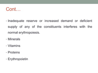
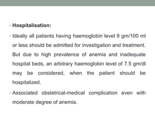
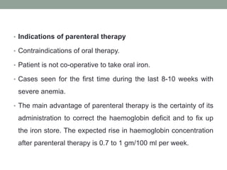
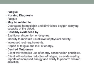
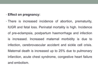
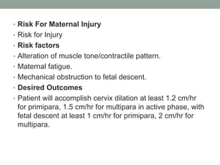
![• Estimation of the total requirement: The manufacturer’s
information is to follow for dose calculation. One such
formula for iron dextran is:
• 0.3xW (100-HIb%) mg of elemental iron. Where W= patient's
weight in pounds. Hb% = observed haemoglobin concentration in
percentage. Additional 50% is to be added for partial replenishment
of the body store iron.
• Example (iron dextran): The total elemental iron required in an
anaemic patient weighing 100 lb with haemoglobin 50% is
calculated as follows :0.3x 100 (100-50) =3/10X 100x 50 1500 mg.
Add 50% = 750 mg. Total elemental iron required
2250 mg.
• Iron (ferrous) Sucrose: Total iron dose (mg) = 2.3x WxD+500 [W =
Weight (kg) before pregnancy; D = Hb (Target Actual) gm/dL; 500
mg for body store]. It is given IV, 100 mg (at a time) in 100 ml
normal saline over 15 minutes.](https://image.slidesharecdn.com/hematologicaldisordersinpregnancy-210615172238/85/Hematological-disorders-in-pregnancy-42-320.jpg)


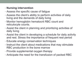
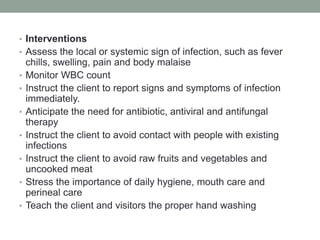

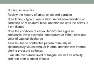
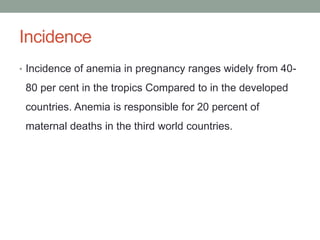
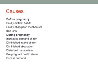
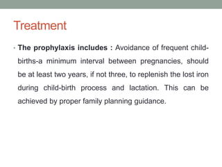
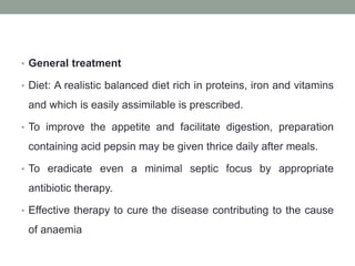
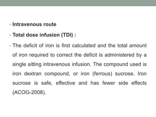
![• Anaemia in pregnancy and associated factors: a
cross sectional study of antenatal attendants at the
Sunyani Municipal Hospital, Ghana
• Out of the 316 participants, 129 (40.8%) were found to be
anaemic (Hb <11.0 g/dl) at the time of their first ANC visit
(mean Hb: 11.21 g/dl, range 6.8–15.1 g/dl). Seventy-nine
(61.2%) of them had mild anemia (Hb 9.0–10.9 g/dl), 48
(37.2%) had moderate anemia (Hb 7.0–8.9 g/dl) whilst 2
(1.6%) had severe anemia (Hb <7.0 g/dl). During their
most recent ANC visit, the prevalence of anaemia was
found to be similar to that of the first visit with 131 (41.5%)
of them being anaemic [mean Hb: 11.24 g/dl, range 8.10–
14.5 g/dl]. The haemoglobin levels however improved
significantly during the most recent visit compared to the
first with none of the women being severely anaemic (Hb
<7.0 g/dl).](https://image.slidesharecdn.com/hematologicaldisordersinpregnancy-210615172238/85/Hematological-disorders-in-pregnancy-117-320.jpg)


This document discusses hematological disorders in pregnancy, focusing on anemia. It notes that anemia is the most common hematological disorder seen in pregnancy. The majority of cases are due to iron, folate or vitamin B12 deficiency, though other conditions like thalassemia can also cause anemia. Anemia is a major public health concern in developing countries, where incidence ranges from 40-80% compared to developed countries. Treatment involves oral iron supplementation, with intravenous iron used for severe or late-presenting cases. Untreated anemia can increase risks for the mother and baby.


![Cells and Organs of immune system [Autosaved].pptx](https://cdn.slidesharecdn.com/ss_thumbnails/cellsandorgansofimmunesystemautosaved-260123152717-ea0cb261-thumbnail.jpg?width=640&height=640&fit=bounds)


