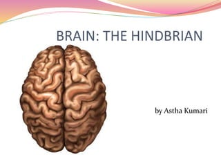
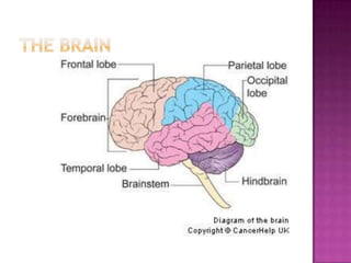
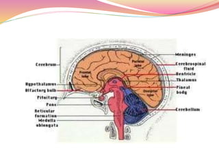
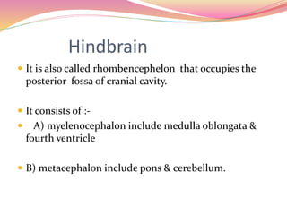
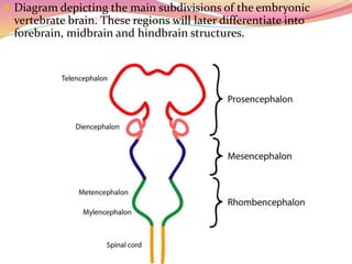
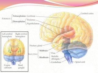
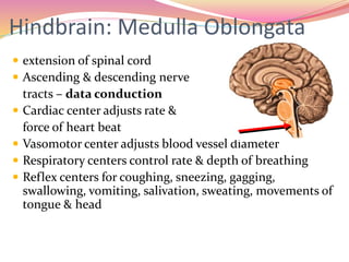
Student ID- 56fd705ce8ce11e9b888dd9b6f57296d
Affiliation - Banaras Hindu University
Course- Academic Writing
BRAIN: THE HINDBRIAN
by Astha Kumari
INTRODUCTION
NERVE
SYSTEM
CENTRAL
NERVE
SYSTEM
BRAIN
SPINAL
CORD
PERIPHERAL
NERVE
SYSTEM
The brain
 Most complex organ
of the body
 Only weighs 1,300
grams
 Contains billions of
neural networks that
interact to create
human behaviour
Cont………
 BRAIN is the most specialized tissue in the body and
it is almost entirely composed of nervous tissue .
 It is enclosed in the protective covering called
meninges.
 It has main three division -hindbrain
-midbrain
-forebrains.
Hindbrain
 It is also called rhombencephelon that occupies the
posterior fossa of cranial cavity.
 It consists of :-
 A) myelenocephalon include medulla oblongata &
fourth ventricle
 B) metacephalon include pons & cerebellum.
 Diagram depicting the main subdivisions of the embryonic
vertebrate brain. These regions will later differentiate into
forebrain, midbrain and hindbrain structures.
I. Medulla oblongata
Hindbrain: Medulla Oblongata
 extension of spinal cord
 Ascending & descending nerve
tracts – data conduction
 Cardiac center adjusts rate &
force of heart beat
 Vasomotor center adjusts blood vessel diameter
 Respiratory centers control rate & depth of breathing
 Reflex centers for coughing, sneezing, gagging,
swallowing, vomiting, salivation, sweating, movements of
tongue & head
Medulla Oblongata
 Axons cross in medulla
 So right side of brain
controls left side of
body & vice versa
Cont……….
 It has three surface such as:-
A) Ventral surface of medulla is marked
by median fissure continues with that of cord & flanked by
longitudinally ridges & pyramids.
 The caudal portion of the fissures(groove) is partially filled
by the decussately of the pyramids.
 Pyramid is the longitudinally band fiber on either side of
the midline between the median fissures & the lateral one .
B)Dorsal surface resemble the half
spinal cord & its rostra hay.
 It forms the rhomboid fossa which is the floor of fourth
ventricle.
Cont….
 The dorsal medial sulcus is the continuation of the
median sulcus of spinal cord & end at apex.
C)lateral surface it is marked by different
origin of cranial nerves. for example glosso pharyngeal
vagus & accessory nerves.
 The medulla oblongata is the most vital part of the
brain because it is the control center for breathing,
blood pressure and heart beat among other
involuntary body functions
Pons
 Bulge in the brainstem, rostral to
the medulla
 Tracts of nerves go through it
 Pathways in & out of cerebellum
 Nuclei concerned with sleep, hearing,
balance, taste, eye movements, facial
expression, facial sensation, respiration,
swallowing, bladder control & posture
II. Pons
 Is a thick transverse fiber tract divided in to bi -laterally
symmetrically halves by a slight midline depression.
 It has three surface such as:-
A) Ventral appear as large ,wide convex
protuberance decreasing in size laterally .
 Marked by a slight depression in midline refered to as basilar
sulcus.
 B) Lateral consider by decrease in width that
continuous with the middle cerebella pedicle which run
caudodorsal direction & disappearing in the cerebellum.
 C) Dorsal corresponding to rostral part of
rhomboid fossa.
Medulla and Pons
Cerebellum
 The cerebellum ((Latin for little brain) is the second largest part
of your brain, and is located near the base of the brain.
Constituent the larg part of hindbrain.
 Is roughly globular having much fissure & located above the
pons medulla.
 Separate from cerebral hemispher by transvers fissure &
tentorium cereblli which contain it.
 It consists of two larger part such as cerebral hemispher narrow
ridge named as vermis.
 The body of cerebellum subdivided to rostral & caudal lobes by
primary fissures.
 The rostral lobe sub divided in to small lingual which is the
rostral part of the central nerve system by numerous fibers
which make up the cerebral pendicels such as -
 Caudal cerebral pendicel that connect medulla & spinal cord
with cerebellum.
Cerebellum
 Muscle coordination, awareness of time, memory and
emotion
 Involved in learning and remembering motor responses
Cont…….
 In the cerebellum the bulk of the gray substance is
arranged as external cortex that in colour the white
substance of medulla.
 The cerebellum is involved in balance, equilibrium, muscle
tone, and the coordination of voluntary motor movement.
 Reticular Formation- Important in arousal and
maintaining consciousness, alertness attention and
Reticular Activating System which controls all cyclic
functions i.e. respiration, circadian rhythm.
Thanks for
yours
attention

Hindbrain

  • 1.
    Student ID- 56fd705ce8ce11e9b888dd9b6f57296d Affiliation- Banaras Hindu University Course- Academic Writing
  • 2.
  • 3.
  • 5.
    The brain  Mostcomplex organ of the body  Only weighs 1,300 grams  Contains billions of neural networks that interact to create human behaviour
  • 6.
    Cont………  BRAIN isthe most specialized tissue in the body and it is almost entirely composed of nervous tissue .  It is enclosed in the protective covering called meninges.  It has main three division -hindbrain -midbrain -forebrains.
  • 12.
    Hindbrain  It isalso called rhombencephelon that occupies the posterior fossa of cranial cavity.  It consists of :-  A) myelenocephalon include medulla oblongata & fourth ventricle  B) metacephalon include pons & cerebellum.
  • 13.
     Diagram depictingthe main subdivisions of the embryonic vertebrate brain. These regions will later differentiate into forebrain, midbrain and hindbrain structures.
  • 16.
  • 17.
    Hindbrain: Medulla Oblongata extension of spinal cord  Ascending & descending nerve tracts – data conduction  Cardiac center adjusts rate & force of heart beat  Vasomotor center adjusts blood vessel diameter  Respiratory centers control rate & depth of breathing  Reflex centers for coughing, sneezing, gagging, swallowing, vomiting, salivation, sweating, movements of tongue & head
  • 18.
    Medulla Oblongata  Axonscross in medulla  So right side of brain controls left side of body & vice versa
  • 19.
    Cont……….  It hasthree surface such as:- A) Ventral surface of medulla is marked by median fissure continues with that of cord & flanked by longitudinally ridges & pyramids.  The caudal portion of the fissures(groove) is partially filled by the decussately of the pyramids.  Pyramid is the longitudinally band fiber on either side of the midline between the median fissures & the lateral one . B)Dorsal surface resemble the half spinal cord & its rostra hay.  It forms the rhomboid fossa which is the floor of fourth ventricle.
  • 20.
    Cont….  The dorsalmedial sulcus is the continuation of the median sulcus of spinal cord & end at apex. C)lateral surface it is marked by different origin of cranial nerves. for example glosso pharyngeal vagus & accessory nerves.  The medulla oblongata is the most vital part of the brain because it is the control center for breathing, blood pressure and heart beat among other involuntary body functions
  • 21.
    Pons  Bulge inthe brainstem, rostral to the medulla  Tracts of nerves go through it  Pathways in & out of cerebellum  Nuclei concerned with sleep, hearing, balance, taste, eye movements, facial expression, facial sensation, respiration, swallowing, bladder control & posture
  • 23.
    II. Pons  Isa thick transverse fiber tract divided in to bi -laterally symmetrically halves by a slight midline depression.  It has three surface such as:- A) Ventral appear as large ,wide convex protuberance decreasing in size laterally .  Marked by a slight depression in midline refered to as basilar sulcus.  B) Lateral consider by decrease in width that continuous with the middle cerebella pedicle which run caudodorsal direction & disappearing in the cerebellum.  C) Dorsal corresponding to rostral part of rhomboid fossa.
  • 24.
  • 25.
    Cerebellum  The cerebellum((Latin for little brain) is the second largest part of your brain, and is located near the base of the brain. Constituent the larg part of hindbrain.  Is roughly globular having much fissure & located above the pons medulla.  Separate from cerebral hemispher by transvers fissure & tentorium cereblli which contain it.  It consists of two larger part such as cerebral hemispher narrow ridge named as vermis.  The body of cerebellum subdivided to rostral & caudal lobes by primary fissures.  The rostral lobe sub divided in to small lingual which is the rostral part of the central nerve system by numerous fibers which make up the cerebral pendicels such as -  Caudal cerebral pendicel that connect medulla & spinal cord with cerebellum.
  • 27.
    Cerebellum  Muscle coordination,awareness of time, memory and emotion  Involved in learning and remembering motor responses
  • 28.
    Cont…….  In thecerebellum the bulk of the gray substance is arranged as external cortex that in colour the white substance of medulla.  The cerebellum is involved in balance, equilibrium, muscle tone, and the coordination of voluntary motor movement.  Reticular Formation- Important in arousal and maintaining consciousness, alertness attention and Reticular Activating System which controls all cyclic functions i.e. respiration, circadian rhythm.
  • 30.

Editor's Notes

  • #6 The brain or the encephalon is possibly the most complex organ to examine within the human body Although only weighing approximately 1,300kg in the average adult, all behaviours, actions, thoughts and feelings originate from billions of neural networks interacting to create what we recognise as human. Without the brain our bodies simply would not function, making it important to have an understanding of its structure and function and the implications of diagnosis and pharmacology associated with mental illness. When looking at the brain, what is distinctive is the numerous folds that give it its wrinkled appearance. This folding together of brain tissue allows for greater amount of cerebral surface area (approx. two thirds of cerebral surface area is locate in the depths of these folds) to be confined within the limited space of the skull, leading to more information being relayed throughout areas of the brain The grooves are called fissures (extend deep into the brain) or sulci (if they are shallower) and the bumps that we see are called Gyri, and serve as markers to identify regions of the brain.