
Recommended
More Related Content
Similar to 719_Pectoral_Region.pptx
Similar to 719_Pectoral_Region.pptx (20)
More from KritikaMishra43
More from KritikaMishra43 (9)
Recently uploaded
Recently uploaded (20)
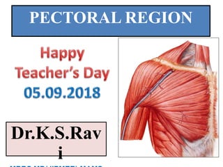
719_Pectoral_Region.pptx
- 2. OBJECTIVES • By the end of the lecture the students should be able to : • Identify and describe the muscles of the pectoral region. Pectoralis major. Pectoralis minor. Subclavius. Serratus anterior. Action of these muscles Clavipectoral Fascia Blood & Nerve Supply • Clinical Relevance
- 3. Pectoral region 1. Bony landmark : Sternum: Jugular notch (body of T2) Sternal angle of Louise (T4-5) Xiphosternal joint (T9) Ribs & costal cartilage
- 4. Scapular: acromian, coracoid process Clavicle: •supraclavicular fossa •infraclavicular fossa Humerus: head
- 5. 2. Superficial structure - skin & derivative of skin (breast) Breast : nipple, areolar, mammary gland (F) 1. Surface Anatomy (position) : Nipple – 4-5” from the midline, intercostal space 4 Breast – between rib 2-7
- 6. 2. Components : - subcutaneous fat, mammary gland - Lactiferous duct - Cooper’s ligament (suspensory ligament) - Retinaculum cutis fascia
- 7. The majority of the breast is in the superficial fascia, except the tail part (Tail of Spence) extends upward laterally into deep fascia at the lower border of pectoralis major. 2/3 of the gland lies on pectoralis major 1/3 of the gland lies on serratus anterior
- 8. Deltopectoral Triangle -deep fascia separating deltoid and pectoralis muscles - Platysma = superficial muscle, thin plate, extends from the mandible to the clavicle
- 9. 3. Muscles of pectoral region a. Pectoralis Major b. Pectoralis Minor c. Serratus Anterior d. Subclavious
- 10. Pectoralismajor Origin Anterior sternalhalf ofthe clavicle; Manubrium andSternum upto sixthcostal cartilages Cartilagesof all the true ribs, Aponeurosisof the abdominal external oblique Insertion Byabilaminar tendon into the lateral lip of the bicipital grooveof the humerus Innervation Medial andlateral pectoral nerves
- 11. Actions Flexionof the humerus, Adductionof the humerus and Medial rotation of the humerus. Clavicular part : flexion, adduction, and medial rotation of the humerus. Sternocostal part extension of theflexed arm asin climbing. It aids indeep inspiration.
- 12. Origin It arisesfrom the uppermargins and outer surfacesof the third, fourth, andfifthribs, Inserted into the medial border and upper surface of the coracoidprocessof the scapula. Innervation Medial andlateral pectoral nerves Actions Protracts the scapulawith serratus anterior Depressesthe shoulder with therhomboids and levator scapulae Important The pectoralisminor muscleiscoveredby the clavipectoralfascia. The medial pectoral nerve piercesthe pectoralisminor . Axillary artery isdivided into three partsby pectoralis minor. Pectoralisminor
- 13. Clavipectoral fascia Enclosessubclavius and Pectoralis Minor. It is pierced by: Lateral pectoral nerve. Thoraco- acromial artery Cephalic vein. Lymphnodesfrom pectoral regionto apicalgroupof axillary lymphnodes
- 14. Clavipectoral fascia / Costocoracoid membrane - deep fascia separating the pectoralis and the subclavious PectoralGirdle : clavicle, scapular, ribs
- 15. Serratusanterior Origin Arises from ribs 1 to 8, to be inserted into the medial border of thescapula. Insertion • Medial border of the scapulabetween the superior and inferiorangles. • 1stand 2n ddigitations to upper angle of scapula.(C5) • 3rdand 4thdigitations to medial border on costal surface upto the inferiorangle. • Lower 4 digitations to inferior angle of scapula. Action Protractionof the scapula alongwith pectoralis minor. • Thefibres inserted on inferior angle rotate scapula laterally and upwardsin overhead abductionwith trapezius. Assistsinrespiration. Innervation long thoracic nerve(Nerve of Bell)
- 17. -Blood supply to the pectoral region 1. Axillary artery 2. Perforating branches of the internal thoracic a.
- 18. 1. Axillary artery : divided into 3 parts First part : Supreme thoracic a. Second part : 1.Thoraco-acromial trunk Acromial branch Pectoral branch Clavicular branch Deltoid branch 2. Lateral thoracic a. Third part : give branches to supply head of humerus and scapular regions
- 19. Venous drainage at the pectoral region 1. Deep veins - axillary v. <= from the muscles
- 20. 2.Superficial veins - cephalic v. (from upper limb) at the deltopectoral triangle, it pierces the clavipectoral fascia (or infraclavicular fossa) into the axillary v. -from mammary gland, it drains into deep veins => internal thoracic v. and lateral thoracic v.
- 21. Nerve Supply of the pectoral region 1. - medial & lateral pectoral nerve (terminal branches from the cords of the Brachial plexus (C5-8 & T1) - nerve to subclavius (a branch from upper trunk of the Brachial plexus) - long thoracic nerve (nerve roots from C5-6-7 of the Brachial plexus)
- 22. Brachial plexus (C5-8 & T1)
- 23. 2.Spinal n. / Sensory (cutaneous) n - Supraclavicular nerve (C3-4, medial, intermediate & lateral branches) - Intercostal nerve T3-7 (anterior & lateral cutaneous branches) *Dermatome at the pectoral region: C3-4, T3-7
- 25. Appliedaspect • • • Serratus anterior is calledthe Boxer’smusclesinceit is responsible for pushing and punching movements. Paralysisof this muscle results in a"wingedscapula",results in protrusion of thescapula on the affected side when the patient is askedto push against the wall with both arms extended. Winged scapulaoccurs in lateral thoracicnerve paralysis
- 26. Paralysis of Serratus anterior muscle.
- 27. Clinical Relevance 1. Chest wall – heart /lung sound 2.Clavipectoral fascia - protection of the vessels and nerves underneath -limit spreading of the abscess from upper limb to the neck 3. Fracture of clavicle -common site is at 1/3 from the lateral -Poland Anomaly -Cardiac Catheterisation- Basilic vein
- 28. 1. Which one of the following muscles performs adduction of the arm ? a. Pectoralis minor. b. Pectoralis major. c. Subclavius. d. Serratus anterior. 2. Serratus anterior is innervated by : a. Thoracodorsal nerve. b. Long thoracic nerve. c. Axillary nerve. d. Radial nerve. 3. Which one of the following muscles contributes in rotation of the scapula above the head? a. Pectoralis major. b. Pectoralis minor. c. Serratus anterior. d. Teres major. 4. Which one of the following do not pierces clavipectoral fascia? a. Lateral Pectoral Nerve. b. Lymph Nodes. c. Cephalic Vein. d. Lateral thoracic artery. 5. Nerve to subclavius is a branch from which part of brachial plexus? a. Roots. b. Divisions. c. Cords. D. Trunks.
- 29. THANK YOU