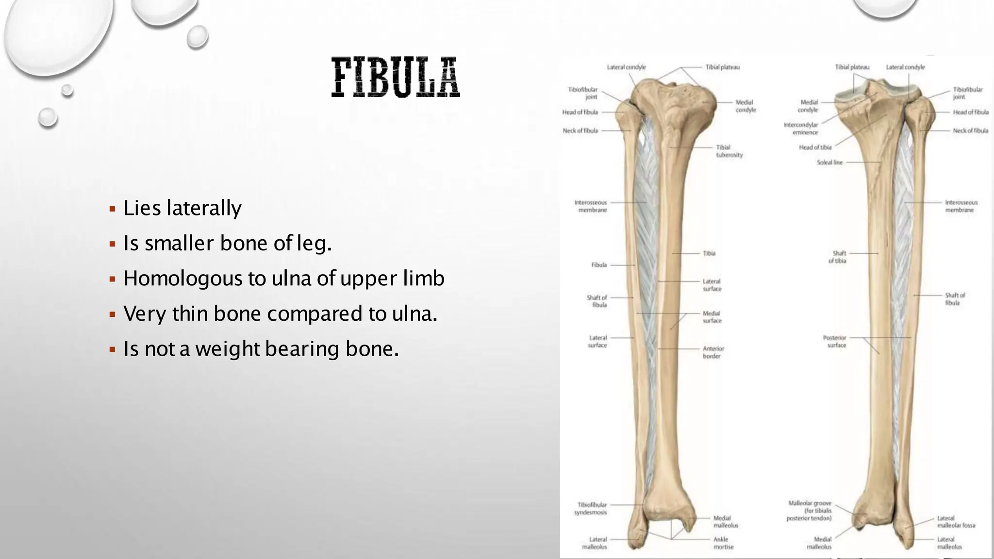
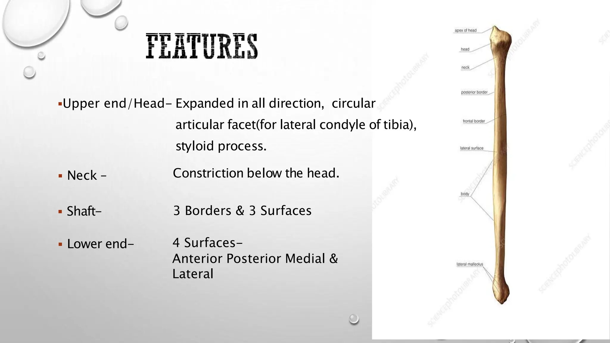
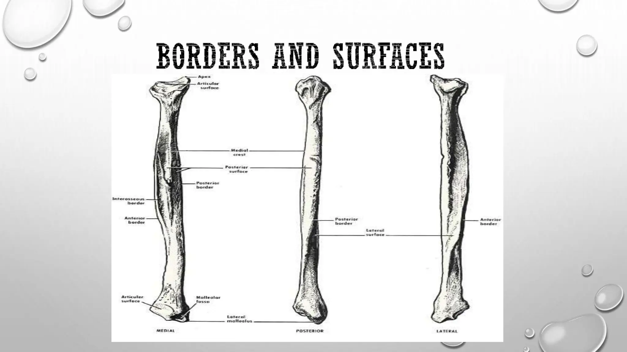
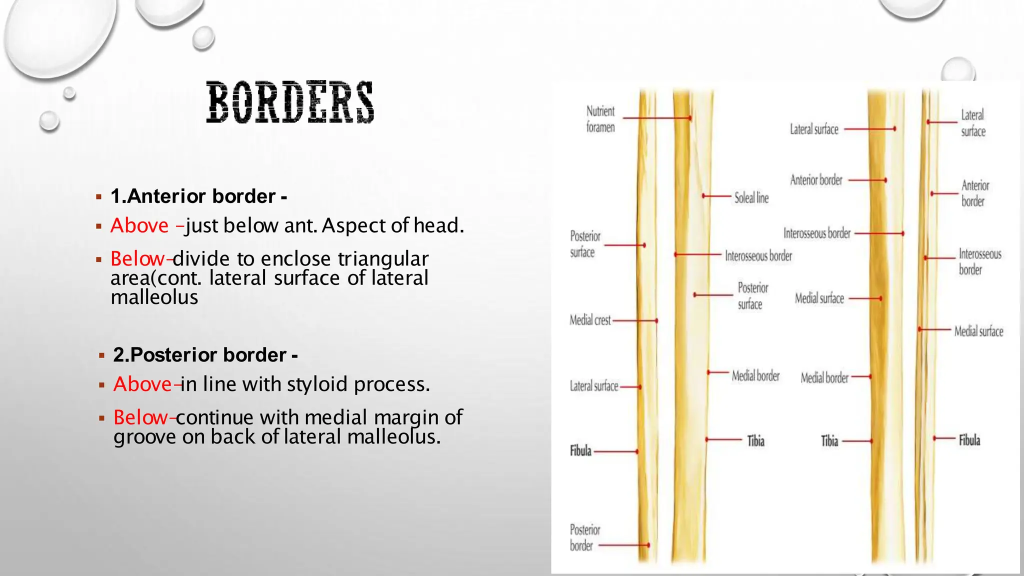
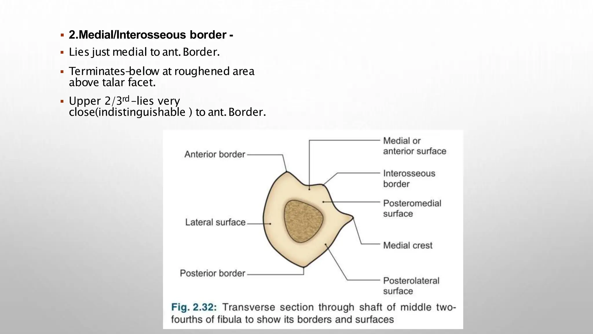
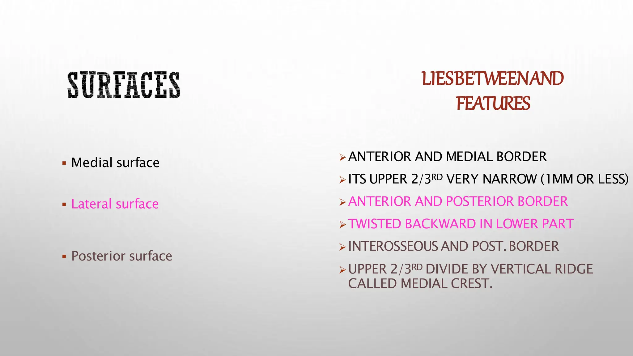
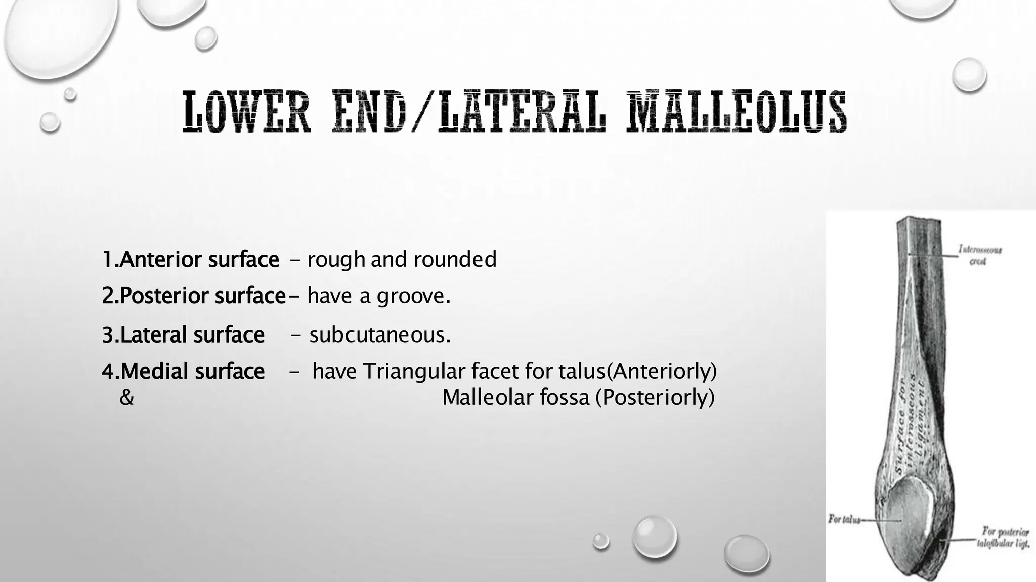
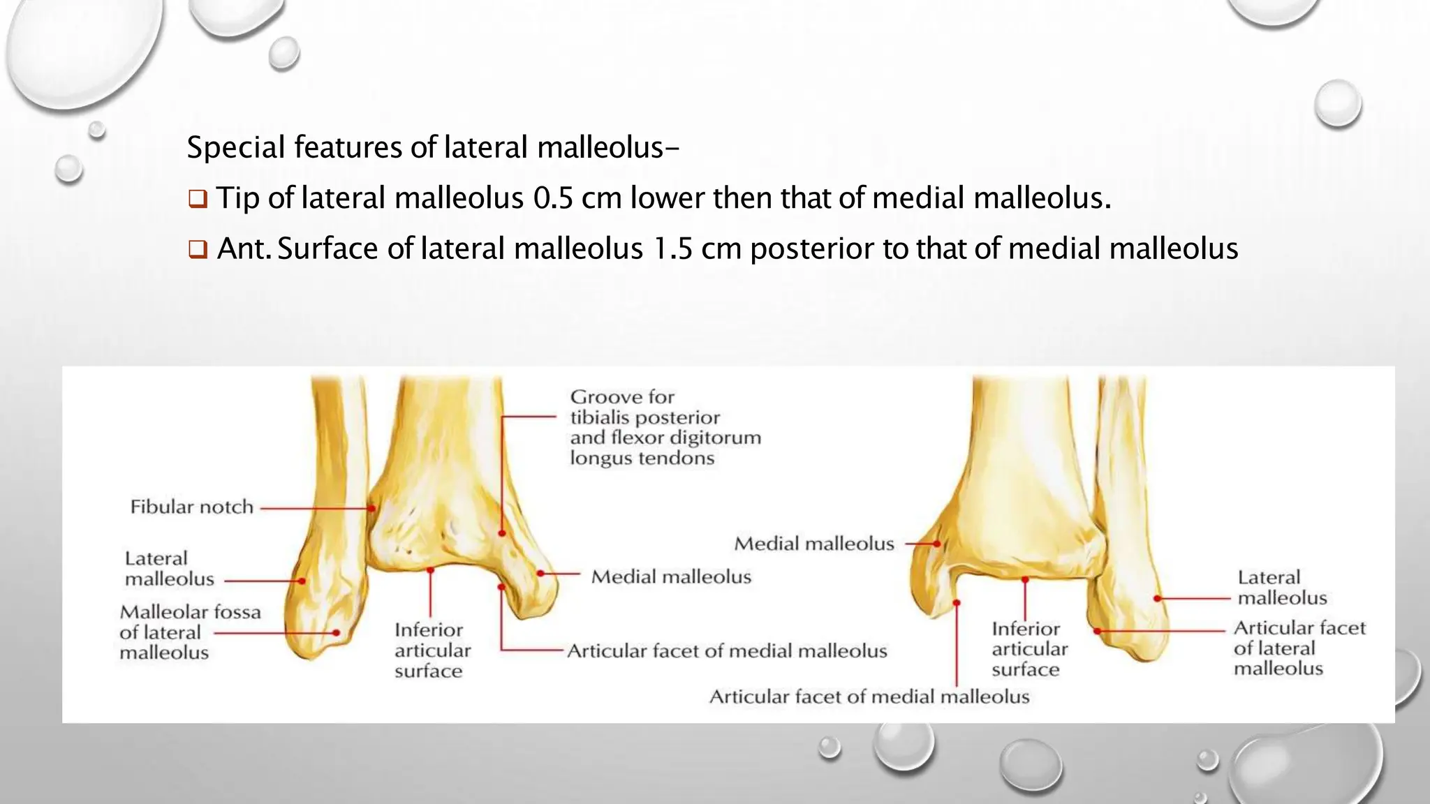
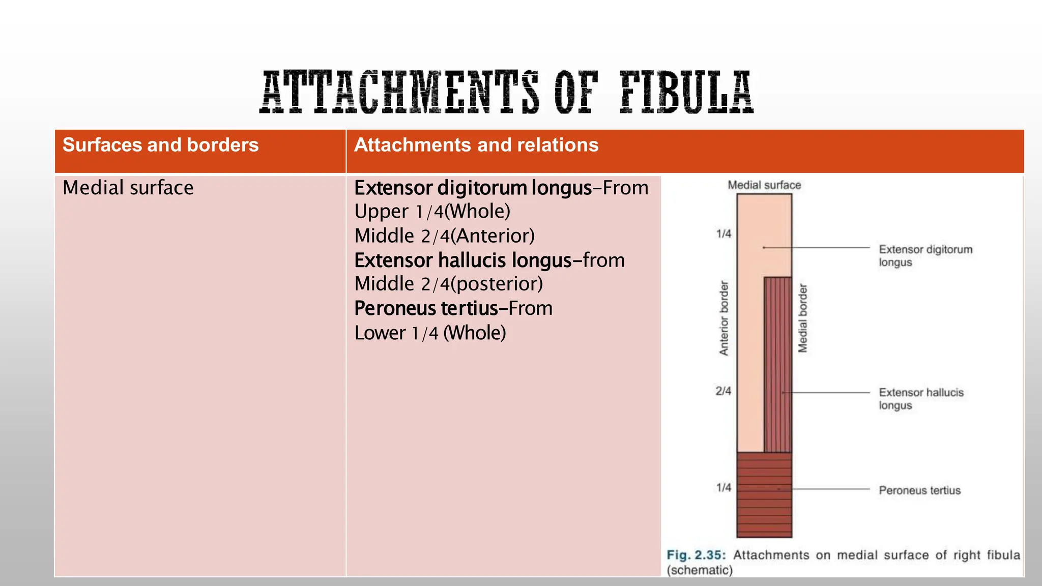
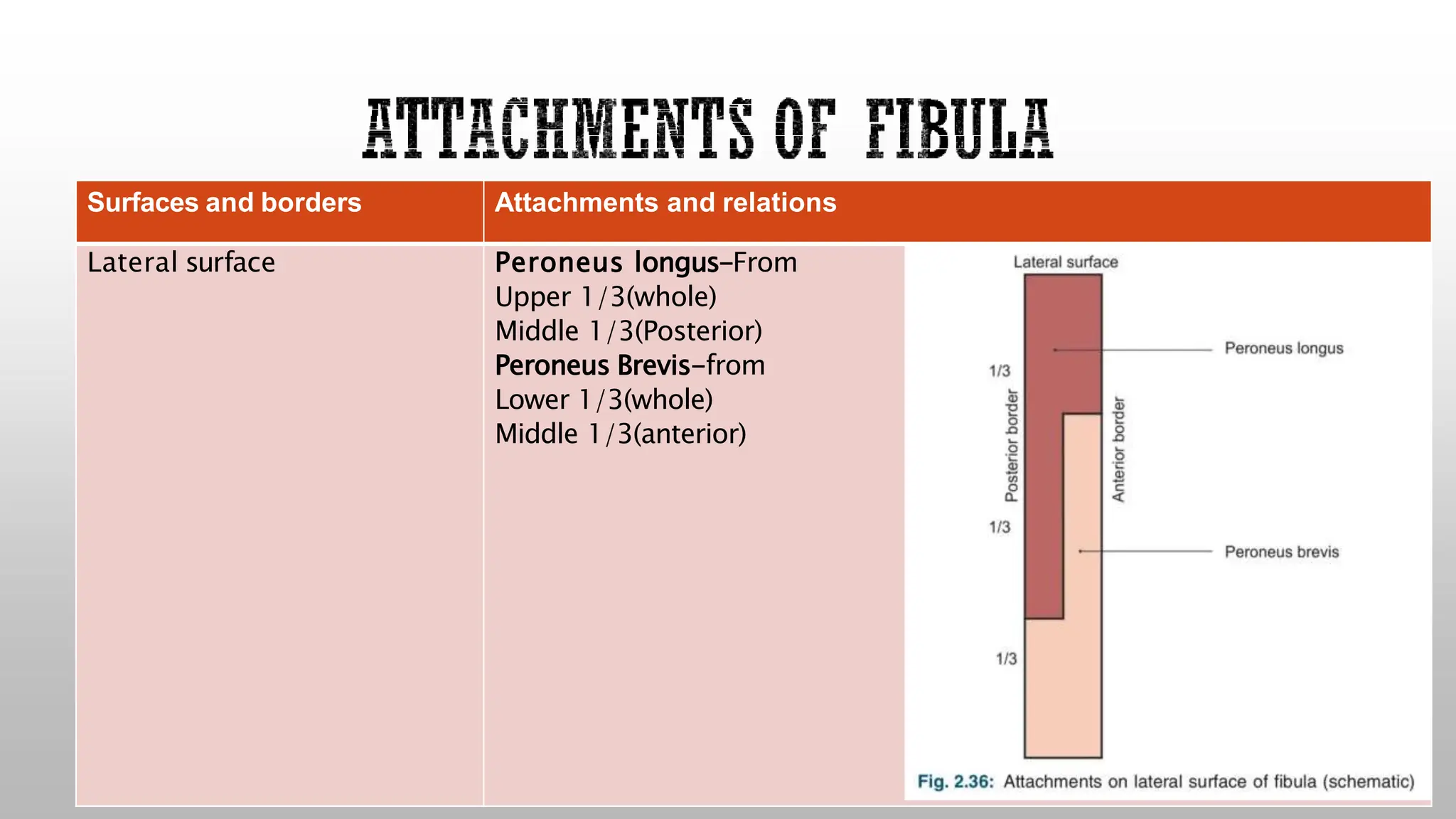
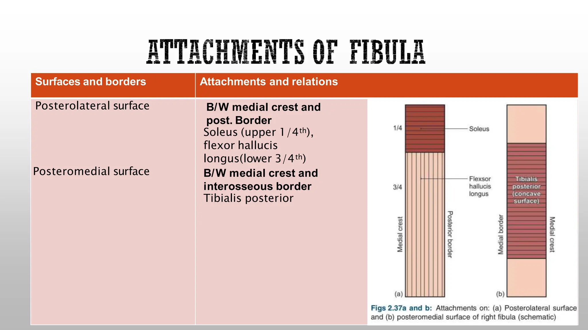
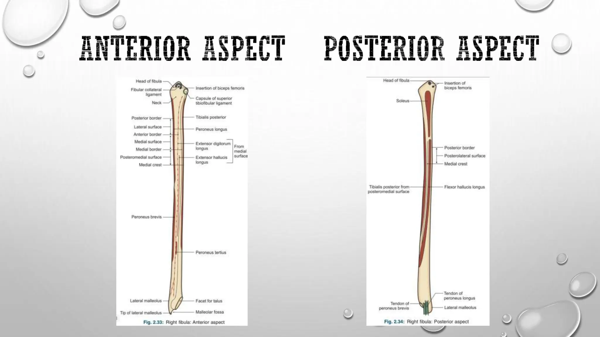
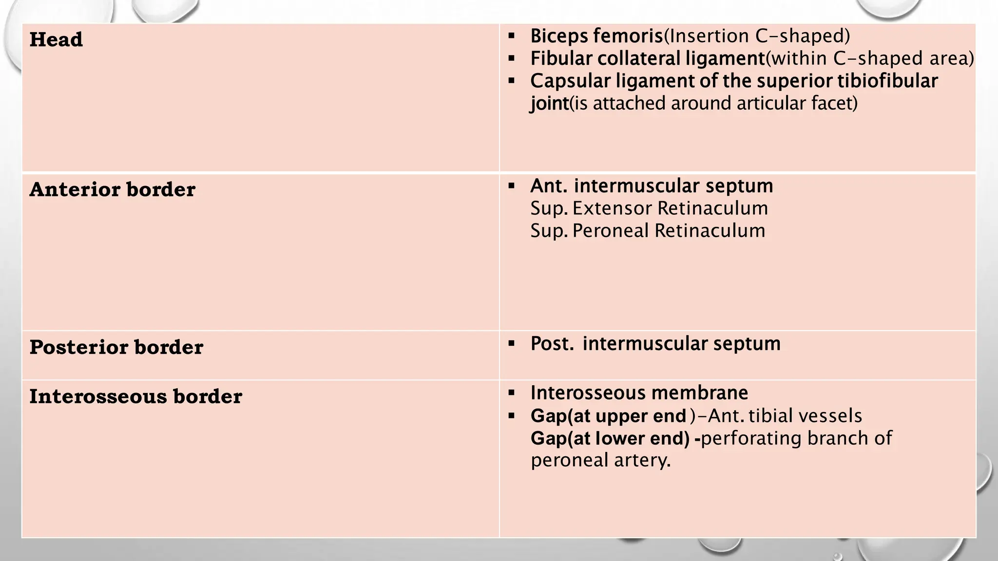
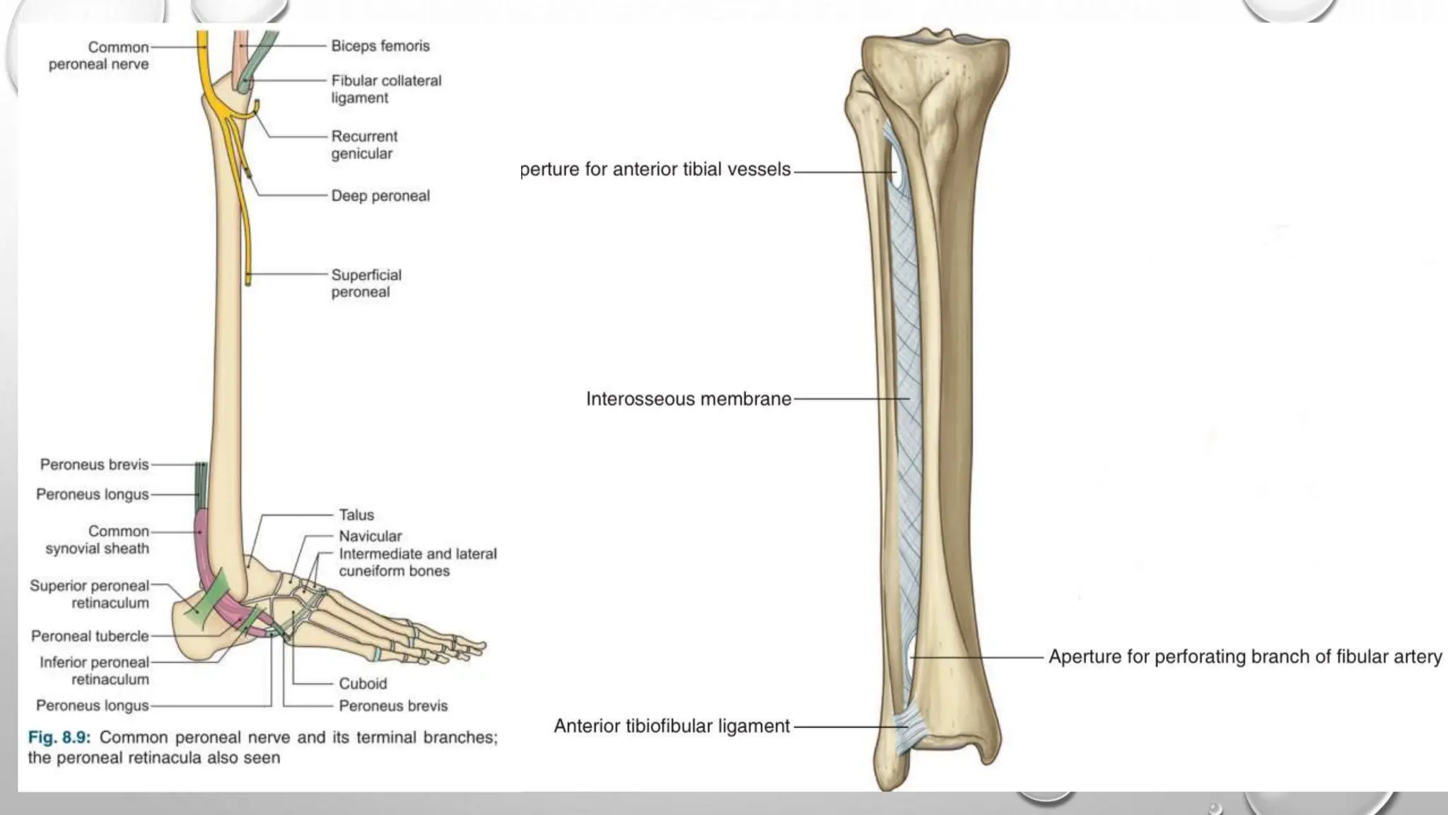
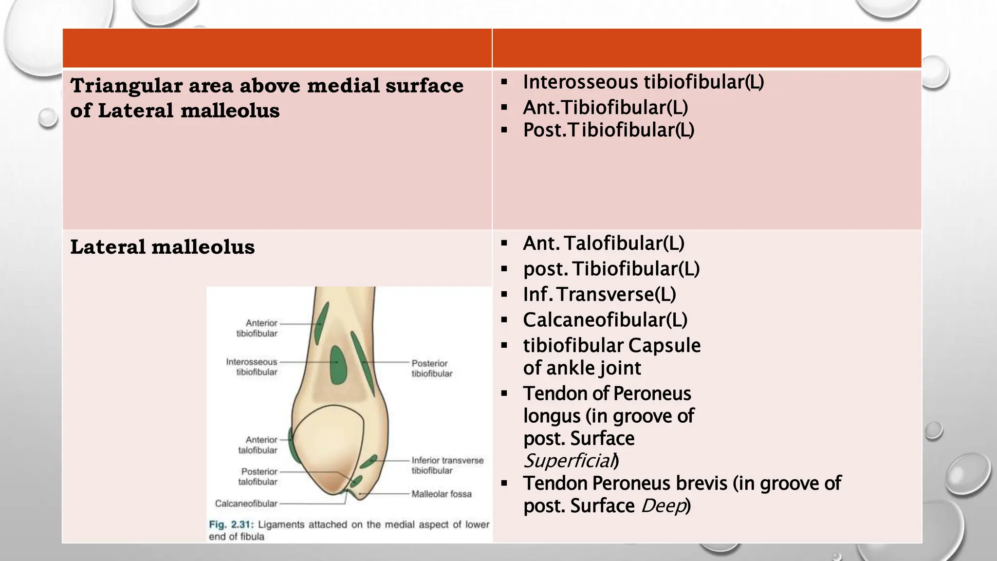
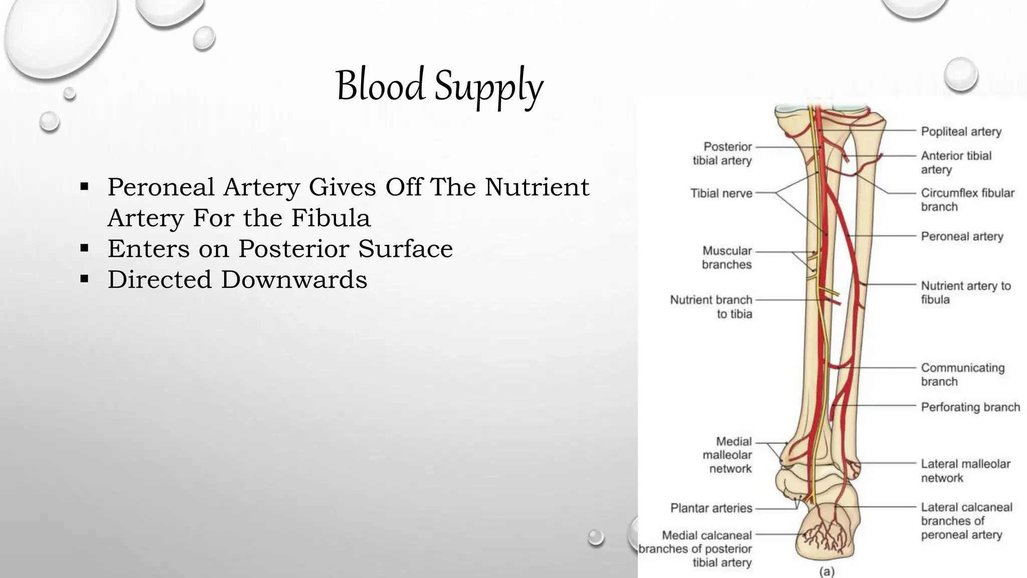
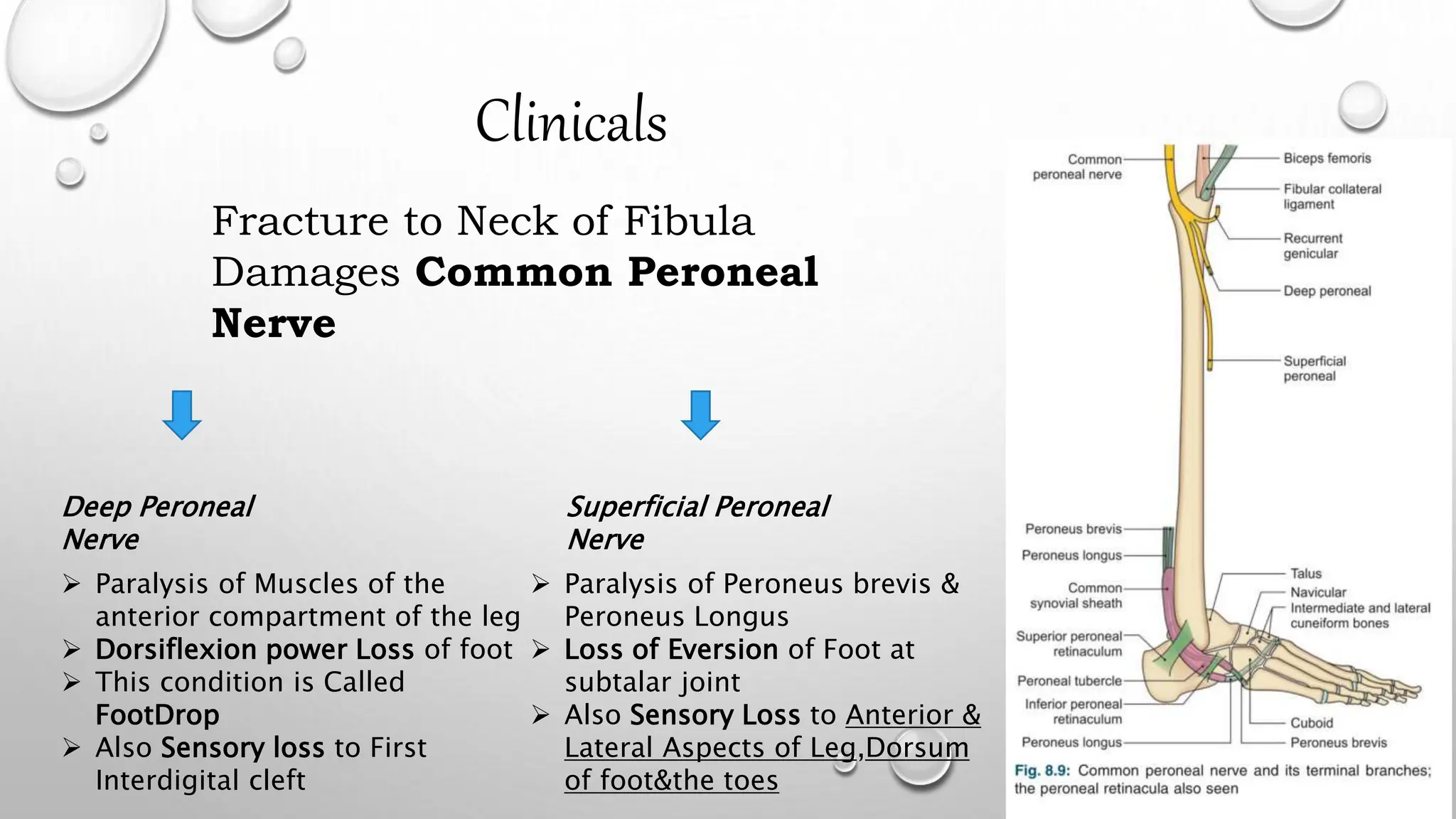
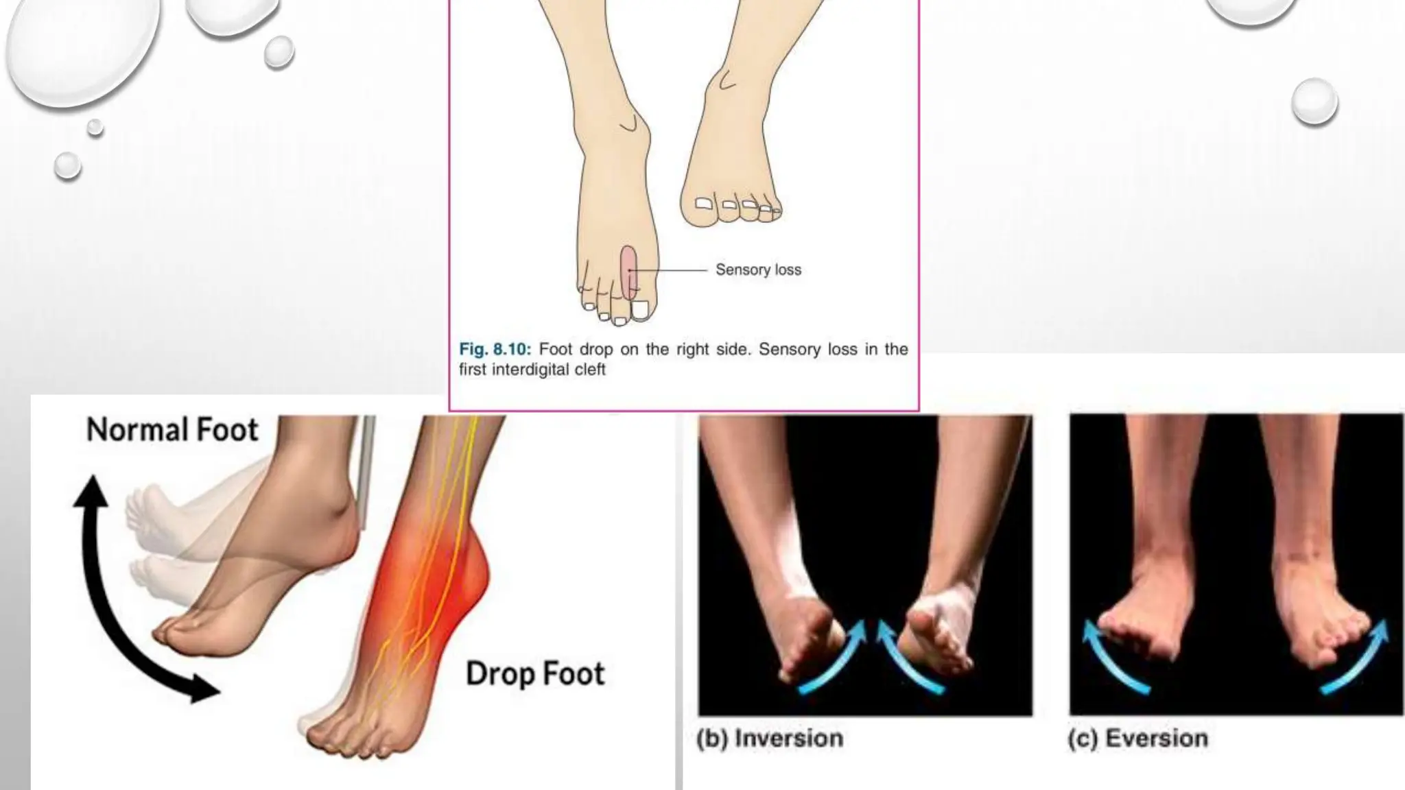
Presented by – Presented to -
Kamlesh Nishad Vd. Sachin
Ravan
Kunal Yadav Vd. Gaurav
Sharma
Gulshan Rawat
Soaib
Parth Priyadarshi
 Identify fibula.
 Demonstrate the different parts, borders and surfaces of fibula.
 Determine the side of the fibula
 Hold the fibula in its anatomical position.
 Demonstrate attachment of joint capsule ,ligaments and muscles on
the fibula.
 Lies laterally
 Is smaller bone of leg.
 Homologous to ulna of upper limb
 Very thin bone compared to ulna.
 Is not a weight bearing bone.
Side Determination
Upper End – Expanded in All Directions
Lower End – Expanded In Anteroposteriorly
Flattened from Side to Side
On Medial Side of Lower End--Triangular Articular
Facet(Anteriorly)
Malleolar Fossa(Posteriorly)
Upper end/Head-
 Neck – Constriction below the head.
 Shaft-
 Lower end-
Expanded in all direction, circular
articular facet(for lateral condyle of tibia),
styloid process.
3 Borders & 3 Surfaces
4 Surfaces-
Anterior Posterior Medial &
Lateral
 1.Anterior border -
 Above –just below ant. Aspect of head.
 Below-
divide to enclose triangular
area(cont. lateral surface of lateral
malleolus
 2.Posterior border -
 Above-
in line with styloid process.
 Below-
continue with medial margin of
groove on back of lateral malleolus.
 2.Medial/Interosseous border -
 Lies just medial to ant.Border.
 Terminates-
below at roughened area
above talar facet.
 Upper 2/3rd–lies very
close(indistinguishable ) to ant.Border.
 Medial surface
 Lateral surface
 Posterior surface
LIESBETWEENAND
FEATURES
ANTERIOR AND MEDIAL BORDER
ITS UPPER 2/3RD VERY NARROW (1MM OR LESS)
ANTERIOR AND POSTERIOR BORDER
TWISTED BACKWARD IN LOWER PART
INTEROSSEOUS AND POST.BORDER
UPPER 2/3RD DIVIDE BY VERTICAL RIDGE
CALLED MEDIAL CREST.
1.Anterior surface - rough and rounded
2.Posterior surface- have a groove.
3.Lateral surface - subcutaneous.
4.Medial surface - have Triangular facet for talus(Anteriorly)
& Malleolar fossa (Posteriorly)
Special features of lateral malleolus-
 Tip of lateral malleolus 0.5 cm lower then that of medial malleolus.
 Ant.Surface of lateral malleolus 1.5 cm posterior to that of medial malleolus
Surfaces and borders Attachments and relations
Medial surface Extensor digitorum longus-From
Upper 1/4(Whole)
Middle 2/4(Anterior)
Extensor hallucis longus-from
Middle 2/4(posterior)
Peroneus tertius-From
Lower 1/4 (Whole)
Surfaces and borders Attachments and relations
Lateral surface Peroneus longus-From
Upper 1/3(whole)
Middle 1/3(Posterior)
Peroneus Brevis-from
Lower 1/3(whole)
Middle 1/3(anterior)
Surfaces and borders Attachments and relations
Posterolateral surface B/W medial crest and
post. Border
Soleus (upper 1/4th),
flexor hallucis
longus(lower 3/4th)
B/W medial crest and
interosseous border
Tibialis posterior
Posteromedial surface
Head  Biceps femoris(Insertion C-shaped)
 Fibular collateral ligament(within C-shaped area)
 Capsular ligament of the superior tibiofibular
joint(is attached around articular facet)
Anterior border  Ant. intermuscular septum
Sup. Extensor Retinaculum
Sup. Peroneal Retinaculum
Posterior border  Post. intermuscular septum
Interosseous border  Interosseous membrane
 Gap(at upper end )-Ant. tibial vessels
Gap(at lower end) -perforating branch of
peroneal artery.
Triangular area above medial surface
of Lateral malleolus
 Interosseous tibiofibular(L)
 Ant.Tibiofibular(L)
 Post.Tibiofibular(L)
Lateral malleolus  Ant. Talofibular(L)
 post. Tibiofibular(L)
 Inf.Transverse(L)
 Calcaneofibular(L)
 tibiofibular Capsule
of ankle joint
 Tendon of Peroneus
longus (in groove of
post. Surface
Superficial)
 Tendon Peroneus brevis (in groove of
post. Surface Deep)
Blood Supply
 Peroneal Artery Gives Off The Nutrient
Artery For the Fibula
 Enters on Posterior Surface
 Directed Downwards
Clinicals
Fracture to Neck of Fibula
Damages Common Peroneal
Nerve
Deep Peroneal
Nerve
Superficial Peroneal
Nerve
 Paralysis of Muscles of the
anterior compartment of the leg
 Dorsiflexion power Loss of foot
 This condition is Called
FootDrop
 Also Sensory loss to First
Interdigital cleft
 Paralysis of Peroneus brevis &
Peroneus Longus
 Loss of Eversion of Foot at
subtalar joint
 Also Sensory Loss to Anterior &
Lateral Aspects of Leg,Dorsum
of foot&the toes
Thank you

Anatomy of Fibula with Muscle Attachments.pptx

  • 1.
    Presented by –Presented to - Kamlesh Nishad Vd. Sachin Ravan Kunal Yadav Vd. Gaurav Sharma Gulshan Rawat Soaib Parth Priyadarshi
  • 2.
     Identify fibula. Demonstrate the different parts, borders and surfaces of fibula.  Determine the side of the fibula  Hold the fibula in its anatomical position.  Demonstrate attachment of joint capsule ,ligaments and muscles on the fibula.
  • 3.
     Lies laterally Is smaller bone of leg.  Homologous to ulna of upper limb  Very thin bone compared to ulna.  Is not a weight bearing bone.
  • 4.
    Side Determination Upper End– Expanded in All Directions Lower End – Expanded In Anteroposteriorly Flattened from Side to Side On Medial Side of Lower End--Triangular Articular Facet(Anteriorly) Malleolar Fossa(Posteriorly)
  • 5.
    Upper end/Head-  Neck– Constriction below the head.  Shaft-  Lower end- Expanded in all direction, circular articular facet(for lateral condyle of tibia), styloid process. 3 Borders & 3 Surfaces 4 Surfaces- Anterior Posterior Medial & Lateral
  • 7.
     1.Anterior border-  Above –just below ant. Aspect of head.  Below- divide to enclose triangular area(cont. lateral surface of lateral malleolus  2.Posterior border -  Above- in line with styloid process.  Below- continue with medial margin of groove on back of lateral malleolus.
  • 8.
     2.Medial/Interosseous border-  Lies just medial to ant.Border.  Terminates- below at roughened area above talar facet.  Upper 2/3rd–lies very close(indistinguishable ) to ant.Border.
  • 9.
     Medial surface Lateral surface  Posterior surface LIESBETWEENAND FEATURES ANTERIOR AND MEDIAL BORDER ITS UPPER 2/3RD VERY NARROW (1MM OR LESS) ANTERIOR AND POSTERIOR BORDER TWISTED BACKWARD IN LOWER PART INTEROSSEOUS AND POST.BORDER UPPER 2/3RD DIVIDE BY VERTICAL RIDGE CALLED MEDIAL CREST.
  • 10.
    1.Anterior surface -rough and rounded 2.Posterior surface- have a groove. 3.Lateral surface - subcutaneous. 4.Medial surface - have Triangular facet for talus(Anteriorly) & Malleolar fossa (Posteriorly)
  • 11.
    Special features oflateral malleolus-  Tip of lateral malleolus 0.5 cm lower then that of medial malleolus.  Ant.Surface of lateral malleolus 1.5 cm posterior to that of medial malleolus
  • 12.
    Surfaces and bordersAttachments and relations Medial surface Extensor digitorum longus-From Upper 1/4(Whole) Middle 2/4(Anterior) Extensor hallucis longus-from Middle 2/4(posterior) Peroneus tertius-From Lower 1/4 (Whole)
  • 13.
    Surfaces and bordersAttachments and relations Lateral surface Peroneus longus-From Upper 1/3(whole) Middle 1/3(Posterior) Peroneus Brevis-from Lower 1/3(whole) Middle 1/3(anterior)
  • 14.
    Surfaces and bordersAttachments and relations Posterolateral surface B/W medial crest and post. Border Soleus (upper 1/4th), flexor hallucis longus(lower 3/4th) B/W medial crest and interosseous border Tibialis posterior Posteromedial surface
  • 16.
    Head  Bicepsfemoris(Insertion C-shaped)  Fibular collateral ligament(within C-shaped area)  Capsular ligament of the superior tibiofibular joint(is attached around articular facet) Anterior border  Ant. intermuscular septum Sup. Extensor Retinaculum Sup. Peroneal Retinaculum Posterior border  Post. intermuscular septum Interosseous border  Interosseous membrane  Gap(at upper end )-Ant. tibial vessels Gap(at lower end) -perforating branch of peroneal artery.
  • 19.
    Triangular area abovemedial surface of Lateral malleolus  Interosseous tibiofibular(L)  Ant.Tibiofibular(L)  Post.Tibiofibular(L) Lateral malleolus  Ant. Talofibular(L)  post. Tibiofibular(L)  Inf.Transverse(L)  Calcaneofibular(L)  tibiofibular Capsule of ankle joint  Tendon of Peroneus longus (in groove of post. Surface Superficial)  Tendon Peroneus brevis (in groove of post. Surface Deep)
  • 20.
    Blood Supply  PeronealArtery Gives Off The Nutrient Artery For the Fibula  Enters on Posterior Surface  Directed Downwards
  • 21.
    Clinicals Fracture to Neckof Fibula Damages Common Peroneal Nerve Deep Peroneal Nerve Superficial Peroneal Nerve  Paralysis of Muscles of the anterior compartment of the leg  Dorsiflexion power Loss of foot  This condition is Called FootDrop  Also Sensory loss to First Interdigital cleft  Paralysis of Peroneus brevis & Peroneus Longus  Loss of Eversion of Foot at subtalar joint  Also Sensory Loss to Anterior & Lateral Aspects of Leg,Dorsum of foot&the toes
  • 23.