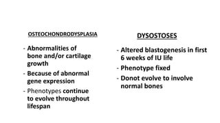
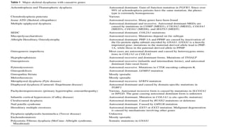
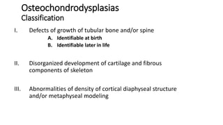
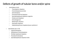
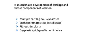
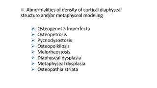
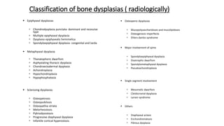
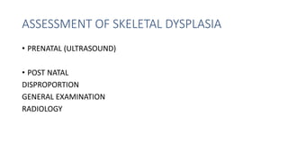
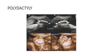
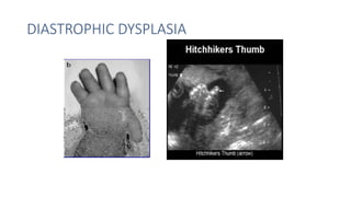
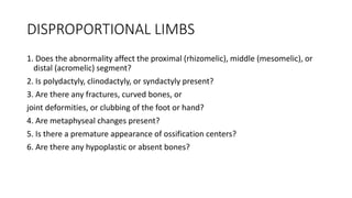
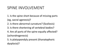
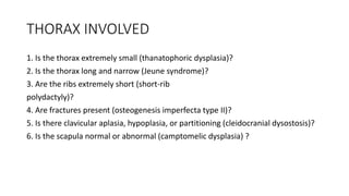
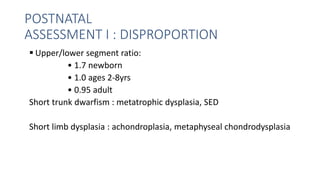
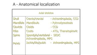
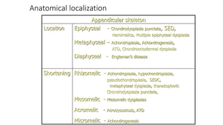
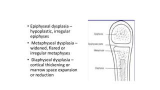
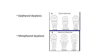
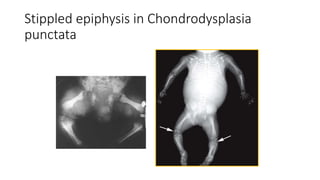
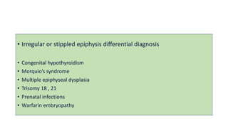
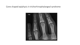
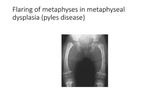
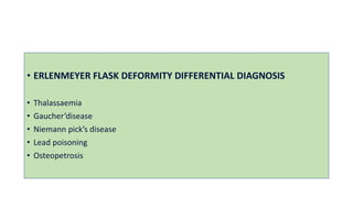
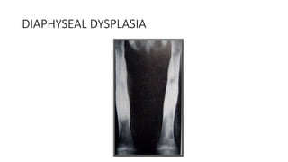
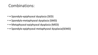
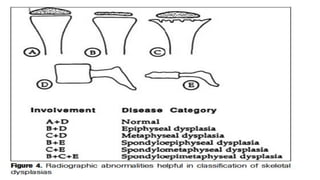
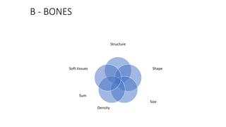
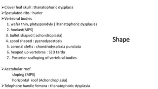
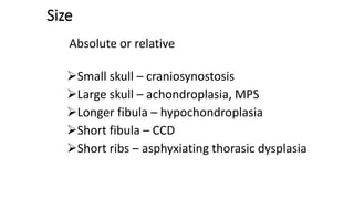
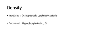
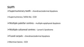
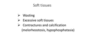
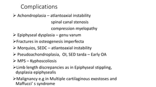
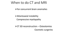
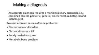
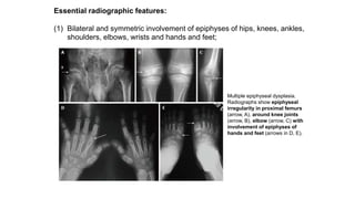
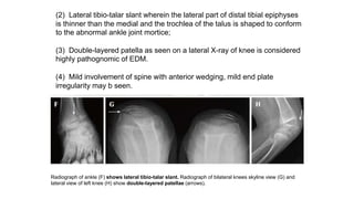
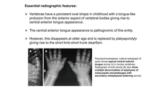
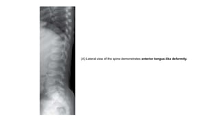
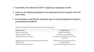
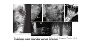
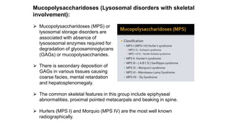
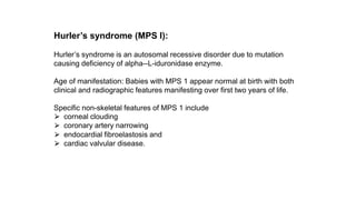
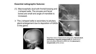
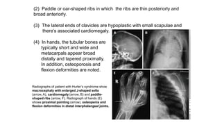
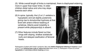
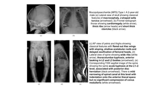
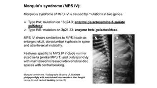
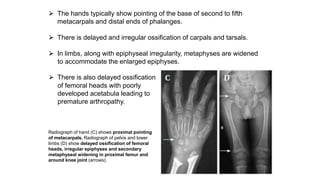
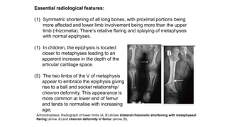
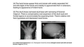
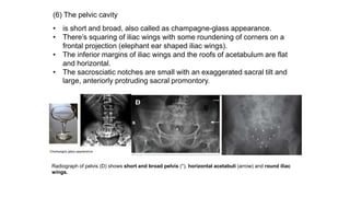
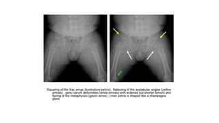
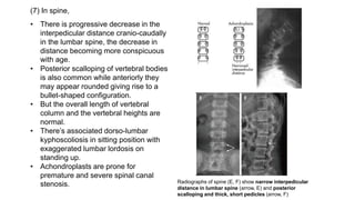
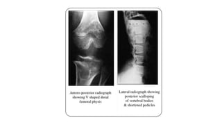
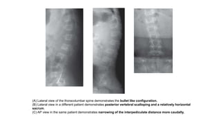
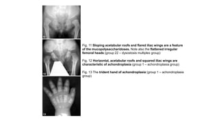
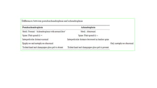
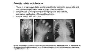
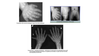
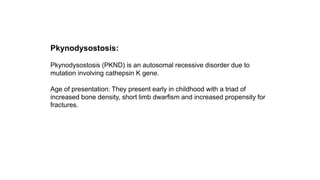
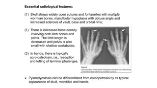
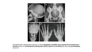
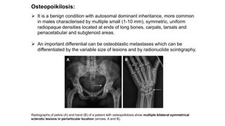
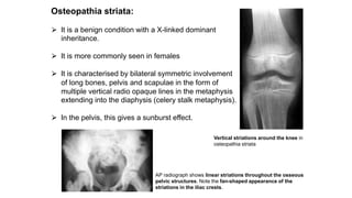
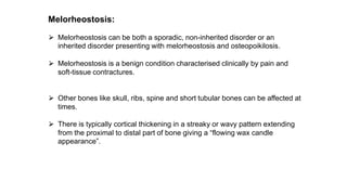
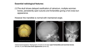
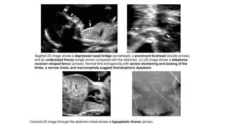
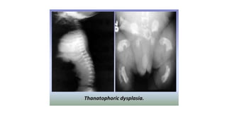
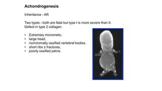
SKELETAL DYSPLASIAS
Moderator : Dr SANJU
(assistant professor )
Presentation by
Dr Navya ,PGY2
Department of radiodiagnosis
MMC,Khammam
INTRODUCTION
•Heterogeneous group of disorders characterised by
abnormal shape, size, growth, number and density of
bones
•Affects the growth and development.
•Disproportionate long bones, spine, and skull
OSTEOCHONDRODYSPLASIA DYSOSTOSES
- Abnormalities of
bone and/or cartilage
growth
- Because of abnormal
gene expression
- Phenotypes continue
to evolve throughout
lifespan
- Altered blastogenesis in first
6 weeks of IU life
- Phenotype fixed
- Donot evolve to involve
normal bones
Osteochondrodysplasias
Classification
I. Defects of growth of tubular bone and/or spine
A. Identifiable at birth
B. Identifiable later in life
II. Disorganized development of cartilage and fibrous
components of skeleton
III. Abnormalities of density of cortical diaphyseal structure
and/or metaphyseal modeling
Defects of growth of tubular bone and/or spine
A. Identifiable at birth
 Thanatophoric dysplasia
 Chondrodysplasia punctata
 Achondroplasia
 Chondroectodermal dysplasia
 Spondyloepiphyseal dysplasia congenita
 Cleidocranial dysplasia
 Achondrogenesis
 Metatrophic dwarfism
 Diastrophic dwarfism
 Asphyxiating thoracic dysplasia (jeune syndrome )
B. Identifiable later in life
 Hypochondroplasia
 Metaphyseal chondrodysplasia
 Multiple epiphyseal dysplasia
 Pseudoachondroplasia
 Spondyloepiphyseal dysplasia tarda
II. Disorganized development of cartilage and
fibrous components of skeleton
 Multiple cartilaginous exostoses
 Enchondromatosis (olliers disease)
 Fibrous dysplasia
 Dysplasia epiphysealis hemimelica
III. Abnormalities of density of cortical diaphyseal
structure and/or metaphyseal modeling
 Osteogenesis Imperfecta
 Osteopetrosis
 Pycnodysostosis
 Osteopoikilosis
 Melorheostosis
 Diaphyseal dysplasia
 Metaphyseal dysplasia
 Osteopathia striata
Classification of bone dysplasias ( radiologically)
 Epiphyseal dysplasias
 Chondrodysplasia punctata- dominant and recessive
type
 Multiple epiphyseal dysplasia
 Dysplasia epiphysealis hemimelica
 Spondyloepiphyseal dysplasia- congenital and tarda
 Metaphyseal dysplasia
 Thanatophoric dwarfism
 Asphyxiating thoracic dysplasia
 Chondroectodermal dysplasia
 Achondroplasia
 Hypochondroplasia
 Hypophosphatasia
 Sclerosing dysplasias
 Osteopetrosis
 Osteopoikilosis
 Osteopathia striata
 Melorheostosis
 Pyknodysostosis
 Progressive diaphyseal dysplasia
 Infantile cortical hyperostosis
 Osteopenic dysplasias
 Mucopolysaccharidoses and mucolipidoses
 Osteogenesis imperfecta
 Ehlers danlos syndrome
 Major involvement of spine
 Spondyloepiphyseal dysplasia
 Diastrophic dwarfism
 Spondylometaphyseal dysplasia
 Pseudoachondroplasia
 Single segment involvement
 Mesomelic dwarfism
 Cleidocranial dysplasia
 Larsen syndrome
 Others
 Diaphyseal aclasis
 Enchondromatosis
 Fibrous dysplasia
ASSESSMENT OF SKELETAL DYSPLASIA
• PRENATAL (ULTRASOUND)
• POST NATAL
DISPROPORTION
GENERAL EXAMINATION
RADIOLOGY
PRENATAL ASSESSMENT
SKULL
• Head circumference and BPD : to exclude macrocephaly
• Shape, mineralization, and degree of ossification
• Interorbital distance measured to exclude hyper- or hypotelorism
• Other features such as micrognathia, short upper lip, abnormally
shaped ears, frontal bossing, and cloverleaf skull should be assessed
CHEST
1.Look for markers of Pulmonary hypoplasia
• Chest circumference less than the 5th percentile
for gestational age
• Chest–trunk length ratio less than 0.32
• Femur length–abdominal circumference ratio less
than 0.16
Hypoplastic thorax occurs in – lethal dysplasia
e.g. Thanatophoric dysplasia
• Achondrogenesis
• Osteogenesis imperfecta
• Camptomelic dysplasia
• Chondroectodermal dysplasia
• Asphyxiating thorasic dysplasia
2. Absent clavicle : CCD
3. Absent scapula : Camptomelic dysplasia
SPINE
• Relative total length
• Curvature : to exclude scoliosis
• Mineralization of vertebral bodies and neural arches
• Look for platyspondyly (flattened vertebral
body shape with reduced distance between the endplates) :
Thanatophoric dysplasia
SPINE : Platyspondyly
SPINE
• Relative total length
• Curvature : to exclude scoliosis
• Mineralization of vertebral bodies and neural arches
• Look for platyspondyly (flattened vertebral
body shape with reduced distance between the endplates) :
Thanatophoric dysplasia
SPINE : Platyspondyly
LONG BONES
- Long bones lengths
- Absence and malformation
- Hypoplasia : Rhizomelia, Mesomelia, Rhizo-mesomelia, Acromelia
- Curvature, degree of mineralization and fractures
- Femur length–foot length ratio (normal = >1, <1
suggests skeletal dysplasia ) (14-40 wks)
HANDS AND FEET
• Pre- or postaxial polydactyly (presence of more
than 5 fingers)
• Syndactyly :soft tissue or bony fusion
• Clinodactyly : Deviation of finger
• Any other deformities : hitchhiker’s thumb, rocker-
bottom feet, and clubbed feet or hands
POLYDACTYLY
DIASTROPHIC DYSPLASIA
PRENATAL ASSESSMENT CONCLUSION
DISPROPORTIONAL LIMBS
1. Does the abnormality affect the proximal (rhizomelic), middle (mesomelic), or
distal (acromelic) segment?
2. Is polydactyly, clinodactyly, or syndactyly present?
3. Are there any fractures, curved bones, or
joint deformities, or clubbing of the foot or hand?
4. Are metaphyseal changes present?
5. Is there a premature appearance of ossification centers?
6. Are there any hypoplastic or absent bones?
SPINE INVOLVEMENT
1. Is the spine short because of missing parts
(eg, sacral agenesis)?
2. Is there abnormal curvature? (Scoliosis)
3. Is there shortening of vertebral bodies?
4. Are all parts of the spine equally affected?
(achondrogenesis)
5. Is platyspondyly present (thanatophoric
dysplasia)?
THORAX INVOLVED
1. Is the thorax extremely small (thanatophoric dysplasia)?
2. Is the thorax long and narrow (Jeune syndrome)?
3. Are the ribs extremely short (short-rib
polydactyly)?
4. Are fractures present (osteogenesis imperfecta type II)?
5. Is there clavicular aplasia, hypoplasia, or partitioning (cleidocranial dysostosis)?
6. Is the scapula normal or abnormal (camptomelic dysplasia) ?
POSTNATAL ASSESSMENT
POSTNATAL
ASSESSMENT I : DISPROPORTION
 Upper/lower segment ratio:
• 1.7 newborn
• 1.0 ages 2-8yrs
• 0.95 adult
Short trunk dwarfism : metatrophic dysplasia, SED
Short limb dysplasia : achondroplasia, metaphyseal chondrodysplasia
• Sitting height : ascertains trunkal shortening
Limb lengths:
• Rhizomelia
(humerus and femur)
• Mesomelia
(radius, ulna, tibia and
fibula)
• Acromelia
(Hands and feet)
Spine: assess for
 scoliosis
 kyphosis
 lordosis
POSTNATAL ASSESSMENT II
GENERAL EXAMINATION
• General examination: facial features, hair quality, dental health, nails
• Systemic features: renal problems, cardiac abnormalities
• Developmental history: Most normal
• Family history
• Ethnicity: SEMD with joint laxity in SA
CLINICAL SETTING
Mental retardation - Chondrodysplasia punctata
Dental deformities - CCD
Disproportionately large head - Achondroplasia ,thanotophoric
dysplasia
Congenital cataract - Chondrodysplasia punctata
Myopia - SED congenita
Renal involvement – Asphyxiating thoracic dysplasia
POSTNATAL ASSESSMENT III
Radiology : skeletal survey
Skull (AP and lateral) to include atlas and axis
Spine (AP and lateral)
Chest (AP)
Pelvis (AP)
One Upper limb (AP)
 One Lower limb (AP)
Hand (bone age) AP
Feet (AP)
Additional views
Lat knee for patella
 Lat foot for calcaneum
In cases with epiphyseal stippling/limb asymmetry- B/L limbs
• In preterm fetuses and stillbirths, babygram i.e. anteroposterior (AP)
and lateral films from head to foot
• Imaging of other family members suspected of having same condition
WHAT TO LOOK FOR
• A – Anatomical localisation : axial / appendicular/ combinations
• B – Bones
• C – Complications
A - Anatomical localization
Axial skeleton
Skull
Mandible
Clavicle
Ribs
Spine
Pelvis
Cranio/cranial - Achondroplasia, CCD
Mandibulo - Pyknodysostosis
Cleido - CCD
Costo - ATD, Thanatophoric
Spondylo/vertebral - SEDC
Achondroplasia, MPS
Ischio/ilio/pubic - Achondroplasia, MPS
Anatomical localization
Appendicular skeleton
Location Epiphyseal - Chondrodysplasia punctata, SED,
Hemimelica, Multiple epiphyseal dysplasia
Metaphyseal – Achondroplasia, Achondrogenesis,
ATD, Chondroectodermal dysplasia
Diaphyseal - Engleman’s disease
Shortening Rhizomelic - Achondroplasia, hypochondroplasia,
pseudochondroplasia, SEDC,
metaphyseal dysplasia, thanatophoric
Chondrodysplasia punctata,
Mesomelic - Mesomelic dysplasias
Acromelic - Acrodysostosis, ATD
Micromelic - Achondrogenesis
• Epiphyseal dysplasia –
hypoplastic, irregular
epiphyses
• Metaphyseal dysplasia –
widened, flared or
irregular metaphyses
• Diaphyseal dysplasia –
cortical thickening or
marrow space expansion
or reduction
• Epiphyseal dysplasia
• Metaphyseal dysplasia
Stippled epiphysis in Chondrodysplasia
punctata
• Irregular or stippled epiphysis differential diagnosis
• Congenital hypothyroidism
• Morquio’s syndrome
• Multiple epiphyseal dysplasia
• Trisomy 18 , 21
• Prenatal infections
• Warfarin embryopathy
Cone shaped epiphysis in trichorhinophalangeal syndrome
Flaring of metaphyses in metaphyseal
dysplasia (pyles disease)
• ERLENMEYER FLASK DEFORMITY DIFFERENTIAL DIAGNOSIS
• Thalassaemia
• Gaucher’disease
• Niemann pick’s disease
• Lead poisoning
• Osteopetrosis
DIAPHYSEAL DYSPLASIA
Combinations:
• • Spondylo-epiphyseal dysplasia (SED)
• • Spondylo-metaphyseal dysplasia (SMD)
• • Metaphyseal-epiphyseal dysplasia (MED)
• • Spondylo-epiphyseal-metaphyseal dysplasia(SEMD)
B - BONES
Structure
Shape
Size
Sum
Density
Soft tissues
Structure
 General appearance
 Tumorous lesions such as exostoses
 Metaphyseal striations : osteopathia
striata
Bone islands e.g. osteopoikilosis
Multiple exostoses
METAPHYSEAL STRIATIONS BONY ISLANDS
Shape
Clover leaf skull : thanatophoric dysplasia
Spatulated ribs : hurler
Vertebral bodies
1. wafer thin, platyspondyly (Thanatophoric dysplasia)
2. hooked(MPS)
3. bullet shaped ( achondroplasia)
4. spool shaped : pycnodysostosis
5. coronal clefts : chondrodysplasia punctata
6. heaped up vertebrae : SED tarda
7. Posterior scalloping of vertebral bodies
Acetabular roof
sloping (MPS)
horizontal roof (Achondroplasia)
Telephone handle femora : thanatophoric dysplasia
Size
Absolute or relative
Small skull – craniosynostosis
Large skull – achondroplasia, MPS
Longer fibula – hypochondroplasia
Short fibula – CCD
Short ribs – asphyxiating thorasic dysplasia
Density
• Increased : Osteopetrosis , pyknodysostosis
• Decreased : Hypophosphatasia , OI
sum
Supernumerary teeth - chondroectodermal dysplasia
Supernumerary / bifid ribs : CCD
Multiple patellar centres - multiple epiphyseal dysplasia
Multiple calcaneal centres - Larsen’s Syndrome
Fused carpals - chondroectodermal dysplasia
Wormian bones - CCD
Soft tissues
 Wasting
 Excessive soft tissues
 Contractures and calcification
(melorheostosis, hypophosphatasia)
Complications
 Achondroplasia – atlantoaxial instability
spinal canal stenosis
compression myelopathy
 Epiphyseal dysplasia – genu varum
Fractures in osteogenesis imperfecta
 Morquios, SEDC – atlantoaxial instability
 Pseudoachondroplasia, OI, SED tarda – Early OA
 MPS – Kyphoscoliosis
Limb length discrepancies as in Epiphyseal stippling,
dysplasia epiphysealis
Malignancy e.g in Multiple cartilaginous exostoses and
Maffucci’ s syndrome
When to do CT and MRI
For concurrent brain anomalies
Atlantoaxial instability
Compressive myelopathy
CT 3D reconstruction – Osteotomies
Cosmetic surgeries
Making a diagnosis
An accurate diagnosis requires a multidisciplinary approach, i.e.,
combined clinical, pediatric, genetic, biochemical, radiological and
pathological.
Rule out acquired causes of bone problems:
• Neuromuscular disorders
• Chronic diseases – JIA
• Poorly healed fractures
• Metabolic bone problem
Non-lethal dysplasias
The groups created based on common X-ray findings include:
(1) GROUP I: Epiphyseal dysplasias with/without spine involvement
(Platyspondyly +/-);
(2) GROUP II: Metaphyseal dysplasias with limb shortening/abnormal limb
length;
(3) GROUP III: Dysplasias with altered bone density; and
(4) GROUP IV: Miscellaneous dysplasias, i.e., those which do not typically
have limb shortening or be clearly bracketed anatomically into sponylo-
epi/metaphyseal dysplasias.
Group I-epiphyseal dysplasias
All dysplasias in this group have common radiological findings of abnormal
epiphyses and epiphyseal irregularity leading to early osteoarthritis and
deformities like coxa vara and genu valga.
In addition, there is secondary metaphyseal flaring and irregularity due to
epiphyseal abnormality.
Within this broad group, there can be
(1) isolated epiphyseal abnormality without platyspondyly as seen in
chondrodysplasia punctata group;
(1) concomitant involvement of spine (platyspondyly) as seen in
spondyloepiphyseal dysplasia congenita and tarda.
(3) concomitant metaphyseal involvement as seen in
spondyl(epi)metaphyseal dysplasias, multiple epiphyseal dysplasia,
pseudoachondroplasia and mucopolysaccharidoses.
Spondyloepiphyseal dysplasia congenita:
⮚ The mode of inheritance is autosomal dominant
affecting Type II collagen protein.
⮚ Age of manifestation: At birth with delayed ossification
of epiphyses as its hallmark.
Essential radiographic features:
(1) Bulbous and pear-shaped vertebrae at birth
which later flatten leading to severe
platyspondyly with thin intervertebral disc
spaces.
(2) Absent pubic bones at birth with horizontal
roofs of acetabula and short and broad iliac
wings;
Spondyloepiphyseal dysplasia congenita. Lateral radiograph of cevical spine show platyspondyly.
Radiograph of pelvis (C) shows small femoral epiphyses (white arrow), horizontal acetabuli (black arrow)
and short iliac wings (a).
Radiograph of skull (D) shows relatively enlarged calvarium (arrow). Radiographs of lower limbs (E, F)
show relatively short femurs and small epiphyses with secondary metaphyseal irregularity (arrow, F).
(3) Absent epiphyses of calcaneum and knee at birth. Later, there’s delay in
the ossification of the heads of femur.
(4) Other features include large and dolicocephalic skull and rhizomelic
shortening of extremities, more in lower than upper limbs and metaphyseal
widening secondary to abnormal epiphyses.
Spondyloepiphyseal dysplasia tarda:
⮚ The classical spondyloepiphyseal dysplasia tarda (SEDT) has a X-
linked recessive inheritance and is noted only in males.
⮚ Age of manifestation: The classic age of presentation is between 5-10
years of age, though it can variable, even first manifesting in the second
decade of life.
⮚ However, the appearance at birth is normal, unlike SEDC.
Essential radiographic features:
(1)Platyspondyly with heaping up and hyperostosis on the posterior two-
thirds of end-plates giving rise to a heaped-up or hump-shaped
appearance;
(2) Small pelvis with mild-to-moderate epiphyseal irregularity leading to
early osteoarthritis at hips, knees and ankles. However hands, feet and
skull are typically not involved; and
(3) Other features: In addition to these
findings, progressive narrowing of
interpedicular distance in lumbar spine
have also been described, similar to
achondroplasia, in the autosomal
recessive forms of SEDT.
Spondyloepiphyseal dysplasia tarda. Lateral radiograph of lumbar spine (A) shows characteristic posterior
hump (arrow). Radiograph of pelvis (B) shows bilateral flattened femoral heads, short necks and
premature degenerative changes (arrow).
Differential diagnoses of SEDT include
(1) SEDC and
(1) Multiple epiphyseal dysplasia/Pseudochondroplasia.
⮚ In SEDT, the disorder manifests predominantly in young boys whereas
in SEDC, the short-trunk-short-limb dwarfism is apparent at birth itself
⮚ In multiple epiphyseal dysplasia/pseudoachondroplasia group,
epiphyses of hands and feet are also involved and platyspondyly is
typically absent EDM or moderate (pseudochondroplasia).
Multiple epiphyseal dysplasia:
⮚ Multiple epiphyseal dysplasia (EDM) is a genetically heterogeneous
entity caused by mutations in multiple genes.
⮚ Most cases of EDM are inherited in autosomal dominant manner while
EDM4 has autosomal recessive inheritance.
⮚ Age of manifestation: Despite the genetic heterogeneity, MED usually
presents after the age of 2-4 years when the child begins to walk.
Essential radiographic features:
(1) Bilateral and symmetric involvement of epiphyses of hips, knees, ankles,
shoulders, elbows, wrists and hands and feet;
Multiple epiphyseal dysplasia.
Radiographs show epiphyseal
irregularity in proximal femurs
(arrow, A), around knee joints
(arrow, B), elbow (arrow, C) with
involvement of epiphyses of
hands and feet (arrows in D, E).
Radiograph of ankle (F) shows lateral tibio-talar slant. Radiograph of bilateral knees skyline view (G) and
lateral view of left knee (H) show double-layered patellae (arrows).
(2) Lateral tibio-talar slant wherein the lateral part of distal tibial epiphyses
is thinner than the medial and the trochlea of the talus is shaped to conform
to the abnormal ankle joint mortice;
(3) Double-layered patella as seen on a lateral X-ray of knee is considered
highly pathognomic of EDM.
(4) Mild involvement of spine with anterior wedging, mild end plate
irregularity may b seen.
Pseudoachondroplasia:
⮚ Pseudoachondroplasia is an autosomal dominantly inherited dysplasia
caused due to mutation affecting genes similar to EDM.
⮚ Since both pseudoachondroplasia and EDM are genotypic alleles, there
is considerable overlap in age of presentation and radiographic
appearance in both entities.
⮚ However, pseudoachondroplasia overall has more severe clinical and
radiographic involvement as compared to EDM.
Essential radiographic features:
⮚ Vertebrae have a persistent oval shape in childhood with a tongue-like
protusion from the anterior aspect of vertebral bodies giving rise to
central anterior tongue appearance.
⮚ The central anterior tongue appearance is pathognomic of this entity.
⮚ However, this disappears at older age and is replaced by platyspondyly
giving rise to the short limb-short trunk dwarfism.
Pseudochondroplasia. Lateral radiograph of
spine shows typical central anterior
tongue (arrow, A) in lumbar vertebrae.
Radiograph of both hands (B) also show
multiple abnormalities of epiphyses of
metacarpals and phalanges with
secondary metaphyseal widening (arrow).
(A) Lateral view of the spine demonstrates anterior tongue-like deformity.
Differential diagnoses of pseudoachondroplasia include
(1) EDM and
(2) achondroplasia.
EDM and pseudoachondroplasia may be differentiated on basis of presence
of central anterior tongue and platyspondyly in former and double layered
patella in EDM.
Secondly, in pseudoachondroplasia, the dwarfism and shortening of the
extremities is quite dramatic as compared to EDM as pseudoachondroplasia
is considered a more severe manifestation of mutation on same gene.
Chondrodysplasia punctata:
Chondrodysplasia punctata (CDP) is a genetically heterogeneous dysplasia.
Rhizomelic type chondrodysplasia punctata (RCDP) associated with
peroxisomal enzyme disorder.
type inheritance
Conradi-hunermann type
(most common)
X-linked dominant
Rhizomelic types of CDP
RCDP1
RCDP2
RCDP3
Autosomal recessive
Brachytelephalangic type of CDP
(uncommon)
X-linked recessive
In addition to genetically inherited forms of CDP, CDP can also be seen in
❖ warfarin embryotoxicity with features similar to Conradi-Hunermann
type of CDP and
❖ in babies born to mothers with auto-immune diseases like SLE who
present with features similar to RCDP.
Stippling can also be seen in Zellweger’s syndrome which is a separate
peroxisomal enzyme biogenesis disorder.
⮚ Essentially, the hallmark of CDP is stippling of epiphyses at birth.
⮚ Later on, the stippling disappears and epiphyses become irregular with limb
asymmetry.
⮚ It is important to identify the radiologic type of chondrodysplasia punctata to
prognosticate the patient.
Chondrodysplasia punctata. radiograph (A) shows coronal clefting (arrow). Radiographs of another patient
show stippling of vertebral bodies, in toes, tarsal bones and in carpals.
Mucopolysaccharidoses (Lysosomal disorders with skeletal
involvement):
⮚ Mucopolysaccharidoses (MPS) or
lysosomal storage disorders are
associated with absence of
lysososomal enzymes required for
degradation of glycosaminoglycans
(GAGs) or mucopolysaccharides.
⮚ There is secondary deposition of
GAGs in various tissues causing
coarse facies, mental retardation
and hepatosplenomegaly.
⮚ The common skeletal features in this group include epiphyseal
abnormalities, proximal pointed metacarpals and beaking in spine.
⮚ Hurlers (MPS I) and Morquio (MPS IV) are the most well known
radiographically.
Hurler’s syndrome (MPS I):
Hurler’s syndrome is an autosomal recessive disorder due to mutation
causing deficiency of alpha--L-iduronidase enzyme.
Age of manifestation: Babies with MPS 1 appear normal at birth with both
clinical and radiographic features manifesting over first two years of life.
Specific non-skeletal features of MPS 1 include
⮚ corneal clouding
⮚ coronary artery narrowing
⮚ endocardial fibroelastosis and
⮚ cardiac valvular disease.
Essential radiographic features:
(1) Macrocephalic skull with frontal bossing and
J-shaped sella. The sinuses and facial
bones are small and angle of mandible is
increased.
⮚ The J-shaped sella is secondary to pituitary
gland enlargement due to deposition of GAGs
in the gland;
Magnified views of lateral skull radiographs. Normal skull,
presenting a regularly shaped sella (a). Skull of a 2-
year-old patient affected by MPS (b); the abnormal J-
shaped sella (white arrow).
(2) Paddle or oar-shaped ribs in which the ribs are thin posteriorly and
broad anteriorly.
(3) The lateral ends of clavicles are hypoplastic with small scapulae and
there’s associated cardiomegaly.
(4) In hands, the tubular bones are
typically short and wide and
metacarpals appear broad
distally and tapered proximally.
In addition, osteoporosis and
flexion deformities are noted.
Radiographs of patient with Hurler’s syndrome show
macrocephaly with enlarged J-shaped sella
(arrow, A), cardiomegaly (arrow, B) and paddle-
shaped ribs (arrow, F). Radiograph of hands (E)
shows proximal pointing (arrow), osteopenia and
flexion deformities in distal interphalangeal joints.
(5) While overall length of limbs is maintained, there is diaphyseal widening,
more in upper than lower limbs. In older
children distal radius and ulna may slope
towards each other.
(6) In spine, typically, the L2 or L1 vertebra is
hypoplastic and set slightly posteriorly
giving rise to dorsolumbar kyphosis at that
level with antero-inferior beaking of
vertebrae. Atlanto-axial instability is
present while platyspondyly is absent.
(7) Other features include flared out iliac
wings with sloping, shallow acetabular
roofs and delayed ossification of femoral
heads.
Radiographs of patient with Hurler’s syndrome. Also note relative diaphyseal widening in humerus (upper
arrow, C) and sloping lower ends or radius and ulna (lower arrow, C). Radiograph of spine (D) shows
hypoplastic L1 and antero-inferior beaking (arrows).
Mucopolysaccharide (MPS) Type I. A 2-year-old
male (a) Lateral view of skull showing classical
features of macrocephaly, J-shaped sella
turcica (arrowhead). (b) Frontal radiograph
thorax showing cardiomegaly (white arrow),
thick ribs (arrow heads) and short thick
clavicles (black arrow).
(c) AP view of pelvis and thighs showing
classical features with flared out iliac wings
with sloping, shallow acetabular roofs and
delayed ossification of femoral heads. (d)
Lateral view of spine showing wide ribs (white
arrow), thoracolumbar kyphosis, anterior
beaking in L2 and L3 bodies (arrowhead). (e)
Corresponding T2W sagittal image of the spine
showing the same acute kyphosis at the L1–2
level, associated with posterior disc
herniation (black arrowhead). There is mild
narrowing of spinal canal at this level with
indentation onto the anterior thecal space
but no significant compression of conus
medullaris (white arrowhead).
Morquio’s syndrome (MPS IV):
Morquio’s syndrome of MPS IV is caused by mutations in two genes.
⮚ Type IVA; mutation on 16q24.3; enzyme galactosamine-6-sulfate
sulfatase
⮚ Type IVB; mutation on 3p21.33; enzyme beta-galactosidase.
MPS IV shows similarities to MPS I such as
enlarged skull, dorsolumbar kyphosis in spine
and atlanto-axial instability.
Features specific to MPS IV include normal
sized sella (unlike MPS 1) and platyspondyly
with maintained/increased intervertebral disc
spaces with central beaking.
Morquio’s syndrome. Radiographs of spine (A, B) show
platyspondyly with maintained intervertebral disc height
(arrow, A) and central beaking (arrow, B).
⮚ The hands typically show pointing of the base of second to fifth
metacarpals and distal ends of phalanges.
⮚ There is delayed and irregular ossification of carpals and tarsals.
⮚ In limbs, along with epiphyseal irregularity, metaphyses are widened
to accommodate the enlarged epiphyses.
⮚ There is also delayed ossification
of femoral heads with poorly
developed acetabula leading to
premature arthropathy.
Radiograph of hand (C) shows proximal pointing
of metacarpals. Radiograph of pelvis and lower
limbs (D) show delayed ossification of femoral
heads, irregular epiphyses and secondary
metaphyseal widening in proximal femur and
around knee joint (arrows).
⮚ Initial diagnosis of MPS is made by qualitiative and quantitative urine
analysis for elevated GAGs and confirmed by decreased enzyme activity
in leucocytes or cultured skin fibroblasts.
⮚ Definitive diagnosis can be made by identifying the underlying genetic
mutation
Group II-metaphyseal dysplasias
In this group, there is
(1) predominant metaphyseal irregularity/widening and
(1) abnormal limb length.
Thus limb shortening can either be
(1) rhizomelic as seen in achondroplasia group comprising of
achondroplasia, hypochondroplasia and thanatophoric dysplasia
(lethal) and metaphyseal chondrodysplasias or
(1) mesomelic or acromelic as seen in chondroectodermal dysplasia
(Ellis-Van-Creveld syndrome), Jeune’s/Asphyxiating Thoracic
Dysplasia (ATD) (lethal) and short rib polydactyly dysplasias.
Achondroplasia:
⮚ Achondroplasia is the most common non-lethal dysplasia and is the
prototype of rhizomelic dwarfism.
⮚ It is inherited in an autosomal dominant fashion, with 80% occurring
sporadically, attributable to spontaneous mutation affecting Fibroblast
Growth Factor Receptor 3 (FGFR3) gene.
⮚ Increased incidence of sporadic mutations
have also been associated with increasing
paternal age.
⮚ Age of manifestation: The typical features of
achondroplasia are obvious at birth.
⮚ The most characteristic changes are found in
the spine, especially in the lumbar region,
pelvis, limbs and skull.
Essential radiological features:
(1) Symmetric shortening of all long bones, with proximal portions being
more affected and lower limb involvement being more than the upper
limb (rhizomelia). There’s relative flaring and splaying of metaphyses
with normal epiphyses.
(1) In children, the epiphysis is located
closer to metaphyses leading to an
apparent increase in the depth of the
articular cartilage space.
(3) The two limbs of the V of metaphysis
appear to embrace the epiphysis giving
rise to a ball and socket relationship/
chevron deformity. This appearance is
more common at lower end of femur
and tends to normalise with increasing
age;
Achondroplasia. Radiograph of lower limbs (A, B) shows bilateral rhizomelic shortening with metaphyseal
flaring (arrow, A) and chevron deformity in femur (arrow, B).
(4) The hand bones appear thick and tubular with widely separated 3rd
and 4rth digits of the hands and inability to approximate them in extension,
leading to appearance of trident hand;
(5) The skull shows narrowed skull base with narrowing of foramen
magnum. There is compensatory over-expansion of the skull vault and
frontal regions to accommodate the expanding brain. There’s relative mid-
face hypoplasia and depressed nasal bones.
Note trident hand appearance in (C). Radiograph of skull (G) shows enlarged cranial vault with narrowed
foramen magnum (arrow).
(6) The pelvic cavity
• is short and broad, also called as champagne-glass appearance.
• There’s squaring of iliac wings with some roundening of corners on a
frontal projection (elephant ear shaped iliac wings).
• The inferior margins of iliac wings and the roofs of acetabulum are flat
and horizontal.
• The sacrosciatic notches are small with an exaggerated sacral tilt and
large, anteriorly protruding sacral promontory.
Radiograph of pelvis (D) shows short and broad pelvis (*), horizontal acetabuli (arrow) and round iliac
wings.
(7) In spine,
• There is progressive decrease in the
interpedicular distance cranio-caudally
in the lumbar spine, the decrease in
distance becoming more conspicuous
with age.
• Posterior scalloping of vertebral bodies
is also common while anteriorly they
may appear rounded giving rise to a
bullet-shaped configuration.
• But the overall length of vertebral
column and the vertebral heights are
normal.
• There’s associated dorso-lumbar
kyphoscoliosis in sitting position with
exaggerated lumbar lordosis on
standing up.
• Achondroplasts are prone for
premature and severe spinal canal
stenosis. Radiographs of spine (E, F) show narrow interpedicular
distance in lumbar spine (arrow, E) and posterior
scalloping and thick, short pedicles (arrow, F)
(A) Lateral view of the thoracolumbar spine demonstrates the bullet like configuration.
(B) Lateral view in a different patient demonstrates posterior vertebral scalloping and a relatively horizontal
sacrum.
(C) AP view in the same patient demonstrates narrowing of the interpediculate distance more caudally.
Fig. 11 Sloping acetabular roofs and flared iliac wings are a feature
of the mucopolysaccharidoses. Note also the flattened irregular
femoral heads (group 22 – dysostosis multiplex group)
Fig. 12 Horizontal, acetabular roofs and squared iliac wings are
characteristic of achondroplasia (group 1 – achondroplasia group)
Fig. 13 The trident hand of achondroplasia (group 1 – achondroplasia
group)
In general there’s little difficulty in diagnosing achondroplasia. At birth, it
must be distinguished from thanatophoric dysplasia and in adulthood
from hypochondroplasia.
Hypochondroplasia:
Hypochondroplasia, a milder form of achondroplasia, is caused due to a
mutation of the same FGF receptor gene.
Age of presentation: It usually manifests after 2-4 years of age as short
stature and limb shortening.
Radiographic features:
• Spine and limb changes are similar to achondroplasia with decreased
interpedicular distance in lumbar spine.
• But other vertebral changes are mild and spinal stenosis is less
common.
• Limbs also show shortening but in addition to rhizomelia, mesomelia
can also be seen.
• In contrast to achondroplasia, the skull, pelvis and hands are essentially
normal.
• There may be slight enlargement of skull in frontal region
(macrocephaly).
• There’s mild symmetric brachydactyly involving
all metacarpals and phalanges whereas in
achondroplasia, the 2nd to 5th metacarpals and
proximal phalanges are more affected.
• Thus the trident hand of achondroplasia is not
seen in hypochondroplasia
Chondroectodermal dysplasia:
• Chondroectodermal dysplasia or Ellis-Van Creveld syndrome (EVC) is an
autosomal recessively inherited dysplasia caused due to mutation
affecting EVC gene.
• Age of presentation: The condition can be noted at birth with dysplastic
nails, teeth, polydactyly and congenital cardiac defects, most common
being common atrium and atrioventricular cushion defects.
Essential radiographic features:
• There is progressive distal shortening of limbs leading to mesomelia and
acromelia with postaxial hexadactyly in hands and feet,
• carpal fusion (syncarpalism) involving capitate and hamate,
• premature ossification of femoral heads and
• narrow thorax with short ribs.
Multiple radiographs of patient with chondroectodermal dysplasia show mesomelia (arrow, A), polydactyly on
ulnar aspect with fused metacarpals (arrow, B), cardiomegaly with right side enlargement due to atrial
septal defect (arrow, C).
Differential diagnoses: Other dysplasias with similar radiological features
include Jeune’s dysplasia and short rib dysplasia with/without polydactyly.
✔ But the combination of non-skeletal involvement of hair, nail, teeth and
cardiac abnormalities with these radiologic findings are diagnostic of CED.
Group III-dysplasias with altered bone density: osteopenic
or osteosclerotic:
⮚ Osteopenic dysplasias are dysplasias with decreased bone density; of
which osteogenesis imperfecta is the prototype.
⮚ Osteosclerotic dysplasias are dysplasias with increased bone density; of
which osteopetrosis is the prototype.
⮚ Both OI and osteopetrosis are genetically heterogeneous diseases,
caused by multiple genetic mutations that phenotypically have a common
appearance of decreased or increased bone density respectively.
• Osteogenesis imperfecta:
• Osteogenesis imperfecta (OI) is an autosomal dominantly or recessively
inherited genetic disorder due to mutations in type 1 procollagen genes,
characterised by decreased bone mass and increased bone fragility.
• Severity varies widely from perinatal lethality (type II) to milder forms with
minimal fractures.
• Extraskeletal manifestations like blue
• sclerae, dentinogenesis imperfecta
• and deafness are also seen.
• Initially, OI was divided into four subtypes based on clinical features and
disease severity: OI type I, with blue sclerae; OI type II, perinatal lethal or
congenital type; OI type III, a progressively deforming form with normal
sclerae; and OI type IV, with normal sclerae which has been further
expanded to eight types.
• The bone fragility increases in severity from type I < type IV< V < VI < VII <
Type III < Type VIII < Type II.
Essential radiological features:
• Radiologically, OI is characterised by a triad of diffuse
osteopenia, pencil-thin cortices, and multiple bony
fractures. The fractures are usually multiple and heal
with exuberant callus formation giving rise to
“pseudotumour” formation. Associated findings include
deformities and pseudoarthrosis;
• The vertebrae are also osteopenic, have a biconcave
“cod-fish vertebrae” appearance with areas of
collapse.
• The skull shows multiple wormian bones, lucent
calvarium, enlarged sinuses and platybasia.
• The pelvis is also abnormal in shape with
deformities like protusio acetabuli and “shepherd
crook” femurs.
Osteogenesis Imperfecta. Infantogram of 1-mo baby shows
diffuse osteopenia with multiple fractures in extremities
(arrow). Radiograph of another patient shows fractures in
bilateral femurs with callus formation (arrow). Radiograph
of spine (C) shows osteopenia with codfish vertebrae.
(A) AP view of the humerus demonstrates the characteristic demineralized, gracile bones of OI. Note the
fracture of the distal diaphysis (arrow).
(B) Lateral view of the thoracolumbar spine demonstrates multiple wedge and biconcave fractures.
(C) Postmortem radiograph shows wavy ribs (black arrow) and irregular deformed long bones (white
arrows) due to multiple fractures.
• Differential diagnoses include battered baby syndrome, hypophosphatasia,
juvenile idiopathic osteoporosis, all of which can be excluded by careful
analysis of X-rays, clinical and biochemical evaluation.
• In addition, multiple new syndromes with congenital brittle bones have
been elucidated which are similar to OI but have additional clinical features
and are due to mutations in other than type 1 procollagen genes.
• These are referred to Syndromes Resembling Osteogenesis Imperfecta
and should not be mistakenly labelled as OI without a complete evaluation.
Osteopetrosis:
• It is characterised by wide clinical and genetic heterogeneity with a
common end-pathway of failure of normal osteoclastic resorption of bone
and increased density in medullary portions of bones with sparing of
cortices.
• The most severe form, termed as autosomal recessive type is
characterised by early onset of symptoms, obliteration of medullary
canals with bone marrow failure leading to anemia, thrombocytopenia,
hepatosplenomegaly and early death.
• On other hand, in dominant form, the onset occurs in adulthood with
variable penetrance. These patients have mild anemia and present more
with fractures and deformities.
Essential radiological features:
(1) There is diffuse sclerosis involving both the skull vault and base with
progressive narrowing of foramina causing cranial nerve
impingement, more so in the recessive type. In addition, there is
prognathism with predisposition to mandibular osteomyelitis;
(1) In limbs, despite increased density, there are multiple fractures.
Fracture healing rate is normal but callus formation is defective
comprising of osteoporotic bone. In addition, there is metaphyseal
flaring leading to Erlenmeyer flask deformity.
(1) “Bone-within-bone” appearance typically noted in spine, pelvis and
short tubular bones. In spine, this is termed as a “sandwich
vertebrae” appearance due to end-plate sclerosis and relative
lucency of centre of body. In pelvis, they appear as multiple dense
white lines parallel to the iliac crest.
Osteopetrosis. Radiograph of skull shows diffusely increased density (A). Radiograph of bilateral femurs show
obliteration of medullary cavity and Erlenmeyer flask deformity (arrow, B). Also note sandwich vertebrae
(arrow, C) bone-within-bone appearance in pelvis (arrow, D) and increased density in hand bones (E).
Sandwich vertebral body is a radiologic appearance in which the endplates are densely sclerotic, giving the
appearance of a sandwich. This term and pattern is distinctive for osteopetrosis. It resembles Rugger-jersey
spine but can be differentiated by being more dense and sharply defined
Rugger-jersey spine describes the prominent subendplate densities at multiple contiguous levels to produce
an alternating dense-lucent-dense appearance. This term and pattern is distinctive for hyperparathyroidism.
The sclerotic bands on the superior and inferior endplates of the vertebral bodies represent accumulations of
excess osteoid and appear opaque because of their increased volume when compared to normal bone.
Picture frame vertebral body is a radiologic appearance in which the cortex of the vertebral body is
thickened. This sign is seen in a patient with Paget disease. This is a result of disorganized new cortical bone
formation after excessive osteoclastic activity.
Pkynodysostosis:
Pkynodysostosis (PKND) is an autosomal recessive disorder due to
mutation involving cathepsin K gene.
Age of presentation: They present early in childhood with a triad of
increased bone density, short limb dwarfism and increased propensity for
fractures.
Essential radiological features:
(1) Skull shows widely open sutures and fontanelles with multiple
wormian bones, mandibular hypoplasia with obtuse angle and
increased sclerosis of vault, base and orbital rims;
(1) There is increased bone density
involving both limb bones and
pelvis. The limb length is
decreased and pelvis is also
small with shallow acetabulae;
(3) In hands, there is typically
acro-osteolysis, i.e., resorption
and tufting of terminal phalanges.
⮚ Pyknodyostosis can be differentiated from osteopetrosis by its typical
appearance of skull, mandible and hands.
Pyknodysostosis. Radiographs of skull (A, C) show hypoplastic mandible, open sutures and increased bone
density (arrows, A). Increased bone desnity also noted in pelvis (B) and hands (D). Also note acroosteolysis
(arrow, D).
Osteopoikilosis:
⮚ It is a benign condition with autosomal dominant inheritance, more common
in males characterised by multiple small (1-10 mm), symmetric, uniform
radiopaque densities located at ends of long bones, carpals, tarsals and
periacetabular and subglenoid areas.
⮚ An important differential can be osteoblastic metastases which can be
differentiated by the variable size of lesions and by radionuclide scintigraphy.
Radiographs of pelvis (A) and hand (B) of a patient with osteopoikilosis show multiple bilateral symmetrical
sclerotic lesions in periarticular location (arrows, A and B).
Osteopathia striata:
⮚ It is a benign condition with a X-linked dominant
inheritance.
⮚ It is more commonly seen in females
⮚ It is characterised by bilateral symmetric involvement
of long bones, pelvis and scapulae in the form of
multiple vertical radio opaque lines in the metaphysis
extending into the diaphysis (celery stalk metaphysis).
⮚ In the pelvis, this gives a sunburst effect.
Vertical striations around the knee in
osteopathia striata
AP radiograph shows linear striations throughout the osseous
pelvic structures. Note the fan-shaped appearance of the
striations in the iliac crests.
Melorheostosis:
⮚ Melorheostosis can be both a sporadic, non-inherited disorder or an
inherited disorder presenting with melorheostosis and osteopoikilosis.
⮚ Melorheostosis is a benign condition characterised clinically by pain and
soft-tissue contractures.
⮚ Other bones like skull, ribs, spine and short tubular bones can be affected at
times.
⮚ There is typically cortical thickening in a streaky or wavy pattern extending
from the proximal to distal part of bone giving a “flowing wax candle
appearance”.
Group IV-miscellaneous entities
Cleidocranial dysplasia:
⮚ is an autosomal dominant dysplasia with predominant membranous bone
involvement.
⮚ Due to its dominant mode of inheritance it can be seen in multiple
members of the same family and can present in childhood to as late as 30
years of life.
Essential radiological features:
(1)The skull shows delayed ossification of calvarium, multiple wormian
bones, persistently open sutures and fontanelles giving a hot cross bun
appearance.
However the mandible is normal with maintained angle.
Cleidocranial dysplasia. Radiographs of skull (A, B) show open fontanelles and wormian bones
(arrows, A) and hot cross bone appearance (arrow, B).
Radiograph of chest(C) shows hypoplastic right clavicle (arrow). Radiograph of hand (D) shows elongated
second digit with an accessory epiphyseal centre (arrow) Radiograph of pelvis (E) shows “chef-hat” shaped
femoral heads (arrow) and widened pubis symphysis.
(2) The clavicles are either absent (10%) or hypoplastic (90%), hypoplasia
affecting the lateral ends more than middle or medial ends. Also the
scapulae may be small and thoracic cage cone-shaped;
(3) In hands and feet, the 2nd digit is elongated due to presence of
accessory epiphyses for the second metacarpal while the distal phalanges
are small and pointed; and
(4) The pelvis is small with widened symphysis pubis and abnormal shape
of femoral heads called “chef-hat” appearance.
Differential diagnosis:
The appearance of skull and hypoplasia of clavicle may be confused with
pyknodysostosis; however the bone density and mandibular angle is
maintained in cleidocranial dysplasia and short stature is absent.
Lethal skeletal dysplasias
Lethal skeletal dysplasias form a heterogeneous group which are commonly
characterized being non survivable for prolonged periods ex-utero.
Thanatophoric dysplasia
Commonest lethal skeletal dysplasia
Fatal, activation of FGFR3.
• Severe micromelic,
• cloverleaf skull (global synostosis) with
• macrocephaly and frontal bossing,
• short ribs with hypoplastic chest,
• bell-shaped abdomen,
• squared iliac wings with horizontal acetabula,
• meataphyseal flaring,
• telephone-receiver femurs (short and curved).
• platyspondyly with U-shaped bodies and narrow interpedicular distance,
widened disc spaces.
Sagittal US image shows a depressed nasal bridge (arrowhead), a prominent forehead (double arrows),
and an undersized thorax (single arrow) compared with the abdomen. (c) US image shows a telephone
receiver–shaped femur (arrows). Normal limb echogenicity with severe shortening and bowing of the
limbs, a narrow chest, and macrocephaly suggest thanatophoric dysplasia
Coronal US image through the abdomen-chest shows a hypoplastic thorax (arrow).
Achondrogenesis
Inheritance - AR
Two types - both are fatal but type I is more severe than II.
Defect in type 2 collagen.
• Extremely micromelic,
• large head,
• no/minimally ossified vertebral bodies,
• short ribs ± fractures,
• poorly ossified pelvis.
Asphyxiating thoracic dysplasia (Jeune syndrome)
Inheritance - AR.
• Very short ribs with bulbous ends,
• pulmonary hypoplasia,
• clavicles project far superior to ribs.
• Acromelic (short middle and distal
phalanges with cone-shaped
epiphyses), metaphyseal irregularity
and beaking.
Most of the mortality is due to respiratory compromise.
SKELETAL DYSPLASIAs ( 2024).pptx imaging

SKELETAL DYSPLASIAs ( 2024).pptx imaging

  • 1.
    SKELETAL DYSPLASIAS Moderator :Dr SANJU (assistant professor ) Presentation by Dr Navya ,PGY2 Department of radiodiagnosis MMC,Khammam
  • 2.
    INTRODUCTION •Heterogeneous group ofdisorders characterised by abnormal shape, size, growth, number and density of bones •Affects the growth and development. •Disproportionate long bones, spine, and skull
  • 3.
    OSTEOCHONDRODYSPLASIA DYSOSTOSES - Abnormalitiesof bone and/or cartilage growth - Because of abnormal gene expression - Phenotypes continue to evolve throughout lifespan - Altered blastogenesis in first 6 weeks of IU life - Phenotype fixed - Donot evolve to involve normal bones
  • 5.
    Osteochondrodysplasias Classification I. Defects ofgrowth of tubular bone and/or spine A. Identifiable at birth B. Identifiable later in life II. Disorganized development of cartilage and fibrous components of skeleton III. Abnormalities of density of cortical diaphyseal structure and/or metaphyseal modeling
  • 6.
    Defects of growthof tubular bone and/or spine A. Identifiable at birth  Thanatophoric dysplasia  Chondrodysplasia punctata  Achondroplasia  Chondroectodermal dysplasia  Spondyloepiphyseal dysplasia congenita  Cleidocranial dysplasia  Achondrogenesis  Metatrophic dwarfism  Diastrophic dwarfism  Asphyxiating thoracic dysplasia (jeune syndrome ) B. Identifiable later in life  Hypochondroplasia  Metaphyseal chondrodysplasia  Multiple epiphyseal dysplasia  Pseudoachondroplasia  Spondyloepiphyseal dysplasia tarda
  • 7.
    II. Disorganized developmentof cartilage and fibrous components of skeleton  Multiple cartilaginous exostoses  Enchondromatosis (olliers disease)  Fibrous dysplasia  Dysplasia epiphysealis hemimelica
  • 8.
    III. Abnormalities ofdensity of cortical diaphyseal structure and/or metaphyseal modeling  Osteogenesis Imperfecta  Osteopetrosis  Pycnodysostosis  Osteopoikilosis  Melorheostosis  Diaphyseal dysplasia  Metaphyseal dysplasia  Osteopathia striata
  • 9.
    Classification of bonedysplasias ( radiologically)  Epiphyseal dysplasias  Chondrodysplasia punctata- dominant and recessive type  Multiple epiphyseal dysplasia  Dysplasia epiphysealis hemimelica  Spondyloepiphyseal dysplasia- congenital and tarda  Metaphyseal dysplasia  Thanatophoric dwarfism  Asphyxiating thoracic dysplasia  Chondroectodermal dysplasia  Achondroplasia  Hypochondroplasia  Hypophosphatasia  Sclerosing dysplasias  Osteopetrosis  Osteopoikilosis  Osteopathia striata  Melorheostosis  Pyknodysostosis  Progressive diaphyseal dysplasia  Infantile cortical hyperostosis  Osteopenic dysplasias  Mucopolysaccharidoses and mucolipidoses  Osteogenesis imperfecta  Ehlers danlos syndrome  Major involvement of spine  Spondyloepiphyseal dysplasia  Diastrophic dwarfism  Spondylometaphyseal dysplasia  Pseudoachondroplasia  Single segment involvement  Mesomelic dwarfism  Cleidocranial dysplasia  Larsen syndrome  Others  Diaphyseal aclasis  Enchondromatosis  Fibrous dysplasia
  • 10.
    ASSESSMENT OF SKELETALDYSPLASIA • PRENATAL (ULTRASOUND) • POST NATAL DISPROPORTION GENERAL EXAMINATION RADIOLOGY
  • 11.
  • 12.
    SKULL • Head circumferenceand BPD : to exclude macrocephaly • Shape, mineralization, and degree of ossification • Interorbital distance measured to exclude hyper- or hypotelorism • Other features such as micrognathia, short upper lip, abnormally shaped ears, frontal bossing, and cloverleaf skull should be assessed
  • 13.
    CHEST 1.Look for markersof Pulmonary hypoplasia • Chest circumference less than the 5th percentile for gestational age • Chest–trunk length ratio less than 0.32 • Femur length–abdominal circumference ratio less than 0.16
  • 14.
    Hypoplastic thorax occursin – lethal dysplasia e.g. Thanatophoric dysplasia • Achondrogenesis • Osteogenesis imperfecta • Camptomelic dysplasia • Chondroectodermal dysplasia • Asphyxiating thorasic dysplasia
  • 15.
    2. Absent clavicle: CCD 3. Absent scapula : Camptomelic dysplasia
  • 16.
    SPINE • Relative totallength • Curvature : to exclude scoliosis • Mineralization of vertebral bodies and neural arches • Look for platyspondyly (flattened vertebral body shape with reduced distance between the endplates) : Thanatophoric dysplasia
  • 17.
  • 18.
    SPINE • Relative totallength • Curvature : to exclude scoliosis • Mineralization of vertebral bodies and neural arches • Look for platyspondyly (flattened vertebral body shape with reduced distance between the endplates) : Thanatophoric dysplasia
  • 19.
  • 20.
    LONG BONES - Longbones lengths - Absence and malformation - Hypoplasia : Rhizomelia, Mesomelia, Rhizo-mesomelia, Acromelia - Curvature, degree of mineralization and fractures
  • 21.
    - Femur length–footlength ratio (normal = >1, <1 suggests skeletal dysplasia ) (14-40 wks)
  • 22.
    HANDS AND FEET •Pre- or postaxial polydactyly (presence of more than 5 fingers) • Syndactyly :soft tissue or bony fusion • Clinodactyly : Deviation of finger • Any other deformities : hitchhiker’s thumb, rocker- bottom feet, and clubbed feet or hands
  • 23.
  • 24.
  • 25.
  • 26.
    DISPROPORTIONAL LIMBS 1. Doesthe abnormality affect the proximal (rhizomelic), middle (mesomelic), or distal (acromelic) segment? 2. Is polydactyly, clinodactyly, or syndactyly present? 3. Are there any fractures, curved bones, or joint deformities, or clubbing of the foot or hand? 4. Are metaphyseal changes present? 5. Is there a premature appearance of ossification centers? 6. Are there any hypoplastic or absent bones?
  • 27.
    SPINE INVOLVEMENT 1. Isthe spine short because of missing parts (eg, sacral agenesis)? 2. Is there abnormal curvature? (Scoliosis) 3. Is there shortening of vertebral bodies? 4. Are all parts of the spine equally affected? (achondrogenesis) 5. Is platyspondyly present (thanatophoric dysplasia)?
  • 28.
    THORAX INVOLVED 1. Isthe thorax extremely small (thanatophoric dysplasia)? 2. Is the thorax long and narrow (Jeune syndrome)? 3. Are the ribs extremely short (short-rib polydactyly)? 4. Are fractures present (osteogenesis imperfecta type II)? 5. Is there clavicular aplasia, hypoplasia, or partitioning (cleidocranial dysostosis)? 6. Is the scapula normal or abnormal (camptomelic dysplasia) ?
  • 29.
  • 30.
    POSTNATAL ASSESSMENT I :DISPROPORTION  Upper/lower segment ratio: • 1.7 newborn • 1.0 ages 2-8yrs • 0.95 adult Short trunk dwarfism : metatrophic dysplasia, SED Short limb dysplasia : achondroplasia, metaphyseal chondrodysplasia
  • 31.
    • Sitting height: ascertains trunkal shortening
  • 32.
    Limb lengths: • Rhizomelia (humerusand femur) • Mesomelia (radius, ulna, tibia and fibula) • Acromelia (Hands and feet)
  • 33.
    Spine: assess for scoliosis  kyphosis  lordosis
  • 34.
    POSTNATAL ASSESSMENT II GENERALEXAMINATION • General examination: facial features, hair quality, dental health, nails • Systemic features: renal problems, cardiac abnormalities • Developmental history: Most normal • Family history • Ethnicity: SEMD with joint laxity in SA
  • 35.
    CLINICAL SETTING Mental retardation- Chondrodysplasia punctata Dental deformities - CCD Disproportionately large head - Achondroplasia ,thanotophoric dysplasia Congenital cataract - Chondrodysplasia punctata Myopia - SED congenita Renal involvement – Asphyxiating thoracic dysplasia
  • 36.
    POSTNATAL ASSESSMENT III Radiology: skeletal survey Skull (AP and lateral) to include atlas and axis Spine (AP and lateral) Chest (AP) Pelvis (AP) One Upper limb (AP)  One Lower limb (AP) Hand (bone age) AP Feet (AP) Additional views Lat knee for patella  Lat foot for calcaneum In cases with epiphyseal stippling/limb asymmetry- B/L limbs
  • 37.
    • In pretermfetuses and stillbirths, babygram i.e. anteroposterior (AP) and lateral films from head to foot • Imaging of other family members suspected of having same condition
  • 38.
    WHAT TO LOOKFOR • A – Anatomical localisation : axial / appendicular/ combinations • B – Bones • C – Complications
  • 39.
    A - Anatomicallocalization Axial skeleton Skull Mandible Clavicle Ribs Spine Pelvis Cranio/cranial - Achondroplasia, CCD Mandibulo - Pyknodysostosis Cleido - CCD Costo - ATD, Thanatophoric Spondylo/vertebral - SEDC Achondroplasia, MPS Ischio/ilio/pubic - Achondroplasia, MPS
  • 40.
    Anatomical localization Appendicular skeleton LocationEpiphyseal - Chondrodysplasia punctata, SED, Hemimelica, Multiple epiphyseal dysplasia Metaphyseal – Achondroplasia, Achondrogenesis, ATD, Chondroectodermal dysplasia Diaphyseal - Engleman’s disease Shortening Rhizomelic - Achondroplasia, hypochondroplasia, pseudochondroplasia, SEDC, metaphyseal dysplasia, thanatophoric Chondrodysplasia punctata, Mesomelic - Mesomelic dysplasias Acromelic - Acrodysostosis, ATD Micromelic - Achondrogenesis
  • 41.
    • Epiphyseal dysplasia– hypoplastic, irregular epiphyses • Metaphyseal dysplasia – widened, flared or irregular metaphyses • Diaphyseal dysplasia – cortical thickening or marrow space expansion or reduction
  • 42.
    • Epiphyseal dysplasia •Metaphyseal dysplasia
  • 43.
    Stippled epiphysis inChondrodysplasia punctata
  • 44.
    • Irregular orstippled epiphysis differential diagnosis • Congenital hypothyroidism • Morquio’s syndrome • Multiple epiphyseal dysplasia • Trisomy 18 , 21 • Prenatal infections • Warfarin embryopathy
  • 45.
    Cone shaped epiphysisin trichorhinophalangeal syndrome
  • 46.
    Flaring of metaphysesin metaphyseal dysplasia (pyles disease)
  • 47.
    • ERLENMEYER FLASKDEFORMITY DIFFERENTIAL DIAGNOSIS • Thalassaemia • Gaucher’disease • Niemann pick’s disease • Lead poisoning • Osteopetrosis
  • 48.
  • 49.
    Combinations: • • Spondylo-epiphysealdysplasia (SED) • • Spondylo-metaphyseal dysplasia (SMD) • • Metaphyseal-epiphyseal dysplasia (MED) • • Spondylo-epiphyseal-metaphyseal dysplasia(SEMD)
  • 51.
  • 52.
    Structure  General appearance Tumorous lesions such as exostoses  Metaphyseal striations : osteopathia striata Bone islands e.g. osteopoikilosis
  • 53.
  • 54.
  • 55.
    Shape Clover leaf skull: thanatophoric dysplasia Spatulated ribs : hurler Vertebral bodies 1. wafer thin, platyspondyly (Thanatophoric dysplasia) 2. hooked(MPS) 3. bullet shaped ( achondroplasia) 4. spool shaped : pycnodysostosis 5. coronal clefts : chondrodysplasia punctata 6. heaped up vertebrae : SED tarda 7. Posterior scalloping of vertebral bodies Acetabular roof sloping (MPS) horizontal roof (Achondroplasia) Telephone handle femora : thanatophoric dysplasia
  • 56.
    Size Absolute or relative Smallskull – craniosynostosis Large skull – achondroplasia, MPS Longer fibula – hypochondroplasia Short fibula – CCD Short ribs – asphyxiating thorasic dysplasia
  • 57.
    Density • Increased :Osteopetrosis , pyknodysostosis • Decreased : Hypophosphatasia , OI
  • 58.
    sum Supernumerary teeth -chondroectodermal dysplasia Supernumerary / bifid ribs : CCD Multiple patellar centres - multiple epiphyseal dysplasia Multiple calcaneal centres - Larsen’s Syndrome Fused carpals - chondroectodermal dysplasia Wormian bones - CCD
  • 59.
    Soft tissues  Wasting Excessive soft tissues  Contractures and calcification (melorheostosis, hypophosphatasia)
  • 60.
    Complications  Achondroplasia –atlantoaxial instability spinal canal stenosis compression myelopathy  Epiphyseal dysplasia – genu varum Fractures in osteogenesis imperfecta  Morquios, SEDC – atlantoaxial instability  Pseudoachondroplasia, OI, SED tarda – Early OA  MPS – Kyphoscoliosis Limb length discrepancies as in Epiphyseal stippling, dysplasia epiphysealis Malignancy e.g in Multiple cartilaginous exostoses and Maffucci’ s syndrome
  • 61.
    When to doCT and MRI For concurrent brain anomalies Atlantoaxial instability Compressive myelopathy CT 3D reconstruction – Osteotomies Cosmetic surgeries
  • 62.
    Making a diagnosis Anaccurate diagnosis requires a multidisciplinary approach, i.e., combined clinical, pediatric, genetic, biochemical, radiological and pathological. Rule out acquired causes of bone problems: • Neuromuscular disorders • Chronic diseases – JIA • Poorly healed fractures • Metabolic bone problem
  • 63.
    Non-lethal dysplasias The groupscreated based on common X-ray findings include: (1) GROUP I: Epiphyseal dysplasias with/without spine involvement (Platyspondyly +/-); (2) GROUP II: Metaphyseal dysplasias with limb shortening/abnormal limb length; (3) GROUP III: Dysplasias with altered bone density; and (4) GROUP IV: Miscellaneous dysplasias, i.e., those which do not typically have limb shortening or be clearly bracketed anatomically into sponylo- epi/metaphyseal dysplasias.
  • 64.
    Group I-epiphyseal dysplasias Alldysplasias in this group have common radiological findings of abnormal epiphyses and epiphyseal irregularity leading to early osteoarthritis and deformities like coxa vara and genu valga. In addition, there is secondary metaphyseal flaring and irregularity due to epiphyseal abnormality. Within this broad group, there can be (1) isolated epiphyseal abnormality without platyspondyly as seen in chondrodysplasia punctata group; (1) concomitant involvement of spine (platyspondyly) as seen in spondyloepiphyseal dysplasia congenita and tarda. (3) concomitant metaphyseal involvement as seen in spondyl(epi)metaphyseal dysplasias, multiple epiphyseal dysplasia, pseudoachondroplasia and mucopolysaccharidoses.
  • 65.
    Spondyloepiphyseal dysplasia congenita: ⮚The mode of inheritance is autosomal dominant affecting Type II collagen protein. ⮚ Age of manifestation: At birth with delayed ossification of epiphyses as its hallmark. Essential radiographic features: (1) Bulbous and pear-shaped vertebrae at birth which later flatten leading to severe platyspondyly with thin intervertebral disc spaces. (2) Absent pubic bones at birth with horizontal roofs of acetabula and short and broad iliac wings; Spondyloepiphyseal dysplasia congenita. Lateral radiograph of cevical spine show platyspondyly. Radiograph of pelvis (C) shows small femoral epiphyses (white arrow), horizontal acetabuli (black arrow) and short iliac wings (a).
  • 66.
    Radiograph of skull(D) shows relatively enlarged calvarium (arrow). Radiographs of lower limbs (E, F) show relatively short femurs and small epiphyses with secondary metaphyseal irregularity (arrow, F). (3) Absent epiphyses of calcaneum and knee at birth. Later, there’s delay in the ossification of the heads of femur. (4) Other features include large and dolicocephalic skull and rhizomelic shortening of extremities, more in lower than upper limbs and metaphyseal widening secondary to abnormal epiphyses.
  • 67.
    Spondyloepiphyseal dysplasia tarda: ⮚The classical spondyloepiphyseal dysplasia tarda (SEDT) has a X- linked recessive inheritance and is noted only in males. ⮚ Age of manifestation: The classic age of presentation is between 5-10 years of age, though it can variable, even first manifesting in the second decade of life. ⮚ However, the appearance at birth is normal, unlike SEDC.
  • 68.
    Essential radiographic features: (1)Platyspondylywith heaping up and hyperostosis on the posterior two- thirds of end-plates giving rise to a heaped-up or hump-shaped appearance; (2) Small pelvis with mild-to-moderate epiphyseal irregularity leading to early osteoarthritis at hips, knees and ankles. However hands, feet and skull are typically not involved; and (3) Other features: In addition to these findings, progressive narrowing of interpedicular distance in lumbar spine have also been described, similar to achondroplasia, in the autosomal recessive forms of SEDT. Spondyloepiphyseal dysplasia tarda. Lateral radiograph of lumbar spine (A) shows characteristic posterior hump (arrow). Radiograph of pelvis (B) shows bilateral flattened femoral heads, short necks and premature degenerative changes (arrow).
  • 69.
    Differential diagnoses ofSEDT include (1) SEDC and (1) Multiple epiphyseal dysplasia/Pseudochondroplasia. ⮚ In SEDT, the disorder manifests predominantly in young boys whereas in SEDC, the short-trunk-short-limb dwarfism is apparent at birth itself ⮚ In multiple epiphyseal dysplasia/pseudoachondroplasia group, epiphyses of hands and feet are also involved and platyspondyly is typically absent EDM or moderate (pseudochondroplasia).
  • 70.
    Multiple epiphyseal dysplasia: ⮚Multiple epiphyseal dysplasia (EDM) is a genetically heterogeneous entity caused by mutations in multiple genes. ⮚ Most cases of EDM are inherited in autosomal dominant manner while EDM4 has autosomal recessive inheritance. ⮚ Age of manifestation: Despite the genetic heterogeneity, MED usually presents after the age of 2-4 years when the child begins to walk.
  • 71.
    Essential radiographic features: (1)Bilateral and symmetric involvement of epiphyses of hips, knees, ankles, shoulders, elbows, wrists and hands and feet; Multiple epiphyseal dysplasia. Radiographs show epiphyseal irregularity in proximal femurs (arrow, A), around knee joints (arrow, B), elbow (arrow, C) with involvement of epiphyses of hands and feet (arrows in D, E).
  • 72.
    Radiograph of ankle(F) shows lateral tibio-talar slant. Radiograph of bilateral knees skyline view (G) and lateral view of left knee (H) show double-layered patellae (arrows). (2) Lateral tibio-talar slant wherein the lateral part of distal tibial epiphyses is thinner than the medial and the trochlea of the talus is shaped to conform to the abnormal ankle joint mortice; (3) Double-layered patella as seen on a lateral X-ray of knee is considered highly pathognomic of EDM. (4) Mild involvement of spine with anterior wedging, mild end plate irregularity may b seen.
  • 73.
    Pseudoachondroplasia: ⮚ Pseudoachondroplasia isan autosomal dominantly inherited dysplasia caused due to mutation affecting genes similar to EDM. ⮚ Since both pseudoachondroplasia and EDM are genotypic alleles, there is considerable overlap in age of presentation and radiographic appearance in both entities. ⮚ However, pseudoachondroplasia overall has more severe clinical and radiographic involvement as compared to EDM.
  • 74.
    Essential radiographic features: ⮚Vertebrae have a persistent oval shape in childhood with a tongue-like protusion from the anterior aspect of vertebral bodies giving rise to central anterior tongue appearance. ⮚ The central anterior tongue appearance is pathognomic of this entity. ⮚ However, this disappears at older age and is replaced by platyspondyly giving rise to the short limb-short trunk dwarfism. Pseudochondroplasia. Lateral radiograph of spine shows typical central anterior tongue (arrow, A) in lumbar vertebrae. Radiograph of both hands (B) also show multiple abnormalities of epiphyses of metacarpals and phalanges with secondary metaphyseal widening (arrow).
  • 75.
    (A) Lateral viewof the spine demonstrates anterior tongue-like deformity.
  • 76.
    Differential diagnoses ofpseudoachondroplasia include (1) EDM and (2) achondroplasia. EDM and pseudoachondroplasia may be differentiated on basis of presence of central anterior tongue and platyspondyly in former and double layered patella in EDM. Secondly, in pseudoachondroplasia, the dwarfism and shortening of the extremities is quite dramatic as compared to EDM as pseudoachondroplasia is considered a more severe manifestation of mutation on same gene.
  • 77.
    Chondrodysplasia punctata: Chondrodysplasia punctata(CDP) is a genetically heterogeneous dysplasia. Rhizomelic type chondrodysplasia punctata (RCDP) associated with peroxisomal enzyme disorder. type inheritance Conradi-hunermann type (most common) X-linked dominant Rhizomelic types of CDP RCDP1 RCDP2 RCDP3 Autosomal recessive Brachytelephalangic type of CDP (uncommon) X-linked recessive
  • 78.
    In addition togenetically inherited forms of CDP, CDP can also be seen in ❖ warfarin embryotoxicity with features similar to Conradi-Hunermann type of CDP and ❖ in babies born to mothers with auto-immune diseases like SLE who present with features similar to RCDP. Stippling can also be seen in Zellweger’s syndrome which is a separate peroxisomal enzyme biogenesis disorder.
  • 79.
    ⮚ Essentially, thehallmark of CDP is stippling of epiphyses at birth. ⮚ Later on, the stippling disappears and epiphyses become irregular with limb asymmetry. ⮚ It is important to identify the radiologic type of chondrodysplasia punctata to prognosticate the patient.
  • 80.
    Chondrodysplasia punctata. radiograph(A) shows coronal clefting (arrow). Radiographs of another patient show stippling of vertebral bodies, in toes, tarsal bones and in carpals.
  • 81.
    Mucopolysaccharidoses (Lysosomal disorderswith skeletal involvement): ⮚ Mucopolysaccharidoses (MPS) or lysosomal storage disorders are associated with absence of lysososomal enzymes required for degradation of glycosaminoglycans (GAGs) or mucopolysaccharides. ⮚ There is secondary deposition of GAGs in various tissues causing coarse facies, mental retardation and hepatosplenomegaly. ⮚ The common skeletal features in this group include epiphyseal abnormalities, proximal pointed metacarpals and beaking in spine. ⮚ Hurlers (MPS I) and Morquio (MPS IV) are the most well known radiographically.
  • 82.
    Hurler’s syndrome (MPSI): Hurler’s syndrome is an autosomal recessive disorder due to mutation causing deficiency of alpha--L-iduronidase enzyme. Age of manifestation: Babies with MPS 1 appear normal at birth with both clinical and radiographic features manifesting over first two years of life. Specific non-skeletal features of MPS 1 include ⮚ corneal clouding ⮚ coronary artery narrowing ⮚ endocardial fibroelastosis and ⮚ cardiac valvular disease.
  • 83.
    Essential radiographic features: (1)Macrocephalic skull with frontal bossing and J-shaped sella. The sinuses and facial bones are small and angle of mandible is increased. ⮚ The J-shaped sella is secondary to pituitary gland enlargement due to deposition of GAGs in the gland; Magnified views of lateral skull radiographs. Normal skull, presenting a regularly shaped sella (a). Skull of a 2- year-old patient affected by MPS (b); the abnormal J- shaped sella (white arrow).
  • 84.
    (2) Paddle oroar-shaped ribs in which the ribs are thin posteriorly and broad anteriorly. (3) The lateral ends of clavicles are hypoplastic with small scapulae and there’s associated cardiomegaly. (4) In hands, the tubular bones are typically short and wide and metacarpals appear broad distally and tapered proximally. In addition, osteoporosis and flexion deformities are noted. Radiographs of patient with Hurler’s syndrome show macrocephaly with enlarged J-shaped sella (arrow, A), cardiomegaly (arrow, B) and paddle- shaped ribs (arrow, F). Radiograph of hands (E) shows proximal pointing (arrow), osteopenia and flexion deformities in distal interphalangeal joints.
  • 85.
    (5) While overalllength of limbs is maintained, there is diaphyseal widening, more in upper than lower limbs. In older children distal radius and ulna may slope towards each other. (6) In spine, typically, the L2 or L1 vertebra is hypoplastic and set slightly posteriorly giving rise to dorsolumbar kyphosis at that level with antero-inferior beaking of vertebrae. Atlanto-axial instability is present while platyspondyly is absent. (7) Other features include flared out iliac wings with sloping, shallow acetabular roofs and delayed ossification of femoral heads. Radiographs of patient with Hurler’s syndrome. Also note relative diaphyseal widening in humerus (upper arrow, C) and sloping lower ends or radius and ulna (lower arrow, C). Radiograph of spine (D) shows hypoplastic L1 and antero-inferior beaking (arrows).
  • 86.
    Mucopolysaccharide (MPS) TypeI. A 2-year-old male (a) Lateral view of skull showing classical features of macrocephaly, J-shaped sella turcica (arrowhead). (b) Frontal radiograph thorax showing cardiomegaly (white arrow), thick ribs (arrow heads) and short thick clavicles (black arrow). (c) AP view of pelvis and thighs showing classical features with flared out iliac wings with sloping, shallow acetabular roofs and delayed ossification of femoral heads. (d) Lateral view of spine showing wide ribs (white arrow), thoracolumbar kyphosis, anterior beaking in L2 and L3 bodies (arrowhead). (e) Corresponding T2W sagittal image of the spine showing the same acute kyphosis at the L1–2 level, associated with posterior disc herniation (black arrowhead). There is mild narrowing of spinal canal at this level with indentation onto the anterior thecal space but no significant compression of conus medullaris (white arrowhead).
  • 87.
    Morquio’s syndrome (MPSIV): Morquio’s syndrome of MPS IV is caused by mutations in two genes. ⮚ Type IVA; mutation on 16q24.3; enzyme galactosamine-6-sulfate sulfatase ⮚ Type IVB; mutation on 3p21.33; enzyme beta-galactosidase. MPS IV shows similarities to MPS I such as enlarged skull, dorsolumbar kyphosis in spine and atlanto-axial instability. Features specific to MPS IV include normal sized sella (unlike MPS 1) and platyspondyly with maintained/increased intervertebral disc spaces with central beaking. Morquio’s syndrome. Radiographs of spine (A, B) show platyspondyly with maintained intervertebral disc height (arrow, A) and central beaking (arrow, B).
  • 88.
    ⮚ The handstypically show pointing of the base of second to fifth metacarpals and distal ends of phalanges. ⮚ There is delayed and irregular ossification of carpals and tarsals. ⮚ In limbs, along with epiphyseal irregularity, metaphyses are widened to accommodate the enlarged epiphyses. ⮚ There is also delayed ossification of femoral heads with poorly developed acetabula leading to premature arthropathy. Radiograph of hand (C) shows proximal pointing of metacarpals. Radiograph of pelvis and lower limbs (D) show delayed ossification of femoral heads, irregular epiphyses and secondary metaphyseal widening in proximal femur and around knee joint (arrows).
  • 89.
    ⮚ Initial diagnosisof MPS is made by qualitiative and quantitative urine analysis for elevated GAGs and confirmed by decreased enzyme activity in leucocytes or cultured skin fibroblasts. ⮚ Definitive diagnosis can be made by identifying the underlying genetic mutation
  • 90.
    Group II-metaphyseal dysplasias Inthis group, there is (1) predominant metaphyseal irregularity/widening and (1) abnormal limb length. Thus limb shortening can either be (1) rhizomelic as seen in achondroplasia group comprising of achondroplasia, hypochondroplasia and thanatophoric dysplasia (lethal) and metaphyseal chondrodysplasias or (1) mesomelic or acromelic as seen in chondroectodermal dysplasia (Ellis-Van-Creveld syndrome), Jeune’s/Asphyxiating Thoracic Dysplasia (ATD) (lethal) and short rib polydactyly dysplasias.
  • 91.
    Achondroplasia: ⮚ Achondroplasia isthe most common non-lethal dysplasia and is the prototype of rhizomelic dwarfism. ⮚ It is inherited in an autosomal dominant fashion, with 80% occurring sporadically, attributable to spontaneous mutation affecting Fibroblast Growth Factor Receptor 3 (FGFR3) gene. ⮚ Increased incidence of sporadic mutations have also been associated with increasing paternal age. ⮚ Age of manifestation: The typical features of achondroplasia are obvious at birth. ⮚ The most characteristic changes are found in the spine, especially in the lumbar region, pelvis, limbs and skull.
  • 92.
    Essential radiological features: (1)Symmetric shortening of all long bones, with proximal portions being more affected and lower limb involvement being more than the upper limb (rhizomelia). There’s relative flaring and splaying of metaphyses with normal epiphyses. (1) In children, the epiphysis is located closer to metaphyses leading to an apparent increase in the depth of the articular cartilage space. (3) The two limbs of the V of metaphysis appear to embrace the epiphysis giving rise to a ball and socket relationship/ chevron deformity. This appearance is more common at lower end of femur and tends to normalise with increasing age; Achondroplasia. Radiograph of lower limbs (A, B) shows bilateral rhizomelic shortening with metaphyseal flaring (arrow, A) and chevron deformity in femur (arrow, B).
  • 93.
    (4) The handbones appear thick and tubular with widely separated 3rd and 4rth digits of the hands and inability to approximate them in extension, leading to appearance of trident hand; (5) The skull shows narrowed skull base with narrowing of foramen magnum. There is compensatory over-expansion of the skull vault and frontal regions to accommodate the expanding brain. There’s relative mid- face hypoplasia and depressed nasal bones. Note trident hand appearance in (C). Radiograph of skull (G) shows enlarged cranial vault with narrowed foramen magnum (arrow).
  • 94.
    (6) The pelviccavity • is short and broad, also called as champagne-glass appearance. • There’s squaring of iliac wings with some roundening of corners on a frontal projection (elephant ear shaped iliac wings). • The inferior margins of iliac wings and the roofs of acetabulum are flat and horizontal. • The sacrosciatic notches are small with an exaggerated sacral tilt and large, anteriorly protruding sacral promontory. Radiograph of pelvis (D) shows short and broad pelvis (*), horizontal acetabuli (arrow) and round iliac wings.
  • 96.
    (7) In spine, •There is progressive decrease in the interpedicular distance cranio-caudally in the lumbar spine, the decrease in distance becoming more conspicuous with age. • Posterior scalloping of vertebral bodies is also common while anteriorly they may appear rounded giving rise to a bullet-shaped configuration. • But the overall length of vertebral column and the vertebral heights are normal. • There’s associated dorso-lumbar kyphoscoliosis in sitting position with exaggerated lumbar lordosis on standing up. • Achondroplasts are prone for premature and severe spinal canal stenosis. Radiographs of spine (E, F) show narrow interpedicular distance in lumbar spine (arrow, E) and posterior scalloping and thick, short pedicles (arrow, F)
  • 98.
    (A) Lateral viewof the thoracolumbar spine demonstrates the bullet like configuration. (B) Lateral view in a different patient demonstrates posterior vertebral scalloping and a relatively horizontal sacrum. (C) AP view in the same patient demonstrates narrowing of the interpediculate distance more caudally.
  • 99.
    Fig. 11 Slopingacetabular roofs and flared iliac wings are a feature of the mucopolysaccharidoses. Note also the flattened irregular femoral heads (group 22 – dysostosis multiplex group) Fig. 12 Horizontal, acetabular roofs and squared iliac wings are characteristic of achondroplasia (group 1 – achondroplasia group) Fig. 13 The trident hand of achondroplasia (group 1 – achondroplasia group)
  • 101.
    In general there’slittle difficulty in diagnosing achondroplasia. At birth, it must be distinguished from thanatophoric dysplasia and in adulthood from hypochondroplasia.
  • 102.
    Hypochondroplasia: Hypochondroplasia, a milderform of achondroplasia, is caused due to a mutation of the same FGF receptor gene. Age of presentation: It usually manifests after 2-4 years of age as short stature and limb shortening. Radiographic features: • Spine and limb changes are similar to achondroplasia with decreased interpedicular distance in lumbar spine. • But other vertebral changes are mild and spinal stenosis is less common. • Limbs also show shortening but in addition to rhizomelia, mesomelia can also be seen.
  • 103.
    • In contrastto achondroplasia, the skull, pelvis and hands are essentially normal. • There may be slight enlargement of skull in frontal region (macrocephaly). • There’s mild symmetric brachydactyly involving all metacarpals and phalanges whereas in achondroplasia, the 2nd to 5th metacarpals and proximal phalanges are more affected. • Thus the trident hand of achondroplasia is not seen in hypochondroplasia
  • 104.
    Chondroectodermal dysplasia: • Chondroectodermaldysplasia or Ellis-Van Creveld syndrome (EVC) is an autosomal recessively inherited dysplasia caused due to mutation affecting EVC gene. • Age of presentation: The condition can be noted at birth with dysplastic nails, teeth, polydactyly and congenital cardiac defects, most common being common atrium and atrioventricular cushion defects.
  • 105.
    Essential radiographic features: •There is progressive distal shortening of limbs leading to mesomelia and acromelia with postaxial hexadactyly in hands and feet, • carpal fusion (syncarpalism) involving capitate and hamate, • premature ossification of femoral heads and • narrow thorax with short ribs. Multiple radiographs of patient with chondroectodermal dysplasia show mesomelia (arrow, A), polydactyly on ulnar aspect with fused metacarpals (arrow, B), cardiomegaly with right side enlargement due to atrial septal defect (arrow, C).
  • 107.
    Differential diagnoses: Otherdysplasias with similar radiological features include Jeune’s dysplasia and short rib dysplasia with/without polydactyly. ✔ But the combination of non-skeletal involvement of hair, nail, teeth and cardiac abnormalities with these radiologic findings are diagnostic of CED.
  • 108.
    Group III-dysplasias withaltered bone density: osteopenic or osteosclerotic: ⮚ Osteopenic dysplasias are dysplasias with decreased bone density; of which osteogenesis imperfecta is the prototype. ⮚ Osteosclerotic dysplasias are dysplasias with increased bone density; of which osteopetrosis is the prototype. ⮚ Both OI and osteopetrosis are genetically heterogeneous diseases, caused by multiple genetic mutations that phenotypically have a common appearance of decreased or increased bone density respectively.
  • 109.
    • Osteogenesis imperfecta: •Osteogenesis imperfecta (OI) is an autosomal dominantly or recessively inherited genetic disorder due to mutations in type 1 procollagen genes, characterised by decreased bone mass and increased bone fragility. • Severity varies widely from perinatal lethality (type II) to milder forms with minimal fractures. • Extraskeletal manifestations like blue • sclerae, dentinogenesis imperfecta • and deafness are also seen. • Initially, OI was divided into four subtypes based on clinical features and disease severity: OI type I, with blue sclerae; OI type II, perinatal lethal or congenital type; OI type III, a progressively deforming form with normal sclerae; and OI type IV, with normal sclerae which has been further expanded to eight types. • The bone fragility increases in severity from type I < type IV< V < VI < VII < Type III < Type VIII < Type II.
  • 110.
    Essential radiological features: •Radiologically, OI is characterised by a triad of diffuse osteopenia, pencil-thin cortices, and multiple bony fractures. The fractures are usually multiple and heal with exuberant callus formation giving rise to “pseudotumour” formation. Associated findings include deformities and pseudoarthrosis; • The vertebrae are also osteopenic, have a biconcave “cod-fish vertebrae” appearance with areas of collapse. • The skull shows multiple wormian bones, lucent calvarium, enlarged sinuses and platybasia. • The pelvis is also abnormal in shape with deformities like protusio acetabuli and “shepherd crook” femurs. Osteogenesis Imperfecta. Infantogram of 1-mo baby shows diffuse osteopenia with multiple fractures in extremities (arrow). Radiograph of another patient shows fractures in bilateral femurs with callus formation (arrow). Radiograph of spine (C) shows osteopenia with codfish vertebrae.
  • 111.
    (A) AP viewof the humerus demonstrates the characteristic demineralized, gracile bones of OI. Note the fracture of the distal diaphysis (arrow). (B) Lateral view of the thoracolumbar spine demonstrates multiple wedge and biconcave fractures. (C) Postmortem radiograph shows wavy ribs (black arrow) and irregular deformed long bones (white arrows) due to multiple fractures.
  • 112.
    • Differential diagnosesinclude battered baby syndrome, hypophosphatasia, juvenile idiopathic osteoporosis, all of which can be excluded by careful analysis of X-rays, clinical and biochemical evaluation. • In addition, multiple new syndromes with congenital brittle bones have been elucidated which are similar to OI but have additional clinical features and are due to mutations in other than type 1 procollagen genes. • These are referred to Syndromes Resembling Osteogenesis Imperfecta and should not be mistakenly labelled as OI without a complete evaluation.
  • 113.
    Osteopetrosis: • It ischaracterised by wide clinical and genetic heterogeneity with a common end-pathway of failure of normal osteoclastic resorption of bone and increased density in medullary portions of bones with sparing of cortices. • The most severe form, termed as autosomal recessive type is characterised by early onset of symptoms, obliteration of medullary canals with bone marrow failure leading to anemia, thrombocytopenia, hepatosplenomegaly and early death. • On other hand, in dominant form, the onset occurs in adulthood with variable penetrance. These patients have mild anemia and present more with fractures and deformities.
  • 114.
    Essential radiological features: (1)There is diffuse sclerosis involving both the skull vault and base with progressive narrowing of foramina causing cranial nerve impingement, more so in the recessive type. In addition, there is prognathism with predisposition to mandibular osteomyelitis; (1) In limbs, despite increased density, there are multiple fractures. Fracture healing rate is normal but callus formation is defective comprising of osteoporotic bone. In addition, there is metaphyseal flaring leading to Erlenmeyer flask deformity. (1) “Bone-within-bone” appearance typically noted in spine, pelvis and short tubular bones. In spine, this is termed as a “sandwich vertebrae” appearance due to end-plate sclerosis and relative lucency of centre of body. In pelvis, they appear as multiple dense white lines parallel to the iliac crest.
  • 115.
    Osteopetrosis. Radiograph ofskull shows diffusely increased density (A). Radiograph of bilateral femurs show obliteration of medullary cavity and Erlenmeyer flask deformity (arrow, B). Also note sandwich vertebrae (arrow, C) bone-within-bone appearance in pelvis (arrow, D) and increased density in hand bones (E).
  • 119.
    Sandwich vertebral bodyis a radiologic appearance in which the endplates are densely sclerotic, giving the appearance of a sandwich. This term and pattern is distinctive for osteopetrosis. It resembles Rugger-jersey spine but can be differentiated by being more dense and sharply defined Rugger-jersey spine describes the prominent subendplate densities at multiple contiguous levels to produce an alternating dense-lucent-dense appearance. This term and pattern is distinctive for hyperparathyroidism. The sclerotic bands on the superior and inferior endplates of the vertebral bodies represent accumulations of excess osteoid and appear opaque because of their increased volume when compared to normal bone. Picture frame vertebral body is a radiologic appearance in which the cortex of the vertebral body is thickened. This sign is seen in a patient with Paget disease. This is a result of disorganized new cortical bone formation after excessive osteoclastic activity.
  • 120.
    Pkynodysostosis: Pkynodysostosis (PKND) isan autosomal recessive disorder due to mutation involving cathepsin K gene. Age of presentation: They present early in childhood with a triad of increased bone density, short limb dwarfism and increased propensity for fractures.
  • 121.
    Essential radiological features: (1)Skull shows widely open sutures and fontanelles with multiple wormian bones, mandibular hypoplasia with obtuse angle and increased sclerosis of vault, base and orbital rims; (1) There is increased bone density involving both limb bones and pelvis. The limb length is decreased and pelvis is also small with shallow acetabulae; (3) In hands, there is typically acro-osteolysis, i.e., resorption and tufting of terminal phalanges. ⮚ Pyknodyostosis can be differentiated from osteopetrosis by its typical appearance of skull, mandible and hands.
  • 122.
    Pyknodysostosis. Radiographs ofskull (A, C) show hypoplastic mandible, open sutures and increased bone density (arrows, A). Increased bone desnity also noted in pelvis (B) and hands (D). Also note acroosteolysis (arrow, D).
  • 123.
    Osteopoikilosis: ⮚ It isa benign condition with autosomal dominant inheritance, more common in males characterised by multiple small (1-10 mm), symmetric, uniform radiopaque densities located at ends of long bones, carpals, tarsals and periacetabular and subglenoid areas. ⮚ An important differential can be osteoblastic metastases which can be differentiated by the variable size of lesions and by radionuclide scintigraphy. Radiographs of pelvis (A) and hand (B) of a patient with osteopoikilosis show multiple bilateral symmetrical sclerotic lesions in periarticular location (arrows, A and B).
  • 124.
    Osteopathia striata: ⮚ Itis a benign condition with a X-linked dominant inheritance. ⮚ It is more commonly seen in females ⮚ It is characterised by bilateral symmetric involvement of long bones, pelvis and scapulae in the form of multiple vertical radio opaque lines in the metaphysis extending into the diaphysis (celery stalk metaphysis). ⮚ In the pelvis, this gives a sunburst effect. Vertical striations around the knee in osteopathia striata AP radiograph shows linear striations throughout the osseous pelvic structures. Note the fan-shaped appearance of the striations in the iliac crests.
  • 125.
    Melorheostosis: ⮚ Melorheostosis canbe both a sporadic, non-inherited disorder or an inherited disorder presenting with melorheostosis and osteopoikilosis. ⮚ Melorheostosis is a benign condition characterised clinically by pain and soft-tissue contractures. ⮚ Other bones like skull, ribs, spine and short tubular bones can be affected at times. ⮚ There is typically cortical thickening in a streaky or wavy pattern extending from the proximal to distal part of bone giving a “flowing wax candle appearance”.
  • 127.
    Group IV-miscellaneous entities Cleidocranialdysplasia: ⮚ is an autosomal dominant dysplasia with predominant membranous bone involvement. ⮚ Due to its dominant mode of inheritance it can be seen in multiple members of the same family and can present in childhood to as late as 30 years of life.
  • 128.
    Essential radiological features: (1)Theskull shows delayed ossification of calvarium, multiple wormian bones, persistently open sutures and fontanelles giving a hot cross bun appearance. However the mandible is normal with maintained angle. Cleidocranial dysplasia. Radiographs of skull (A, B) show open fontanelles and wormian bones (arrows, A) and hot cross bone appearance (arrow, B).
  • 129.
    Radiograph of chest(C)shows hypoplastic right clavicle (arrow). Radiograph of hand (D) shows elongated second digit with an accessory epiphyseal centre (arrow) Radiograph of pelvis (E) shows “chef-hat” shaped femoral heads (arrow) and widened pubis symphysis. (2) The clavicles are either absent (10%) or hypoplastic (90%), hypoplasia affecting the lateral ends more than middle or medial ends. Also the scapulae may be small and thoracic cage cone-shaped; (3) In hands and feet, the 2nd digit is elongated due to presence of accessory epiphyses for the second metacarpal while the distal phalanges are small and pointed; and (4) The pelvis is small with widened symphysis pubis and abnormal shape of femoral heads called “chef-hat” appearance.
  • 131.
    Differential diagnosis: The appearanceof skull and hypoplasia of clavicle may be confused with pyknodysostosis; however the bone density and mandibular angle is maintained in cleidocranial dysplasia and short stature is absent.
  • 132.
    Lethal skeletal dysplasias Lethalskeletal dysplasias form a heterogeneous group which are commonly characterized being non survivable for prolonged periods ex-utero. Thanatophoric dysplasia Commonest lethal skeletal dysplasia Fatal, activation of FGFR3. • Severe micromelic, • cloverleaf skull (global synostosis) with • macrocephaly and frontal bossing, • short ribs with hypoplastic chest, • bell-shaped abdomen, • squared iliac wings with horizontal acetabula, • meataphyseal flaring, • telephone-receiver femurs (short and curved). • platyspondyly with U-shaped bodies and narrow interpedicular distance, widened disc spaces.
  • 133.
    Sagittal US imageshows a depressed nasal bridge (arrowhead), a prominent forehead (double arrows), and an undersized thorax (single arrow) compared with the abdomen. (c) US image shows a telephone receiver–shaped femur (arrows). Normal limb echogenicity with severe shortening and bowing of the limbs, a narrow chest, and macrocephaly suggest thanatophoric dysplasia Coronal US image through the abdomen-chest shows a hypoplastic thorax (arrow).
  • 135.
    Achondrogenesis Inheritance - AR Twotypes - both are fatal but type I is more severe than II. Defect in type 2 collagen. • Extremely micromelic, • large head, • no/minimally ossified vertebral bodies, • short ribs ± fractures, • poorly ossified pelvis.
  • 136.
    Asphyxiating thoracic dysplasia(Jeune syndrome) Inheritance - AR. • Very short ribs with bulbous ends, • pulmonary hypoplasia, • clavicles project far superior to ribs. • Acromelic (short middle and distal phalanges with cone-shaped epiphyses), metaphyseal irregularity and beaking. Most of the mortality is due to respiratory compromise.