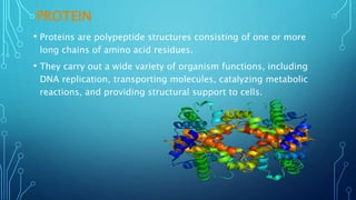
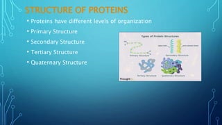
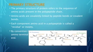
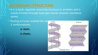
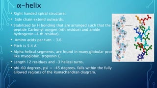
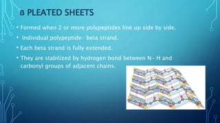
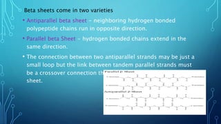
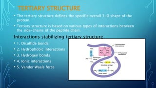
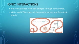
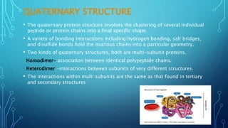
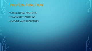
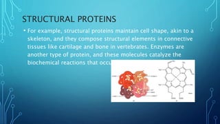
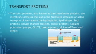
PROTEIN STRUCTURE
AND FUNCTION
PRESENTED BY
MD MOBARAK
HOSSAIN
DEPT.OF
PHARMACEUTICAL
TECHNOLOGY
JADAVPUR
UNIVERSITY
GUIDED BY –
Dr. KUNAL ROY SIR
CONTENT
• INTRODUCTION
• AMINO ACID
• PEPTIDE BOND FORMATION
• PROTEIN
• STRUCTURE OF PROTEIN
PRIMARY STRUCTURE
SECONDARY STRUCTURE
TERTIARY STRUCTURE
QUATERNARY STRUCTURE
FUNCTION OF PROTEIN
STRUCTURAL PROTEINS
TRANSPORT PROTEINS
ENZYME AND RECEPTORS
INTRODUCTION
• Proteins are the most abundant organic molecules of the living
system.
• They constitute about 50% of the cellular dry weight.
• A They constitute the fundamental basis of structure and
function of life.
• In 1839, Dutch chemist GJ. Mulder was first to describe about
proteins.
• The term protein is derived from a Greek word proteios,
meaning first place.
• The proteins are nitrogenous macromolecules that are
composed of many amino acids.
AMINO ACID
• Amino acids are a group of organic compounds containing two
functional groups – amino and carboxyl.
• The amino group [ -NH₂] is basic while the carboxyl group [- COOH] is
acidic in nature.
• There are about 300 amino acids occur in nature. Only 20 of them occur
in proteins.
PEPTIDE BOND FORMATION
• a-carboxyl group of one amino acid (with side
chain R1) forms a covalent peptide bond with a-
amino group of another amino acid (with side
chain R2) by removal of a molecule of water.
• The result is. Dipeptide.
• The dipeptide can then form a second peptide
bond with a third amino acid( with side chain R3)
to give tripeptide.
• petition of this process generates a polypeptide or
protein of specific aminoacid sequence.
• Has 40% double bond character, caused by
resonance.
PROTEIN
• Proteins are polypeptide structures consisting of one or more
long chains of amino acid residues.
• They carry out a wide variety of organism functions, including
DNA replication, transporting molecules, catalyzing metabolic
reactions, and providing structural support to cells.
STRUCTURE OF PROTEINS
• Proteins have different levels of organization
• Primary Structure
• Secondary Structure
• Tertiary Structure
• Quaternary Structure
PRIMARY STRUCTURE
• The primary structure of protein refers to the sequence of
amino acids present in the polypeptide chain.
• Amino acids are covalently linked by peptide bonds or covalent
bonds.
• Each component amino acid in a polypeptide is called a
"residue" or moiety.
• By convention the primary structure of protein starts from the
amino terminal (N) end and ends in the carboxyl terminal (C)
end.
SECONDARY STRUCTURE
• It is a local, regularly occurring structure in proteins and is
mainly formed through hydrogen bonds between backbone
atoms.
• Pauling & Corey studied the secondary structures and proposed
2 conformations.
α-helix.
ẞ sheets.
α-helix
• Right handed spiral structure.
• Side chain extend outwards.
• Stabilized by H bonding that are arranged such that the
peptide Carbonyl oxygen (nth residue) and amide
hydrogen(n+4 th residue).
• Amino acids per turn - 3.6
• Pitch is 5.4 A°
• Alpha helical segments, are found in many globular proteins
like myoglobin, troponin C.
• Length 12 residues and -3 helical turns.
• phi-60 degrees, psi = -45 degrees. falls within the fully
allowed regions of the Ramachandran diagram.
ẞ PLEATED SHEETS
• Formed when 2 or more polypeptides line up side by side.
• Individual polypeptide- beta strand.
• Each beta strand is fully extended.
• They are stabilized by hydrogen bond between N- H and
carbonyl groups of adjacent chains.
Beta sheets come in two varieties
• Antiparallel beta sheet - neighboring hydrogen bonded
polypeptide chains run in opposite direction.
• Parallel beta Sheet - hydrogen bonded chains extend in the
same direction.
• The connection between two antiparallel strands may be just a
small loop but the link between tandem parallel strands must
be a crossover connection that is out of the plane of the ẞ
sheet.
TERTIARY STRUCTURE
• The tertiary structure defines the specific overall 3-D shape of the
protein.
• Tertiary structure is based on various types of interactions between
the side-chains of the peptide chain.
Interactions stabilizing tertiary structure :
• 1. Disulfide bonds
• 2. Hydrophobic interactions
• 3. Hydrogen bonds
• 4. Ionic interactions
• 5. Vander Waals force
. DISULFIDE BONDS
• Covalent bond between sulfur atoms on two cysteine amino
acids.
HYDROPHOBIC INTERACTIONS
• Close attraction of non-polar R groups through dispersion
forces.
• They are non attractive interactions, but results from the
inability of water to form hydrogen bonds with certain side
chains.
• Very weak but collective interactions over large area stabilize
structure.
• Repel polar and charged molecules/particles.
HYDROGEN BONDS
• H bonds are weak which allows to be broken and reformed
easily.
• Allows structural change and produces ‘functional molecules’
IONIC INTERACTIONS
• lons on R groups form salt bridges through ionic bonds.
• NH3+ and COO- areas of the protein attract and form ionic
bonds.
VANDER WAALS FORCE
Van der Waals (dispersion) forces contribute to interactions of proteins
with other molecules or with surfaces, but because of the structural
complexity of protein molecules, the magnitude of these effects is usually
estimated based on idealized models of the molecular geometry, e.g.,
spheres or spheroids.
• The quaternary protein structure involves the clustering of several individual
peptide or protein chains into a final specific shape.
• A variety of bonding interactions including hydrogen bonding, salt bridges,
and disulfide bonds hold the nvarious chains into a particular geometry.
• Two kinds of quaternary structures, both are multi-subunit proteins.
Homodimer- association between identical polypeptide chains.
Heterodimer -interactions between subunits of very different structures.
• The interactions within multi subunits are the same as that found in tertiary
and secondary structures
QUATERNARY STRUCTURE
PROTEIN FUNCTION
• STRUCTURAL PROTEINS
• TRANSPORT PROTEINS
• ENZYME AND RECEPTORS
STRUCTURAL PROTEINS
• For example, structural proteins maintain cell shape, akin to a
skeleton, and they compose structural elements in connective
tissues like cartilage and bone in vertebrates. Enzymes are
another type of protein, and these molecules catalyze the
biochemical reactions that occur in cells.
TRANSPORT PROTEINS
• Transport proteins, also known as transmembrane proteins, are
membrane proteins that aid in the facilitated diffusion or active
transport of ions across the hydrophobic lipid bilayer. Such
proteins include channel proteins, carrier proteins, sodium-
potassium pumps, GLUT1, proton pump, calcium ATPase, and
others.
ENZYME AND RECEPTORS
Enzyme linked receptors or catalytic receptors are a group of
multisubunit transmembrane protein and the second major type
of cell surface receptor. They promote several processes in
animal cells such as cell proliferation, differentiation, cell growth,
cell survival once recognized by specific ligands.
PROTEIN STRUCTURE AND FUNCTION PPT(MD MOBARAK HOSSAIN).pptx

PROTEIN STRUCTURE AND FUNCTION PPT(MD MOBARAK HOSSAIN).pptx

  • 1.
    PROTEIN STRUCTURE AND FUNCTION PRESENTEDBY MD MOBARAK HOSSAIN DEPT.OF PHARMACEUTICAL TECHNOLOGY JADAVPUR UNIVERSITY GUIDED BY – Dr. KUNAL ROY SIR
  • 2.
    CONTENT • INTRODUCTION • AMINOACID • PEPTIDE BOND FORMATION • PROTEIN • STRUCTURE OF PROTEIN PRIMARY STRUCTURE SECONDARY STRUCTURE TERTIARY STRUCTURE QUATERNARY STRUCTURE FUNCTION OF PROTEIN STRUCTURAL PROTEINS TRANSPORT PROTEINS ENZYME AND RECEPTORS
  • 3.
    INTRODUCTION • Proteins arethe most abundant organic molecules of the living system. • They constitute about 50% of the cellular dry weight. • A They constitute the fundamental basis of structure and function of life. • In 1839, Dutch chemist GJ. Mulder was first to describe about proteins. • The term protein is derived from a Greek word proteios, meaning first place. • The proteins are nitrogenous macromolecules that are composed of many amino acids.
  • 4.
    AMINO ACID • Aminoacids are a group of organic compounds containing two functional groups – amino and carboxyl. • The amino group [ -NH₂] is basic while the carboxyl group [- COOH] is acidic in nature. • There are about 300 amino acids occur in nature. Only 20 of them occur in proteins.
  • 6.
    PEPTIDE BOND FORMATION •a-carboxyl group of one amino acid (with side chain R1) forms a covalent peptide bond with a- amino group of another amino acid (with side chain R2) by removal of a molecule of water. • The result is. Dipeptide. • The dipeptide can then form a second peptide bond with a third amino acid( with side chain R3) to give tripeptide. • petition of this process generates a polypeptide or protein of specific aminoacid sequence. • Has 40% double bond character, caused by resonance.
  • 7.
    PROTEIN • Proteins arepolypeptide structures consisting of one or more long chains of amino acid residues. • They carry out a wide variety of organism functions, including DNA replication, transporting molecules, catalyzing metabolic reactions, and providing structural support to cells.
  • 8.
    STRUCTURE OF PROTEINS •Proteins have different levels of organization • Primary Structure • Secondary Structure • Tertiary Structure • Quaternary Structure
  • 9.
    PRIMARY STRUCTURE • Theprimary structure of protein refers to the sequence of amino acids present in the polypeptide chain. • Amino acids are covalently linked by peptide bonds or covalent bonds. • Each component amino acid in a polypeptide is called a "residue" or moiety. • By convention the primary structure of protein starts from the amino terminal (N) end and ends in the carboxyl terminal (C) end.
  • 10.
    SECONDARY STRUCTURE • Itis a local, regularly occurring structure in proteins and is mainly formed through hydrogen bonds between backbone atoms. • Pauling & Corey studied the secondary structures and proposed 2 conformations. α-helix. ẞ sheets.
  • 11.
    α-helix • Right handedspiral structure. • Side chain extend outwards. • Stabilized by H bonding that are arranged such that the peptide Carbonyl oxygen (nth residue) and amide hydrogen(n+4 th residue). • Amino acids per turn - 3.6 • Pitch is 5.4 A° • Alpha helical segments, are found in many globular proteins like myoglobin, troponin C. • Length 12 residues and -3 helical turns. • phi-60 degrees, psi = -45 degrees. falls within the fully allowed regions of the Ramachandran diagram.
  • 12.
    ẞ PLEATED SHEETS •Formed when 2 or more polypeptides line up side by side. • Individual polypeptide- beta strand. • Each beta strand is fully extended. • They are stabilized by hydrogen bond between N- H and carbonyl groups of adjacent chains.
  • 13.
    Beta sheets comein two varieties • Antiparallel beta sheet - neighboring hydrogen bonded polypeptide chains run in opposite direction. • Parallel beta Sheet - hydrogen bonded chains extend in the same direction. • The connection between two antiparallel strands may be just a small loop but the link between tandem parallel strands must be a crossover connection that is out of the plane of the ẞ sheet.
  • 14.
    TERTIARY STRUCTURE • Thetertiary structure defines the specific overall 3-D shape of the protein. • Tertiary structure is based on various types of interactions between the side-chains of the peptide chain. Interactions stabilizing tertiary structure : • 1. Disulfide bonds • 2. Hydrophobic interactions • 3. Hydrogen bonds • 4. Ionic interactions • 5. Vander Waals force
  • 15.
    . DISULFIDE BONDS •Covalent bond between sulfur atoms on two cysteine amino acids.
  • 16.
    HYDROPHOBIC INTERACTIONS • Closeattraction of non-polar R groups through dispersion forces. • They are non attractive interactions, but results from the inability of water to form hydrogen bonds with certain side chains. • Very weak but collective interactions over large area stabilize structure. • Repel polar and charged molecules/particles.
  • 17.
    HYDROGEN BONDS • Hbonds are weak which allows to be broken and reformed easily. • Allows structural change and produces ‘functional molecules’
  • 18.
    IONIC INTERACTIONS • lonson R groups form salt bridges through ionic bonds. • NH3+ and COO- areas of the protein attract and form ionic bonds.
  • 19.
    VANDER WAALS FORCE Vander Waals (dispersion) forces contribute to interactions of proteins with other molecules or with surfaces, but because of the structural complexity of protein molecules, the magnitude of these effects is usually estimated based on idealized models of the molecular geometry, e.g., spheres or spheroids.
  • 20.
    • The quaternaryprotein structure involves the clustering of several individual peptide or protein chains into a final specific shape. • A variety of bonding interactions including hydrogen bonding, salt bridges, and disulfide bonds hold the nvarious chains into a particular geometry. • Two kinds of quaternary structures, both are multi-subunit proteins. Homodimer- association between identical polypeptide chains. Heterodimer -interactions between subunits of very different structures. • The interactions within multi subunits are the same as that found in tertiary and secondary structures QUATERNARY STRUCTURE
  • 21.
    PROTEIN FUNCTION • STRUCTURALPROTEINS • TRANSPORT PROTEINS • ENZYME AND RECEPTORS
  • 22.
    STRUCTURAL PROTEINS • Forexample, structural proteins maintain cell shape, akin to a skeleton, and they compose structural elements in connective tissues like cartilage and bone in vertebrates. Enzymes are another type of protein, and these molecules catalyze the biochemical reactions that occur in cells.
  • 23.
    TRANSPORT PROTEINS • Transportproteins, also known as transmembrane proteins, are membrane proteins that aid in the facilitated diffusion or active transport of ions across the hydrophobic lipid bilayer. Such proteins include channel proteins, carrier proteins, sodium- potassium pumps, GLUT1, proton pump, calcium ATPase, and others.
  • 24.
    ENZYME AND RECEPTORS Enzymelinked receptors or catalytic receptors are a group of multisubunit transmembrane protein and the second major type of cell surface receptor. They promote several processes in animal cells such as cell proliferation, differentiation, cell growth, cell survival once recognized by specific ligands.