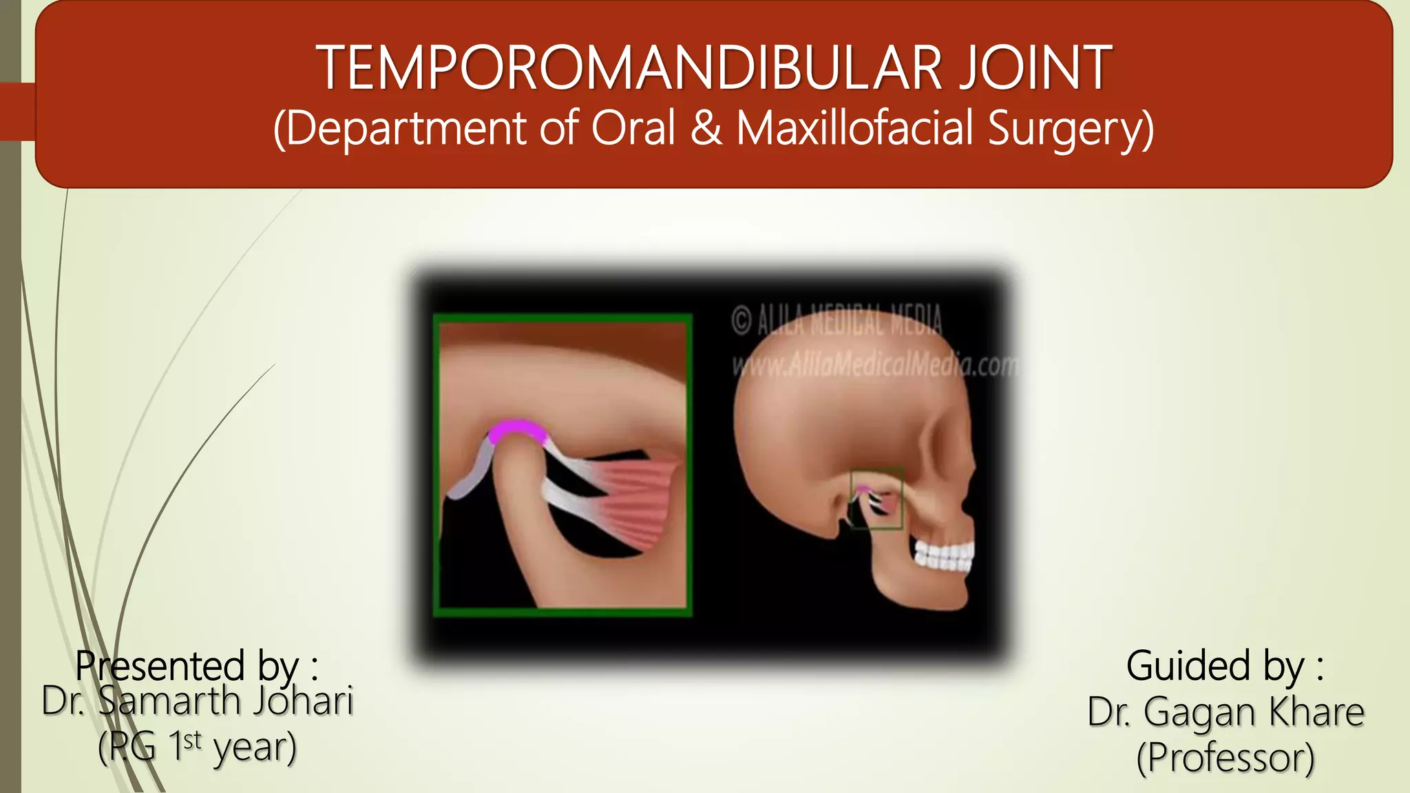
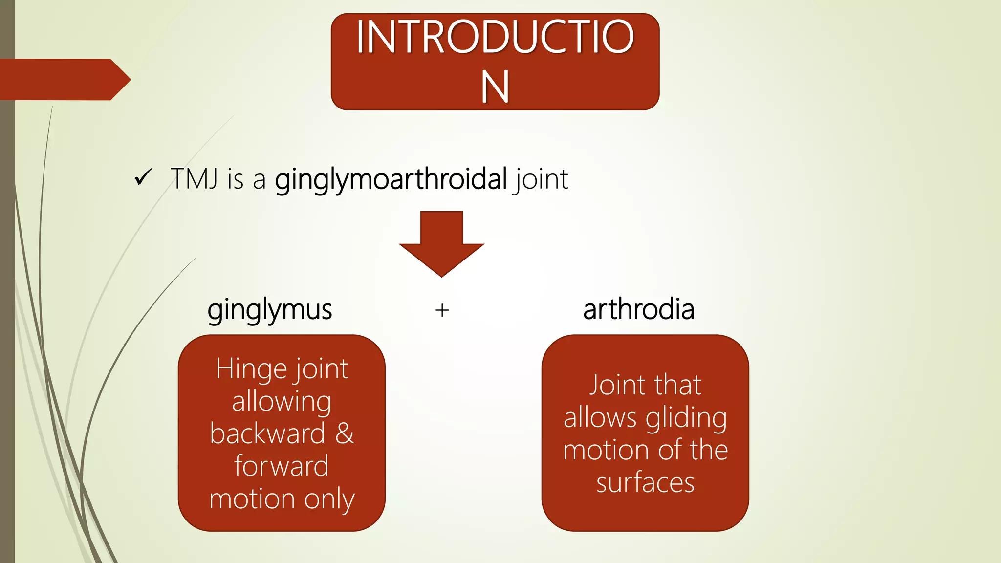
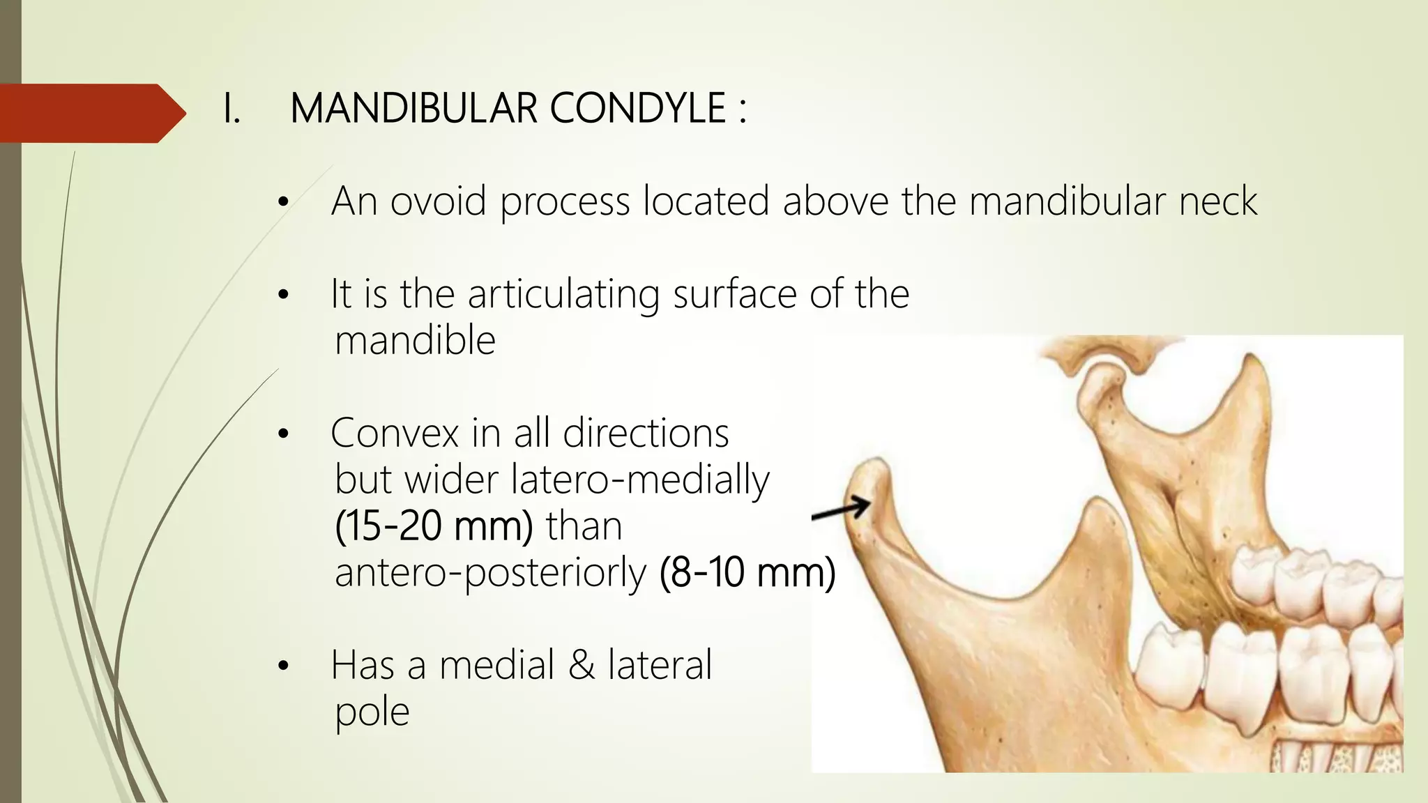
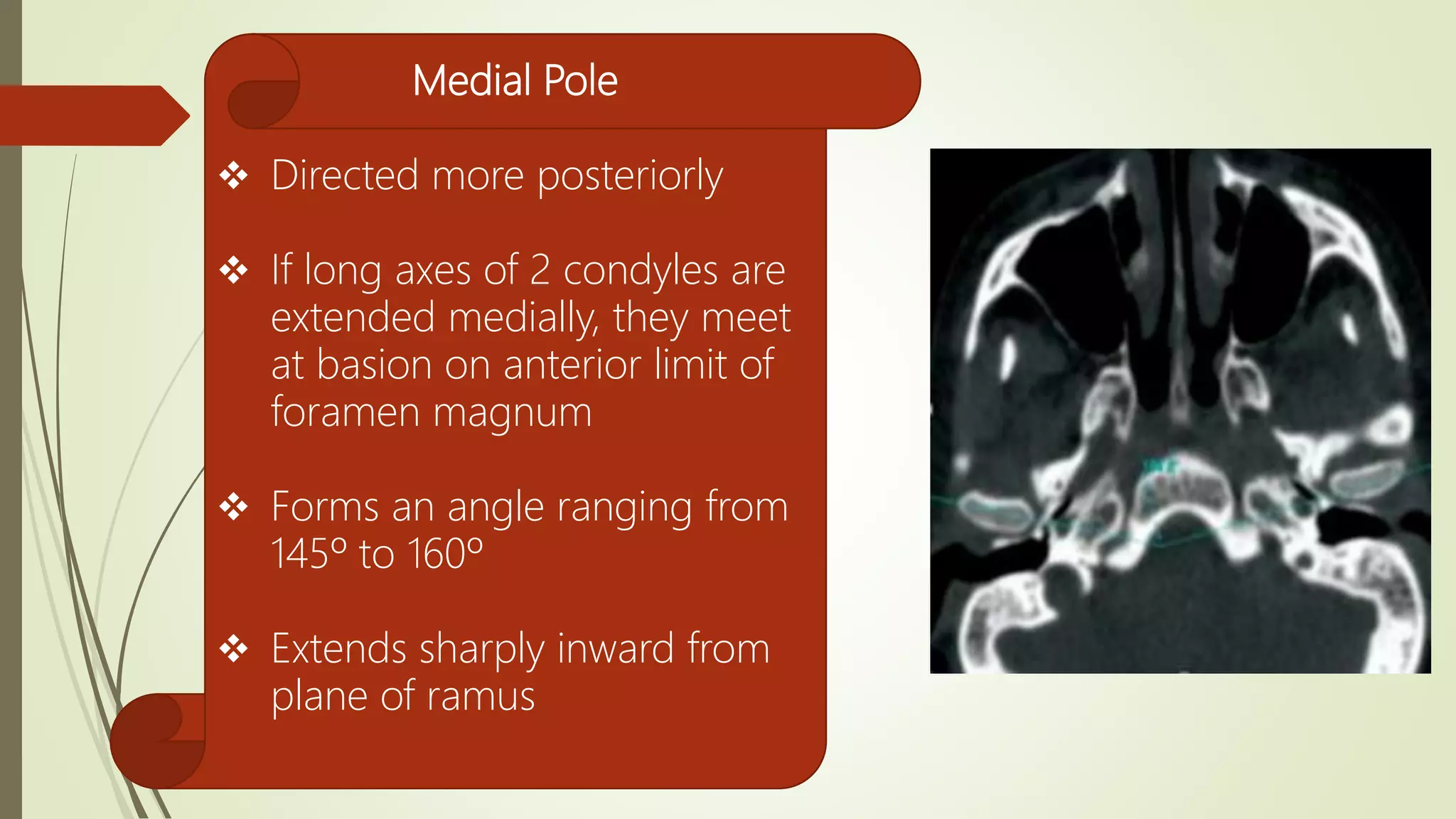
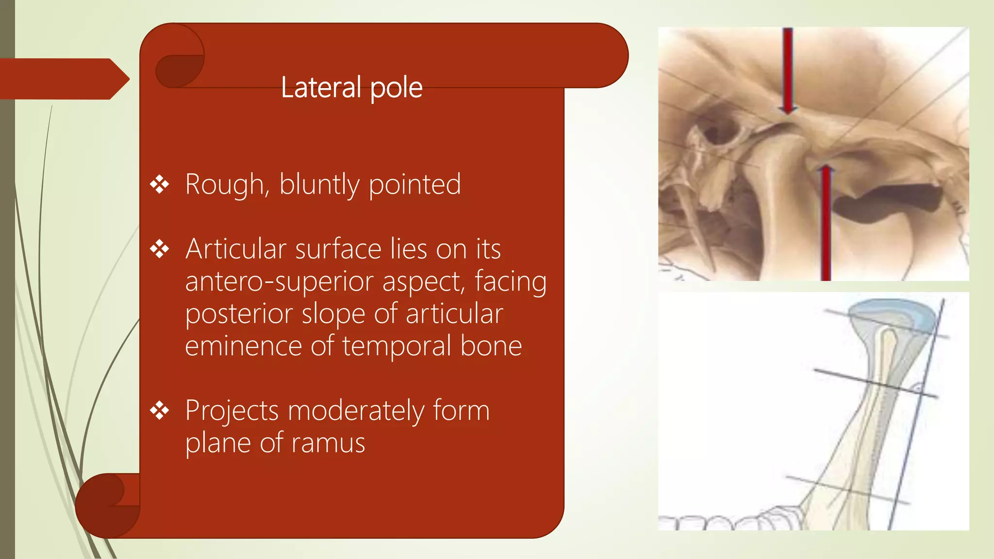
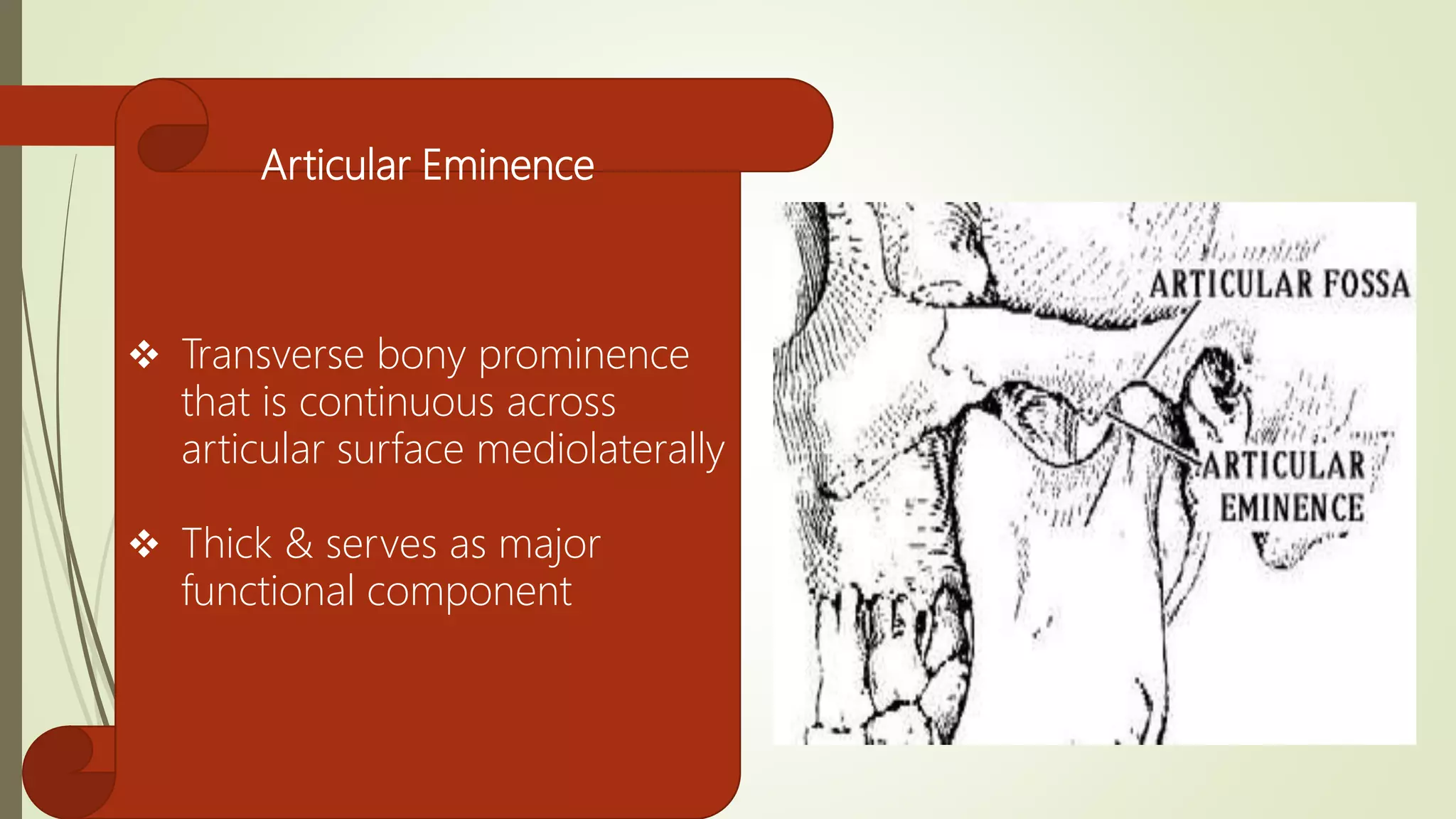
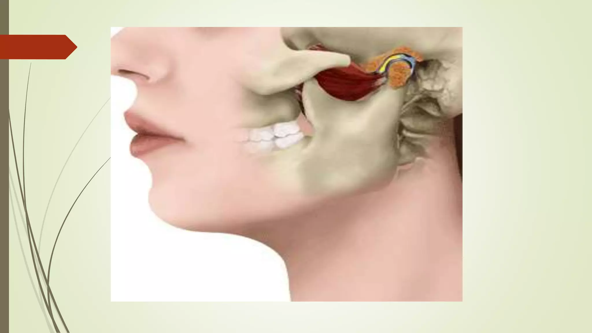
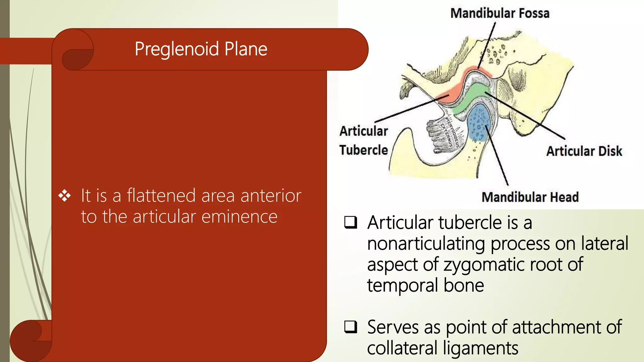
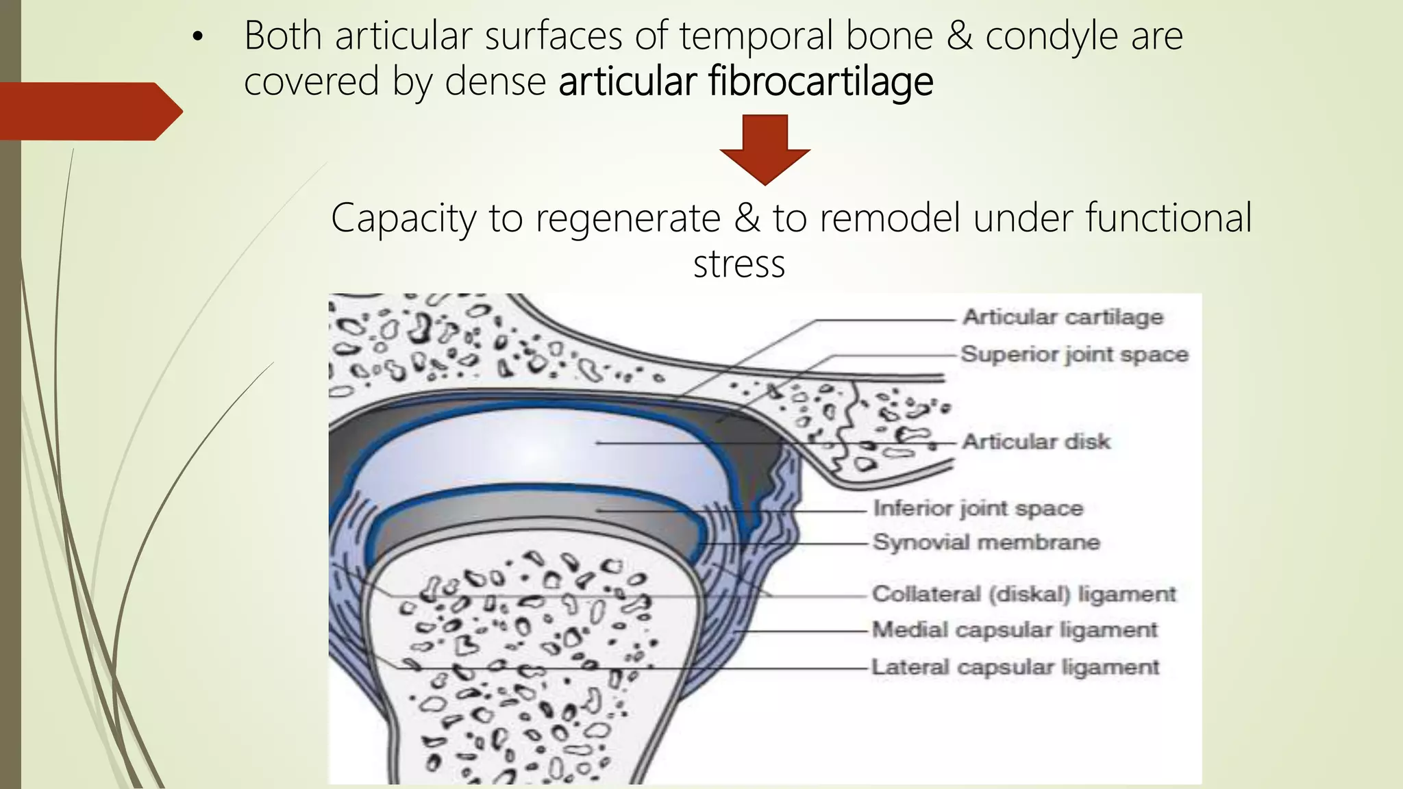
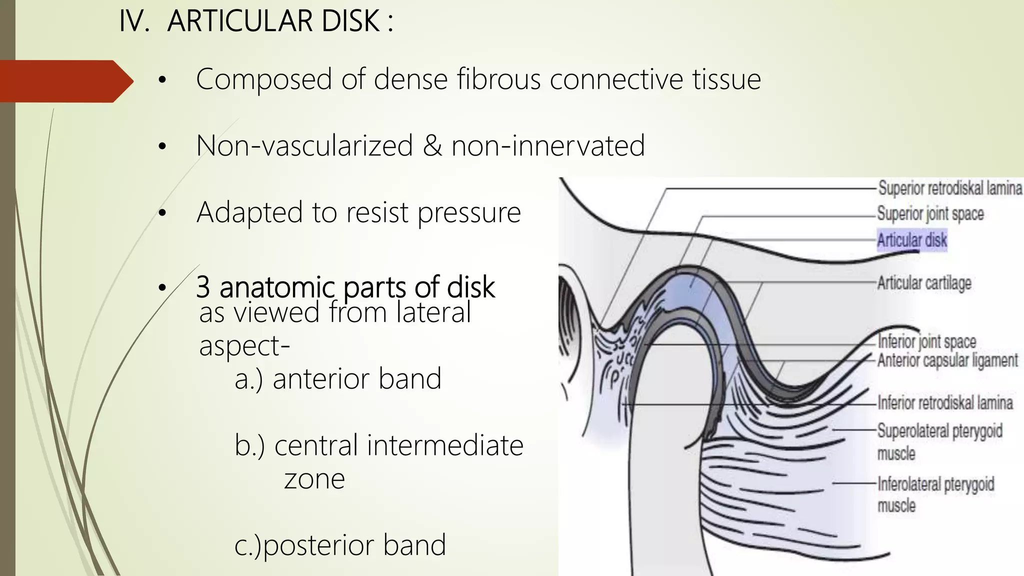
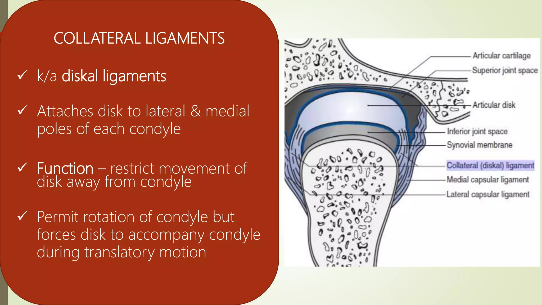
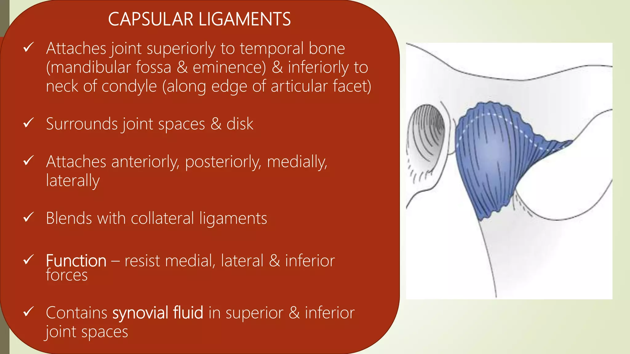
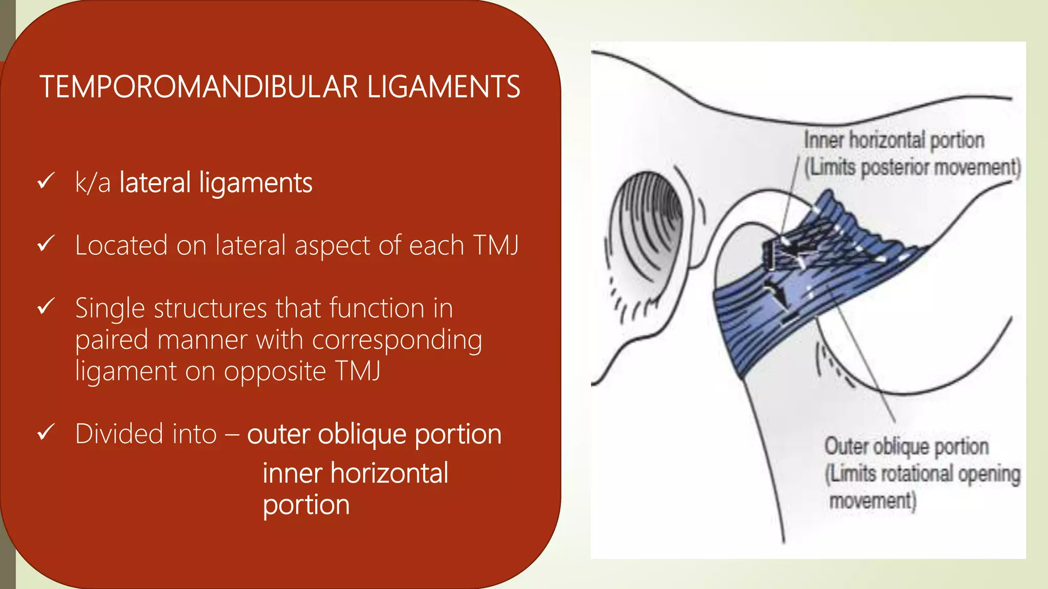
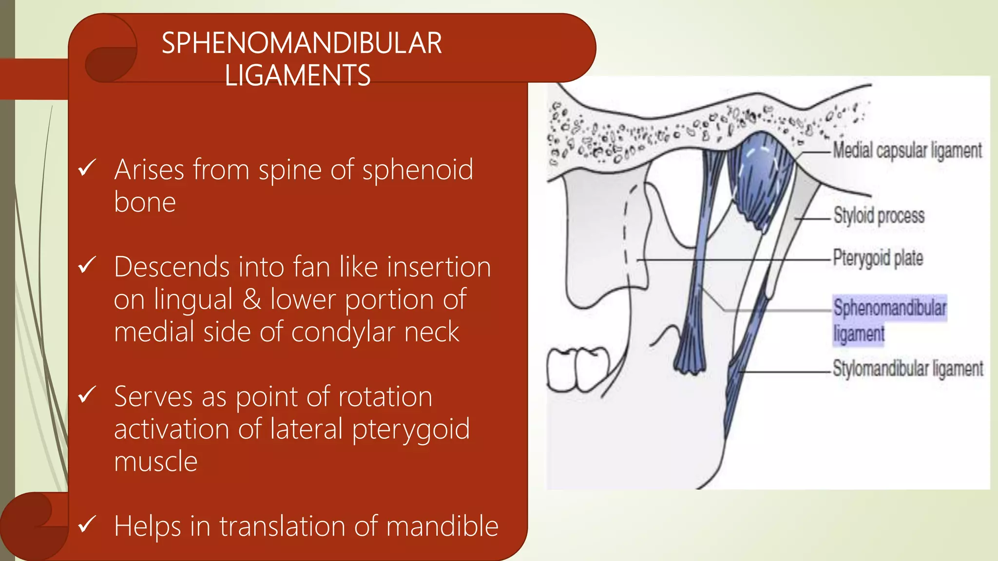
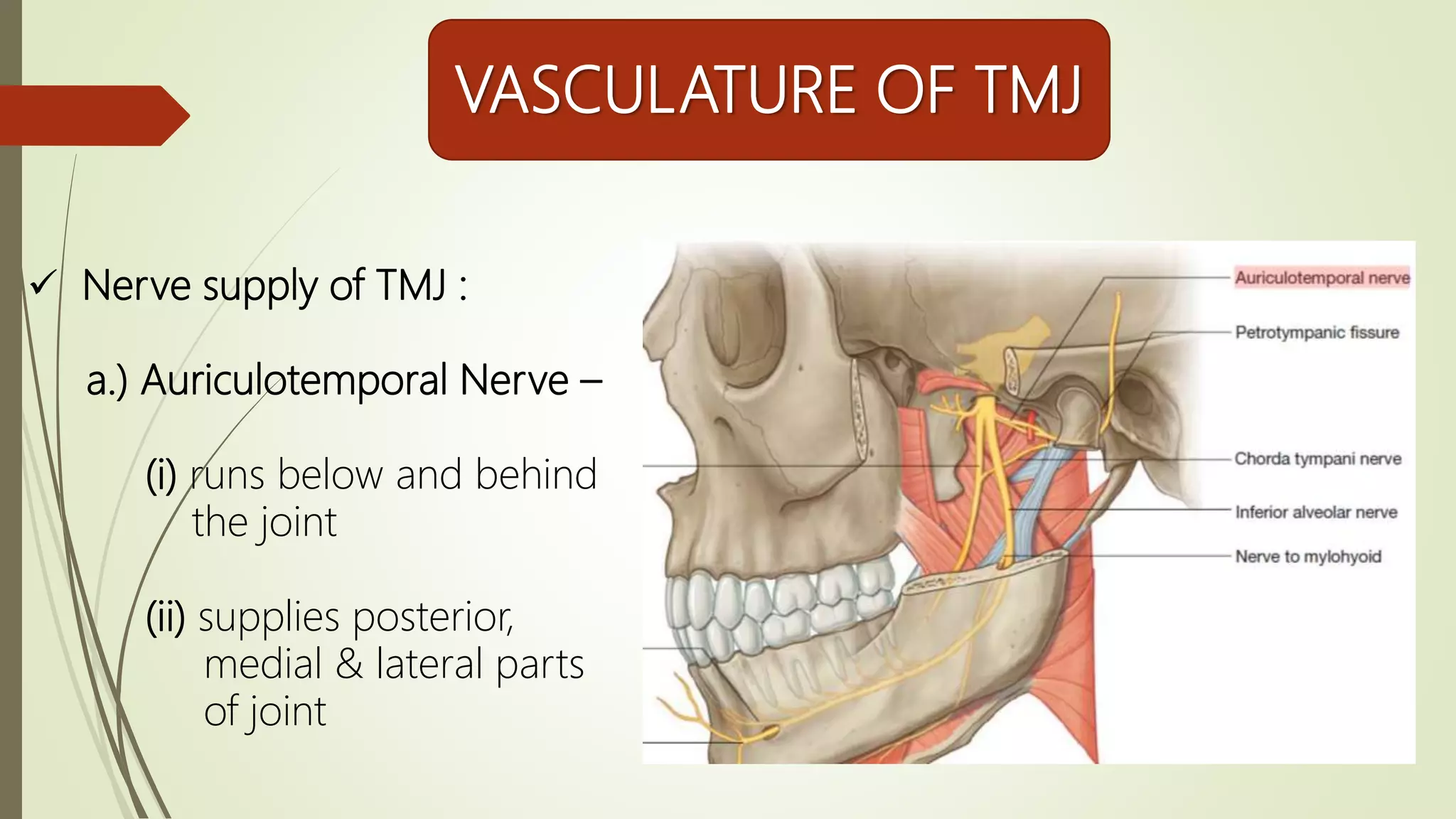
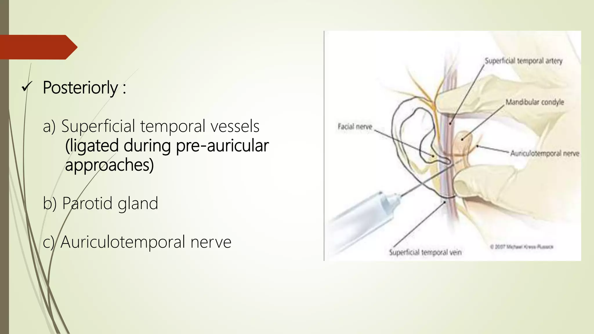
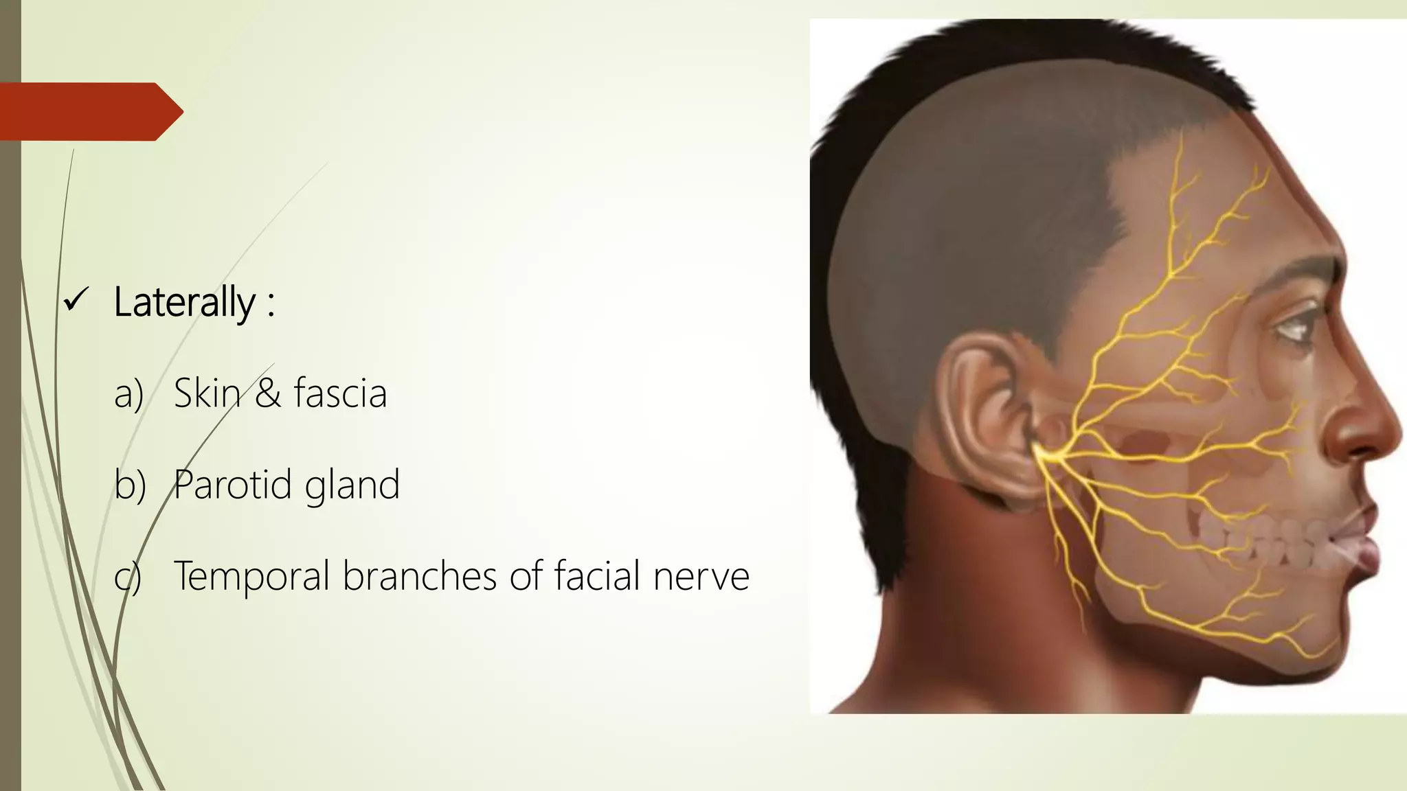
GOOD MORNING
Presented by :
Dr. Samarth Johari
(P.G 1st year)
Guided by :
Dr. Gagan Khare
(Professor)
TEMPOROMANDIBULAR JOINT
(Department of Oral & Maxillofacial Surgery)
CONTENTS
Introduction
Unique Features of TMJ
Components Of TMJ
Vasculature Of TMJ
Relation Of TMJ
Surgical Approaches To TMJ
Conclusion
References
INTRODUCTIO
N
 TMJ is a ginglymoarthroidal joint
ginglymus + arthrodia
Hinge joint
allowing
backward &
forward
motion only
Joint that
allows gliding
motion of the
surfaces
TMJ as a Compound Joint
Anatomically Functionally
• Diarthroidal joint
• Synovial joint
• Has fibrous connective
tissue capsule tightly
attached to bones
• Compound joint
composed of 4
articulating surfaces
 Anatomically –
• Diarthroidal Joint :
 Discontinuous articulation of 2 bones
Permitting free movement produced by
associated muscles & limited by
Ligaments
• Synovial Joint :
 Lined by synovial membrane on
inner aspect which secretes synovial fluid
Acts as a lubricant and provides metabolic & nutritional needs
non vascularized internal joint structures
 Funtionally –
• Composed of 4 articulating surfaces :
 Articular facets of temporal bone
 Mandibular condyle
 Superior surface of articular disc
Articular Disc
 Inferior surface of articular disc
Divides the joint into 2 compartments
1. Lower compartment – permits hinge motion &
rotation (ginglymoid)
2. Superior compartment – permits sliding/transltory
motion (arthroidal)
Ginglymoarthroidal
joint
UNIQUE FEATURES OF
TMJ
 Bilateral diarthrosis – right & left function together
 Articular surfaces covered by fibrocartilage instead of hyaline
cartilage
 Only joint of human body having rigid endpoint of closure (teeth
making occlusal contact)
 Last joint to start developing (in 7th week of I.U life)
COMPONENTS
OF TMJ
I. Mandibular Condyle
II. Articular Surfaces Of Temporal Bone
III. Cartilage and Synovium
IV. Articular Disk
V. Muscular Components
VI. Ligaments
I. MANDIBULAR CONDYLE :
• An ovoid process located above the mandibular neck
• It is the articulating surface of the
mandible
• Convex in all directions
but wider latero-medially
(15-20 mm) than
antero-posteriorly (8-10 mm)
• Has a medial & lateral
pole
Medial Pole
 Directed more posteriorly
 If long axes of 2 condyles are
extended medially, they meet
at basion on anterior limit of
foramen magnum
 Forms an angle ranging from
145º to 160º
 Extends sharply inward from
plane of ramus
Lateral pole
 Rough, bluntly pointed
 Articular surface lies on its
antero-superior aspect, facing
posterior slope of articular
eminence of temporal bone
 Projects moderately form
plane of ramus
II. ARTICULAR SURFACE OF TEMPORAL BONE :
• Situated on inferior aspect of temporal squama anterior to
tympanic plate
• Comopsed of 3 parts –
a.) Articular fossa or mandibular fossa
b.) Articular eminence
c.) Preglenoid plane
Mandibular Fossa
 Largest
 Concave structure extending
from posterior slope of
articular eminence to
psotglenoid fossa (ridge
between fossa & external
acoustic meatus)
 Surface is thin
 Not a major stress bearing
area
Articular Eminence
 Transverse bony prominence
that is continuous across
articular surface mediolaterally
 Thick & serves as major
functional component
 It is a flattened area anterior
to the articular eminence
Preglenoid Plane
 Articular tubercle is a
nonarticulating process on lateral
aspect of zygomatic root of
temporal bone
 Serves as point of attachment of
collateral ligaments
III. CARTILAGE AND SYNOVIUM :
• Inner aspect of the joint is lined by two types of tissues :
a.) Articular Cartilage
b.) Synovium
• Space bound by these two structures
synovial cavity
filled with synovial fluid
• Both articular surfaces of temporal bone & condyle are
covered by dense articular fibrocartilage
Capacity to regenerate & to remodel under functional
stress
• Lining of capsular ligament – synovial membrane
thin, smooth, richly innervated vascular tissue
without epithelium
Synovium is capable of rapid & complete regeneration following
injury
• Synovial fluid contains high concentration of hyaluronic acid
(reason for viscous nature of fluid)
• Other contents of fluid – high % of albumin & low % of -2-
globulin, alkaline phosphatase, leucocytes (<200/cubic mm)
Functions Of Synovial Fluid
 Lubrication of joint
 Phagocytosis of particulate debris
 Nourishment of articular cartilage
IV. ARTICULAR DISK :
• Composed of dense fibrous connective tissue
• Non-vascularized & non-innervated
• Adapted to resist pressure
• 3 anatomic parts of disk
as viewed from lateral
aspect-
a.) anterior band
b.) central intermediate
zone
c.)posterior band
• Intermediate zone is thinnest & generally the area of
function between mandibular condyle and temporal bone
• Disk is flexible & adapts to functional demands of articular
surfaces
• Attached to capsular ligament anteriorly, posteriorly, medially
& laterally
• Some fibres of superior head of lateral pterygoid muscle
insert on the disk at its medial aspect
This stabilizes the disk to mandibular condyle during function
V. MUSCULAR COMPONENTS :
• Total 12 bilateral muscles influence mandibular motion & are
divided into 2 groups by anatomic position -
SUPRAMANDIBULAR
GROUP
 Attaches to ramus & condylar neck
 Consists of :
a.) temporalis
b.) masseter
c.) medial pterygoid
d.) lateral pterygoid
 Predominantly acts as elevator, however,
lateral pterygoid has depressor action also
INFRAMANDIBULAR
GROUP
 Attaches to body & symphyseal area &
hyoid bone
 Consists of :
I. suprahyoid muscles (attach to both
hyoid bone & mandible) -
a.) digastric
b.) geniohyoid
c.) mylohyoid
d.) stylohyoid
II. Infrahyoid muscles –
a.) sternohyoid
b.) omohyoid
c.) sternothyroid
d.) thyrohyoid
Suprahyoid muscles depress
mandible when hyoid bone is
fixed & elevate hyoid bone
when mandible is fixed
Infrahyoid muscles fix hyoid
bone during depressive
movements of mandible
LATERAL PTERYGOID & MEDIAL
PTERYGOID MUSCLES
MASSETER MUSCLE TEMPORALIS MUSCLE
VI. LIGAMENTS :
• Composed of collagen
• Act as restraints to motion of condyle & disk
2 sets of
ligaments
Functional Accessory
FUNCTIONAL LIGAMENTS
 Serve as major anatomic
components of joints
 Types of functional ligaments
:
• Collateral
• Capsular
• Temporomandibular
ACCESSORY LIGAMENTS
 Serve as passive restraints
on mandibular motion
 Types of accessory
ligaments :
• Sphenomandibular
• Stylomandibular
COLLATERAL LIGAMENTS
 k/a diskal ligaments
 Attaches disk to lateral & medial
poles of each condyle
 Function – restrict movement of
disk away from condyle
 Permit rotation of condyle but
forces disk to accompany condyle
during translatory motion
CAPSULAR LIGAMENTS
 Attaches joint superiorly to temporal bone
(mandibular fossa & eminence) & inferiorly to
neck of condyle (along edge of articular facet)
 Surrounds joint spaces & disk
 Attaches anteriorly, posteriorly, medially,
laterally
 Blends with collateral ligaments
 Function – resist medial, lateral & inferior
forces
 Contains synovial fluid in superior & inferior
joint spaces
TEMPOROMANDIBULAR LIGAMENTS
 k/a lateral ligaments
 Located on lateral aspect of each TMJ
 Single structures that function in
paired manner with corresponding
ligament on opposite TMJ
 Divided into – outer oblique portion
inner horizontal
portion
SPHENOMANDIBULAR
LIGAMENTS
 Arises from spine of sphenoid
bone
 Descends into fan like insertion
on lingual & lower portion of
medial side of condylar neck
 Serves as point of rotation
activation of lateral pterygoid
muscle
 Helps in translation of mandible
STYLOMANDIBULAR
LIGAMENTS
 Descends from styloid process
to posterior border of angle of
mandible
 Blends with fascia of medial
pterygoid muscle
 Functions similar to
sphenomandibular ligament
 Limits excessive protrusion of
mandible
VASCULATURE OF TMJ
 Nerve supply of TMJ :
a.) Auriculotemporal Nerve –
(i) runs below and behind
the joint
(ii) supplies posterior,
medial & lateral parts
of joint
b.) Nerve To Masseter –
sends a twig to the joint
c.) Deep Temporal Nerve –
supplies anterior parts of
the joint
 Arterial Supply Of TMJ :
only superficial blood supply of
the joint is present
no blood supply inside the
capsule
takes nourishment from
synovial fluid
superficial blood supply is by
branches of external carotid
artery –
a.) Superficial Temporal Artery b.) Deep auricular Artery
c.) Anterior Tympanic Artery d.) Maxillary Artery
e.) Ascending Pharyngeal Artery
 Venous Supply Of TMJ :
a.) Maxillary Vein
b.) Transverse Facial Vein
c.) Superficial Temporal
vein
RELATION OF TMJ
 Anteriorly :
a) Mandibular notch
b) Lateral pterygoid
c) Masseteric nerve & artery
Applied Anatomy Of Masseteric Nerve
A careful dissection of 16 intact human
cadaveric head specimens revealed The
location of the masseteric artery was then
determined in relation to 3 points process:
1) the anterior-superior aspect of the
condylar neck = 10.3 mm;
2) the most inferior aspect of the articular
tubercle = 11.4 mm;
3) the inferior aspect of the sigmoid notch =
3mm
Journal of Oral and Maxillofacial Surgery.
2009;67 (2) : 369–371
 Posteriorly :
a) Superficial temporal vessels
(ligated during pre-auricular
approaches)
b) Parotid gland
c) Auriculotemporal nerve
 Laterally :
a) Skin & fascia
b) Parotid gland
c) Temporal branches of facial nerve
Applied Anatomy Of Facial Nerve
 Exits skull at stylomastoid formen
 Incise the superficial layer of
temporalis fascia & periosteum
over arch within 8mm boundary,
to prevent damage to branches of
upper trunk
Classic Article By Alkayat & Bramely
(1980)
 Medially :
a) Tympanic plate (separates from ICA)
b) Spine of sphenoid
c) Auriculotemporal & chorda
tympani nerve
a) Middle meningeal artery
Applied Anatomy Of
Auriculotemporal Nerve
 1st branch of 3rd division of
trigeminal nerve
 Exits from foramen ovale
 Runs medial to lateral behind
neck of condyle
 Sensory supply to skin in
temporal & preauricular region &
innervates capsule of TMJ
 Can be damaged while
approaching to TMJ
SURGICAL APPROACHES TO TMJ
Operator
• Humphrey
• Ricdel
• Annandale
Procedure
• Condylectomy
• Meiscectomy
• Disk Repositioning
Year
• 1856
• 1883
• 1887
 Preauricular
 Endaural
 Postauricular
 Rhytidectomal
 Retromandibular
 Intraoral
Types Of Surgical Approaches To TMJ
 Basic incision given by
Dingman in 1951
 No extensive shaving is
required
 Margin of only 1cm from
superior aspect of incision is
adequate
 Inverted hockey stick
incision that follows natural
crease in front of tragus
Preauricular Approach
 Cosmetic modification or
preauricular approach
 Incision is placed on
prominence of tragus itself
 Tragal cartilage has to be
protected to prevent
perichondritis
Endaural Approach
 Popularised by Walter’s &
Geist in 1983
 Disadvantages :
(i) Auricular Stenosis
(ii) Can not be used in
cases of joint infection &
chronic otitis externa
 Advantages :
(i) Excellent exposure to
complete joint
(ii) Ability to camouflage
scar
Postauricular Approach
 Used in cases where extensive
joint exposure is required e.g
major tumor resections
 Endaural incision is extended
in curvilinear fashion around
mastoid tip & S - shaped
incision ending in
submandibular incision
 Allows access to posterior
border of mandible & allows
to locate main trunk of facial
nerve
Rhytidectomy Approach
 Used for neck of condyle &
ramus region
 Supplemets different TMJ
approaches for tunneling
through soft tissue for placing
grafts
 Marginal mandibular branch of
facial nerve & retromandibular
vein have to be protected
Retromandibular Approach
CONCLUSION
The applied anatomy of
temporomandibular joint has many
significant applications in maxillofacial
surgery. Understanding these
important anatomic relations-
variations enables surgeons to perform
the surgical procedures safely.
Knowledge of these concepts helps us
to recognize the problems and
complications as and when they occur
and manage them accordingly.
REFERENCES
 Colour Atlas Of Temporomandibular Joint Surgery by Peter D. Quinn
 Oral Anatomy by Sicher and DuBrul
 Gray’s Anatomy
 Oral & Maxillofacial Surgery by Fonseca
 Atlas of Operative Maxillofacial Trauma Surgery by Springer
 Peterson’s Principles Of Oral & Maxillofacial Surgery
Applied anatomy of tmj

Applied anatomy of tmj

  • 1.
  • 2.
    Presented by : Dr.Samarth Johari (P.G 1st year) Guided by : Dr. Gagan Khare (Professor) TEMPOROMANDIBULAR JOINT (Department of Oral & Maxillofacial Surgery)
  • 3.
    CONTENTS Introduction Unique Features ofTMJ Components Of TMJ Vasculature Of TMJ Relation Of TMJ Surgical Approaches To TMJ Conclusion References
  • 4.
    INTRODUCTIO N  TMJ isa ginglymoarthroidal joint ginglymus + arthrodia Hinge joint allowing backward & forward motion only Joint that allows gliding motion of the surfaces
  • 5.
    TMJ as aCompound Joint Anatomically Functionally • Diarthroidal joint • Synovial joint • Has fibrous connective tissue capsule tightly attached to bones • Compound joint composed of 4 articulating surfaces
  • 6.
     Anatomically – •Diarthroidal Joint :  Discontinuous articulation of 2 bones Permitting free movement produced by associated muscles & limited by Ligaments • Synovial Joint :  Lined by synovial membrane on inner aspect which secretes synovial fluid Acts as a lubricant and provides metabolic & nutritional needs non vascularized internal joint structures
  • 7.
     Funtionally – •Composed of 4 articulating surfaces :  Articular facets of temporal bone  Mandibular condyle  Superior surface of articular disc Articular Disc  Inferior surface of articular disc Divides the joint into 2 compartments 1. Lower compartment – permits hinge motion & rotation (ginglymoid) 2. Superior compartment – permits sliding/transltory motion (arthroidal) Ginglymoarthroidal joint
  • 9.
    UNIQUE FEATURES OF TMJ Bilateral diarthrosis – right & left function together  Articular surfaces covered by fibrocartilage instead of hyaline cartilage  Only joint of human body having rigid endpoint of closure (teeth making occlusal contact)  Last joint to start developing (in 7th week of I.U life)
  • 10.
    COMPONENTS OF TMJ I. MandibularCondyle II. Articular Surfaces Of Temporal Bone III. Cartilage and Synovium IV. Articular Disk V. Muscular Components VI. Ligaments
  • 11.
    I. MANDIBULAR CONDYLE: • An ovoid process located above the mandibular neck • It is the articulating surface of the mandible • Convex in all directions but wider latero-medially (15-20 mm) than antero-posteriorly (8-10 mm) • Has a medial & lateral pole
  • 12.
    Medial Pole  Directedmore posteriorly  If long axes of 2 condyles are extended medially, they meet at basion on anterior limit of foramen magnum  Forms an angle ranging from 145º to 160º  Extends sharply inward from plane of ramus
  • 13.
    Lateral pole  Rough,bluntly pointed  Articular surface lies on its antero-superior aspect, facing posterior slope of articular eminence of temporal bone  Projects moderately form plane of ramus
  • 14.
    II. ARTICULAR SURFACEOF TEMPORAL BONE : • Situated on inferior aspect of temporal squama anterior to tympanic plate • Comopsed of 3 parts – a.) Articular fossa or mandibular fossa b.) Articular eminence c.) Preglenoid plane
  • 15.
    Mandibular Fossa  Largest Concave structure extending from posterior slope of articular eminence to psotglenoid fossa (ridge between fossa & external acoustic meatus)  Surface is thin  Not a major stress bearing area
  • 16.
    Articular Eminence  Transversebony prominence that is continuous across articular surface mediolaterally  Thick & serves as major functional component
  • 18.
     It isa flattened area anterior to the articular eminence Preglenoid Plane  Articular tubercle is a nonarticulating process on lateral aspect of zygomatic root of temporal bone  Serves as point of attachment of collateral ligaments
  • 19.
    III. CARTILAGE ANDSYNOVIUM : • Inner aspect of the joint is lined by two types of tissues : a.) Articular Cartilage b.) Synovium • Space bound by these two structures synovial cavity filled with synovial fluid
  • 20.
    • Both articularsurfaces of temporal bone & condyle are covered by dense articular fibrocartilage Capacity to regenerate & to remodel under functional stress
  • 21.
    • Lining ofcapsular ligament – synovial membrane thin, smooth, richly innervated vascular tissue without epithelium Synovium is capable of rapid & complete regeneration following injury • Synovial fluid contains high concentration of hyaluronic acid (reason for viscous nature of fluid) • Other contents of fluid – high % of albumin & low % of -2- globulin, alkaline phosphatase, leucocytes (<200/cubic mm)
  • 22.
    Functions Of SynovialFluid  Lubrication of joint  Phagocytosis of particulate debris  Nourishment of articular cartilage
  • 23.
    IV. ARTICULAR DISK: • Composed of dense fibrous connective tissue • Non-vascularized & non-innervated • Adapted to resist pressure • 3 anatomic parts of disk as viewed from lateral aspect- a.) anterior band b.) central intermediate zone c.)posterior band
  • 24.
    • Intermediate zoneis thinnest & generally the area of function between mandibular condyle and temporal bone • Disk is flexible & adapts to functional demands of articular surfaces • Attached to capsular ligament anteriorly, posteriorly, medially & laterally • Some fibres of superior head of lateral pterygoid muscle insert on the disk at its medial aspect This stabilizes the disk to mandibular condyle during function
  • 25.
    V. MUSCULAR COMPONENTS: • Total 12 bilateral muscles influence mandibular motion & are divided into 2 groups by anatomic position - SUPRAMANDIBULAR GROUP  Attaches to ramus & condylar neck  Consists of : a.) temporalis b.) masseter c.) medial pterygoid d.) lateral pterygoid  Predominantly acts as elevator, however, lateral pterygoid has depressor action also
  • 26.
    INFRAMANDIBULAR GROUP  Attaches tobody & symphyseal area & hyoid bone  Consists of : I. suprahyoid muscles (attach to both hyoid bone & mandible) - a.) digastric b.) geniohyoid c.) mylohyoid d.) stylohyoid II. Infrahyoid muscles – a.) sternohyoid b.) omohyoid c.) sternothyroid d.) thyrohyoid Suprahyoid muscles depress mandible when hyoid bone is fixed & elevate hyoid bone when mandible is fixed Infrahyoid muscles fix hyoid bone during depressive movements of mandible
  • 27.
    LATERAL PTERYGOID &MEDIAL PTERYGOID MUSCLES
  • 28.
  • 30.
    VI. LIGAMENTS : •Composed of collagen • Act as restraints to motion of condyle & disk 2 sets of ligaments Functional Accessory
  • 31.
    FUNCTIONAL LIGAMENTS  Serveas major anatomic components of joints  Types of functional ligaments : • Collateral • Capsular • Temporomandibular ACCESSORY LIGAMENTS  Serve as passive restraints on mandibular motion  Types of accessory ligaments : • Sphenomandibular • Stylomandibular
  • 32.
    COLLATERAL LIGAMENTS  k/adiskal ligaments  Attaches disk to lateral & medial poles of each condyle  Function – restrict movement of disk away from condyle  Permit rotation of condyle but forces disk to accompany condyle during translatory motion
  • 33.
    CAPSULAR LIGAMENTS  Attachesjoint superiorly to temporal bone (mandibular fossa & eminence) & inferiorly to neck of condyle (along edge of articular facet)  Surrounds joint spaces & disk  Attaches anteriorly, posteriorly, medially, laterally  Blends with collateral ligaments  Function – resist medial, lateral & inferior forces  Contains synovial fluid in superior & inferior joint spaces
  • 34.
    TEMPOROMANDIBULAR LIGAMENTS  k/alateral ligaments  Located on lateral aspect of each TMJ  Single structures that function in paired manner with corresponding ligament on opposite TMJ  Divided into – outer oblique portion inner horizontal portion
  • 35.
    SPHENOMANDIBULAR LIGAMENTS  Arises fromspine of sphenoid bone  Descends into fan like insertion on lingual & lower portion of medial side of condylar neck  Serves as point of rotation activation of lateral pterygoid muscle  Helps in translation of mandible
  • 36.
    STYLOMANDIBULAR LIGAMENTS  Descends fromstyloid process to posterior border of angle of mandible  Blends with fascia of medial pterygoid muscle  Functions similar to sphenomandibular ligament  Limits excessive protrusion of mandible
  • 38.
    VASCULATURE OF TMJ Nerve supply of TMJ : a.) Auriculotemporal Nerve – (i) runs below and behind the joint (ii) supplies posterior, medial & lateral parts of joint
  • 39.
    b.) Nerve ToMasseter – sends a twig to the joint
  • 40.
    c.) Deep TemporalNerve – supplies anterior parts of the joint
  • 41.
     Arterial SupplyOf TMJ : only superficial blood supply of the joint is present no blood supply inside the capsule takes nourishment from synovial fluid superficial blood supply is by branches of external carotid artery – a.) Superficial Temporal Artery b.) Deep auricular Artery c.) Anterior Tympanic Artery d.) Maxillary Artery e.) Ascending Pharyngeal Artery
  • 42.
     Venous SupplyOf TMJ : a.) Maxillary Vein b.) Transverse Facial Vein c.) Superficial Temporal vein
  • 43.
    RELATION OF TMJ Anteriorly : a) Mandibular notch b) Lateral pterygoid c) Masseteric nerve & artery
  • 44.
    Applied Anatomy OfMasseteric Nerve A careful dissection of 16 intact human cadaveric head specimens revealed The location of the masseteric artery was then determined in relation to 3 points process: 1) the anterior-superior aspect of the condylar neck = 10.3 mm; 2) the most inferior aspect of the articular tubercle = 11.4 mm; 3) the inferior aspect of the sigmoid notch = 3mm Journal of Oral and Maxillofacial Surgery. 2009;67 (2) : 369–371
  • 45.
     Posteriorly : a)Superficial temporal vessels (ligated during pre-auricular approaches) b) Parotid gland c) Auriculotemporal nerve
  • 46.
     Laterally : a)Skin & fascia b) Parotid gland c) Temporal branches of facial nerve
  • 47.
    Applied Anatomy OfFacial Nerve  Exits skull at stylomastoid formen  Incise the superficial layer of temporalis fascia & periosteum over arch within 8mm boundary, to prevent damage to branches of upper trunk Classic Article By Alkayat & Bramely (1980)
  • 48.
     Medially : a)Tympanic plate (separates from ICA) b) Spine of sphenoid c) Auriculotemporal & chorda tympani nerve a) Middle meningeal artery
  • 49.
    Applied Anatomy Of AuriculotemporalNerve  1st branch of 3rd division of trigeminal nerve  Exits from foramen ovale  Runs medial to lateral behind neck of condyle  Sensory supply to skin in temporal & preauricular region & innervates capsule of TMJ  Can be damaged while approaching to TMJ
  • 50.
    SURGICAL APPROACHES TOTMJ Operator • Humphrey • Ricdel • Annandale Procedure • Condylectomy • Meiscectomy • Disk Repositioning Year • 1856 • 1883 • 1887
  • 51.
     Preauricular  Endaural Postauricular  Rhytidectomal  Retromandibular  Intraoral Types Of Surgical Approaches To TMJ
  • 52.
     Basic incisiongiven by Dingman in 1951  No extensive shaving is required  Margin of only 1cm from superior aspect of incision is adequate  Inverted hockey stick incision that follows natural crease in front of tragus Preauricular Approach
  • 53.
     Cosmetic modificationor preauricular approach  Incision is placed on prominence of tragus itself  Tragal cartilage has to be protected to prevent perichondritis Endaural Approach
  • 54.
     Popularised byWalter’s & Geist in 1983  Disadvantages : (i) Auricular Stenosis (ii) Can not be used in cases of joint infection & chronic otitis externa  Advantages : (i) Excellent exposure to complete joint (ii) Ability to camouflage scar Postauricular Approach
  • 55.
     Used incases where extensive joint exposure is required e.g major tumor resections  Endaural incision is extended in curvilinear fashion around mastoid tip & S - shaped incision ending in submandibular incision  Allows access to posterior border of mandible & allows to locate main trunk of facial nerve Rhytidectomy Approach
  • 56.
     Used forneck of condyle & ramus region  Supplemets different TMJ approaches for tunneling through soft tissue for placing grafts  Marginal mandibular branch of facial nerve & retromandibular vein have to be protected Retromandibular Approach
  • 57.
    CONCLUSION The applied anatomyof temporomandibular joint has many significant applications in maxillofacial surgery. Understanding these important anatomic relations- variations enables surgeons to perform the surgical procedures safely. Knowledge of these concepts helps us to recognize the problems and complications as and when they occur and manage them accordingly.
  • 58.
    REFERENCES  Colour AtlasOf Temporomandibular Joint Surgery by Peter D. Quinn  Oral Anatomy by Sicher and DuBrul  Gray’s Anatomy  Oral & Maxillofacial Surgery by Fonseca  Atlas of Operative Maxillofacial Trauma Surgery by Springer  Peterson’s Principles Of Oral & Maxillofacial Surgery