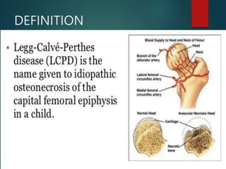
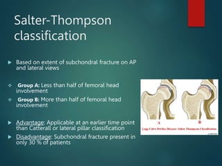
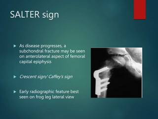
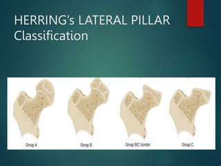
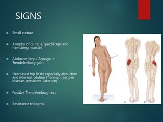
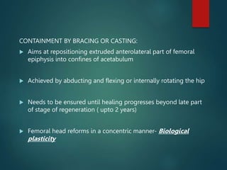
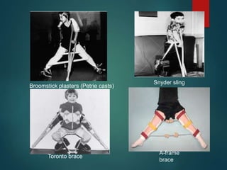
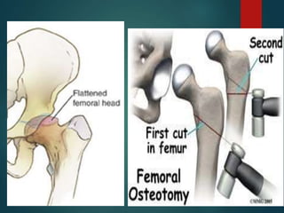
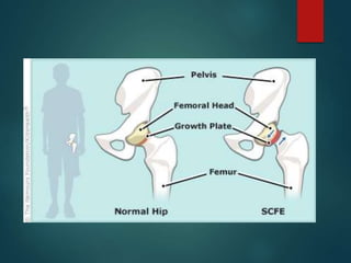
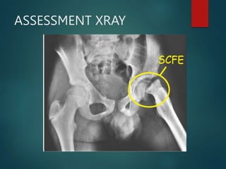
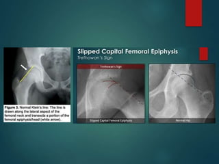
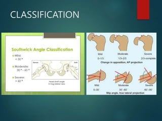
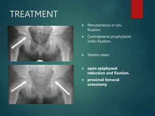
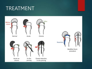
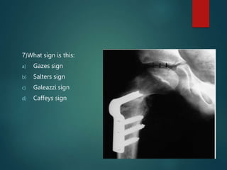
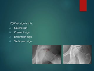
3. HISTORY
Arthur Legg described prominent
characteristics of the disorder: onset
between 5 and 8 years, history of
trauma, painless limp, minimal or no
spasm or shortening of affected limb.
Jacques Calve noted that affected
individuals had minimal atrophy of
the leg and no palpable hip swelling;
abnormal or delayed bone formation
4. Georg Perthes observed the disorder as “a
self limiting, non inflammatory condition,
affecting the capital femoral epiphysis with
stages of degeneration and regeneration,
leading to restoration of the bone nucleus”
Henning Waldenstrom reported radiographic
changes associated with the disorder in
1909; thought the disease was a form of
Tuberculosis.
5. Epidemiology
Incidence 1-4/10,000
Age 4 - 10years; average 7 yrs
As early as 2yrs as late as teens
Boys : girls 4:1
Bilateral 10-12%
No evidence of inheritance
Common in Caucasians; rare in black races
6.
7. ETIOLOGY
1. Vascular supply: Medial circumflex artery is missing or obliterated and obturator
artery or lateral epiphyseal artery also affected.
2. Increased intra-articular pressure
3. Increased intraosseous pressure: Impaired venous drainage in femoral head
4. Coagulation disorder: Absence of factor C or S, increase in serum levels of
lipoproteins, thrombogenic substance, Factor V Leiden mutation.
5. Growth hormones- Reduced levels of growth hormones, somatomedin A and C.
8. 6. Social conditions- Lower socioeconomic status, dietary and environmental
factors.
7. Trauma: Risk of vascular interruption increased due to narrow passage and
thick cartilage of femoral head penetrated by lateral epiphyseal arteries
8. Abnormal growth and development: Bone age is lower than chronological
age by 1-3 yrs (Radiological pause). Usually shorter than their peers.
9. Genetic factors
- Inheritance 2-20%; inconsistent pattern.
- Low birth weight, abnormal birth presentations.
- Ratio of affected 1st, 2nd, 3rd and 4th degree relatives to general
population of the same set – 35: 4: 4:1
9. Pathophysiology
Rapid growth occurs in relation to devlopment of blood supply
Interruption of blood supply results in necrosis, removal of necrotic
tissue, and its replacement with new bone.
Bone replacement may be so complete and perfect that completely
normal bone may result
The adequacy of bone replacement depends on
Age of the patient
Congruity of the involved joint
10. Sources of blood supply
Up to 4years
Metaphyseal vessels
Retinacular vessels
Ligamentum teres – scanty
4 to 7 years
Metaphyseal vessels ceases
Above 7years
Vessels in ligamentum teres have developed
11. Pathology
Goes through stages which may last 3 to 4 years
Stage1
Ischaemia and bone death, cartilage thickens
Stage 2
Revascularization and repair
Dead marrow replaced by granulation tissue
Bone revascularized and new bone laid down
Dead bone resorbed, replaced by fibrous tissue, fragmentation
Stage 3
Distortion and remodelling
Restoration of femoral archtecture or collapse
Femoral head displaces laterally in relation to acetabulum
16. Stage Ia Stage Ib Stage IIa Stage IIb
Stage IIIa Stage
IIIb
Stage
IV
17. Catterall classification
Group I: Only anterior portion of epiphysis
affected
Group II: More of anterior segment involved;
central sequestrum present
Group III: Most of the epiphysis sequestrated,
with unaffected portions located medial and
lateral to central segment
Group IV: Entire epiphysis sequestrated
BENIGN
PROGNOSIS
REQUIRE
TREATMENT
20. HEAD-AT-RISK FACTORS
Lateral subluxation of femoral head
Radiolucent V in the lateral aspect of
epiphysis (Gage sign)
Calcification lateral to epiphysis
Horizontal physeal line
Metaphyseal cyst
21. Salter-Thompson
classification
Based on extent of subchondral fracture on AP
and lateral views
Group A: Less than half of femoral head
involvement
Group B: More than half of femoral head
involvement
Advantage: Applicable at an earlier time point
than Catterall or lateral pillar classification
Disadvantage: Subchondral fracture present in
only 30 % of patients
22. SALTER sign
As disease progresses, a
subchondral fracture may be seen
on anterolateral aspect of femoral
capital epiphysis
Crescent sign/ Caffey’s sign
Early radiographic feature best
seen on frog leg lateral view
25. Bilateral involvement
More severe than unilateral
Boys and girls equally affected
Independent event
Bone age delayed in perthes disease
26. ClInical features
Incidence: higher latitude, western coastal region of South India
Onset: 18 months to skeletal maturity (Most prevalent: 4-8 years)
Male sex prevalence: Boys 4-5 times more susceptible
Involvement: Bilateral in 10-12 %
28. SIGNS
Small stature
Atrophy of gluteus, quadriceps and
hamstring muscles
Abductor limp ( Antalgic +
Trendelenburg gait)
Decreased hip ROM especially abduction
and internal rotation (Transient early in
disease, persistent later on)
Positive Trendelenburg test
Resistance to logroll
29. Investigations
Blood tests
haemogram, ESR, CRP
Imaging
Plain X-rays
Hip U/S
Bone scintigraphy
MRI
Dynamic arthrography
Assess spherity of femoral head
Hinge abduction
Bilateral perthes
Skeletal survey as part of work-up
30. MRI
Accurate for early diagnosis and for
visualising configuration of femoral head
and acetabulum
More reliable information about true
extent of femoral head necrosis than
radiography or scintigraphy
Gadolinium enhanced subtraction MRI as
effective as scintigraphy in delineating
epiphyseal necrosis early
De Sanctis classification:
Group A: < 50 % head necrosis
Group B: > 50 % head necrosis
B0- B3 based on degrees of lateral
extrusion and physeal disruption
31. TREATMENT OPTIONS
1. SYMPTOMATIC THERAPY
Bed rest
Non weight bearing on affected limb
Short term use of NSAIDs
Traction:
Simple longitudinal traction
Balanced suspension and traction
Russell traction
“Slings and springs”
32.
33. CONTAINMENT BY BRACING OR CASTING:
Aims at repositioning extruded anterolateral part of femoral
epiphysis into confines of acetabulum
Achieved by abducting and flexing or internally rotating the hip
Needs to be ensured until healing progresses beyond late part
of stage of regeneration ( upto 2 years)
Femoral head reforms in a concentric manner- Biological
plasticity
35. Treatment: Two main choices
Conservative
Pain control
Gentle exercises
Regular re-assessment
Avoid sport and strenous activities
Containment
Hold hips widely abducted in cast/brace >1yr
Operation
Varus osteotomy of femur
Innominate osteotomy of pelvis
Both
36.
37. Herring Guidelines to treatment
Children <6years
Symptomatic treatment
Children >6years; bone age more imp than chronological age
Bone age at or <6yrs
Lateral pillar A or B/ caterall I and II
Symptomatic treatment
Lateral pillar C/ Caterall III and IV
Bone over 6years
Herring A and B/Caterall I and II
Abduction brace or osteotomy
Herring C/Caterall III and IV
Outcome unaffected by treatment
Children 9yrs and older
Except in very mild cases, operative containment is the treatment of choice
38. Prognostic features
Age
<6yrs; good regardless of treatment
6-9years; not always satisfactory with containment
>10yrs; questionable benefit from containment, poor prognosis
Gender
Girls have worse prognosis
Classification grade
Herrings lateral pillar classification
Salter and thompson grade B worse prognosis
Caterral classification grade
Caterral “head-at-risk” signs
The five signs carry worse prognosis
Others
Body weight, decreased ROM
40. SCFE
Slipped capital femoral epiphysis (SCFE) is an condition of the proximal
femoral physis that leads to slippage of the metaphysis relative to the
epiphysis, and is most commonly seen in adolescent obese males
41. EPIDEMIOLOGY
Most common disorder affecting adolescent hips
Found in 10 per 100,000
More common in
Obese children, associated with puberty
Males (male to female ratio is 2:1.4)
Specific ethnicities - African Americans, Pacific islanders, Latinos
During period of rapid growth (10-16 years of age)
location
left hip is more common
bilateral in 17% to 50% (~25%)
42. RISK FACTORS
Obesity
Single greatest risk factor
Recent data shows trend towards younger age and increased frequency of
bilaterality at presentation, possibly related to increased rates of
childhood obesity
Acetabular retroversion and femoral retroversion
Increased mechanical shearing forces at the physis
History of previous radiation therapy to the femoral head region
43.
44. PATHOPHYSIOLOGY
Due mechanical forces acting on a susceptible physis
Pathoanatomy
Slippage occurs though the hypertrophic zone of the physis
45. PATHOPHYSIOLOGY
Perichondrial ring thins and weakens
Undulating mammillary processes in physis unlocks, further
destabilizing the physis
Physis is still vertical in this age group (160° at birth to 125° at skeletal
maturity), resulting in increased shearing forces
Epiphyseal tubercle can provide a rotational pivot point
Anatomic structure in the posterior superior epiphysis that shrinks with
skeletal maturity
Cartilage in the hypertrophic zone acts as a weak spot
46. PATHOPHYSIOLOGY
Angulation
Metaphysis translates anterior
and externally rotates
Epiphysis remains in the
acetabulum, lies posterior to the
translated metaphysis
Similar to Salter-Harris type
I fracture
48. CLINICAL FEATURES
Abnormal gait / limp
Antalgic, waddling, externally rotated
gait or Trendelenburg gait
Decreased hip motion
Obligatory external rotation during
passive flexion of hip (Drehmann sign)
Due to a combination of synovitis and
impingement of the displaced anterior-
lateral femoral metaphysis on the
acetabular rim
49. CLINICAL FEATURES
Loss of hip internal rotation, abduction, and flexion
Abnormal leg alignment
Externally rotated foot progression angle
Weakness
Thigh atrophy
60. 6)Perthes disease is a self limiting disease, it ,ay last for:
a) 2-3 months
b) 1-2y
c) 3-4y
d) 7-8y
61. 7)What sign is this:
a) Gazes sign
b) Salters sign
c) Galeazzi sign
d) Caffeys sign
62. 8)Which of the following is false with respect to SCFE:
a) Common in males
b) Seen in obese children
c) Not associated with puberty
d) Incidence : Left > bilateral > right
63. 9)In SCFE, slippage occurs through which zone of physis:
a) Proliferative
b) Reserve
c) Hypertrophic
d) Spongiosa
64. 10)What sign is this:
a) Salters sign
b) Crescent sign
c) Drehmann sign
d) Trethowan sign