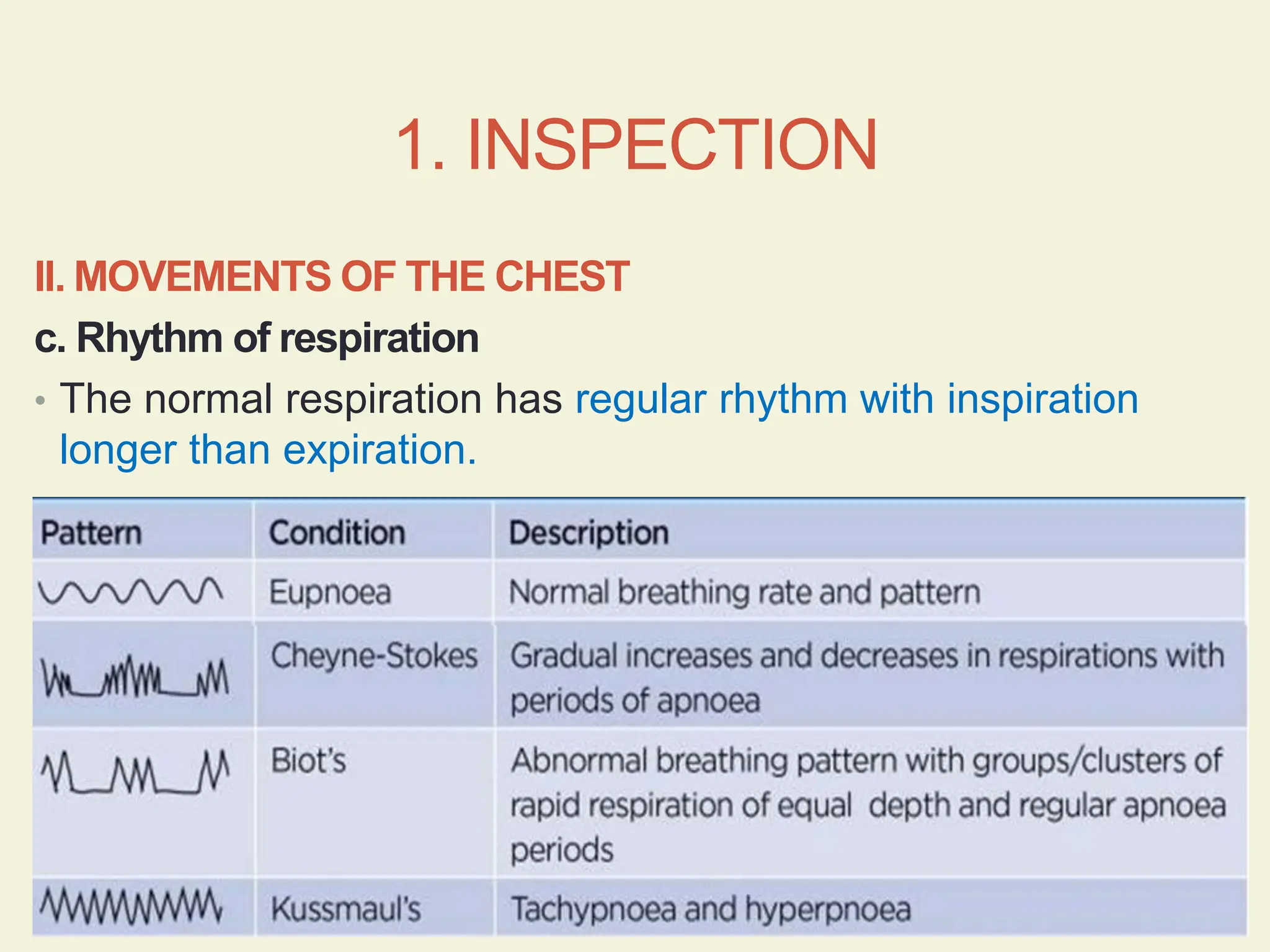
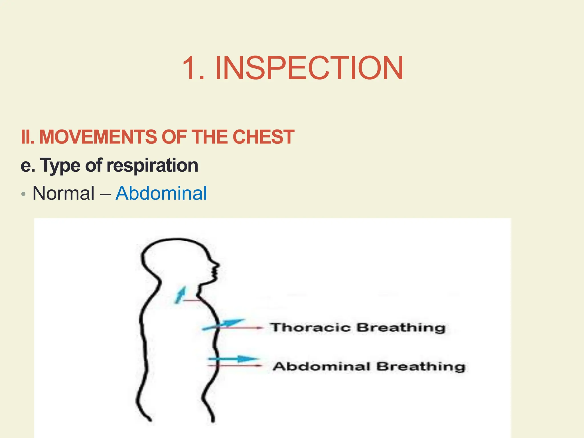
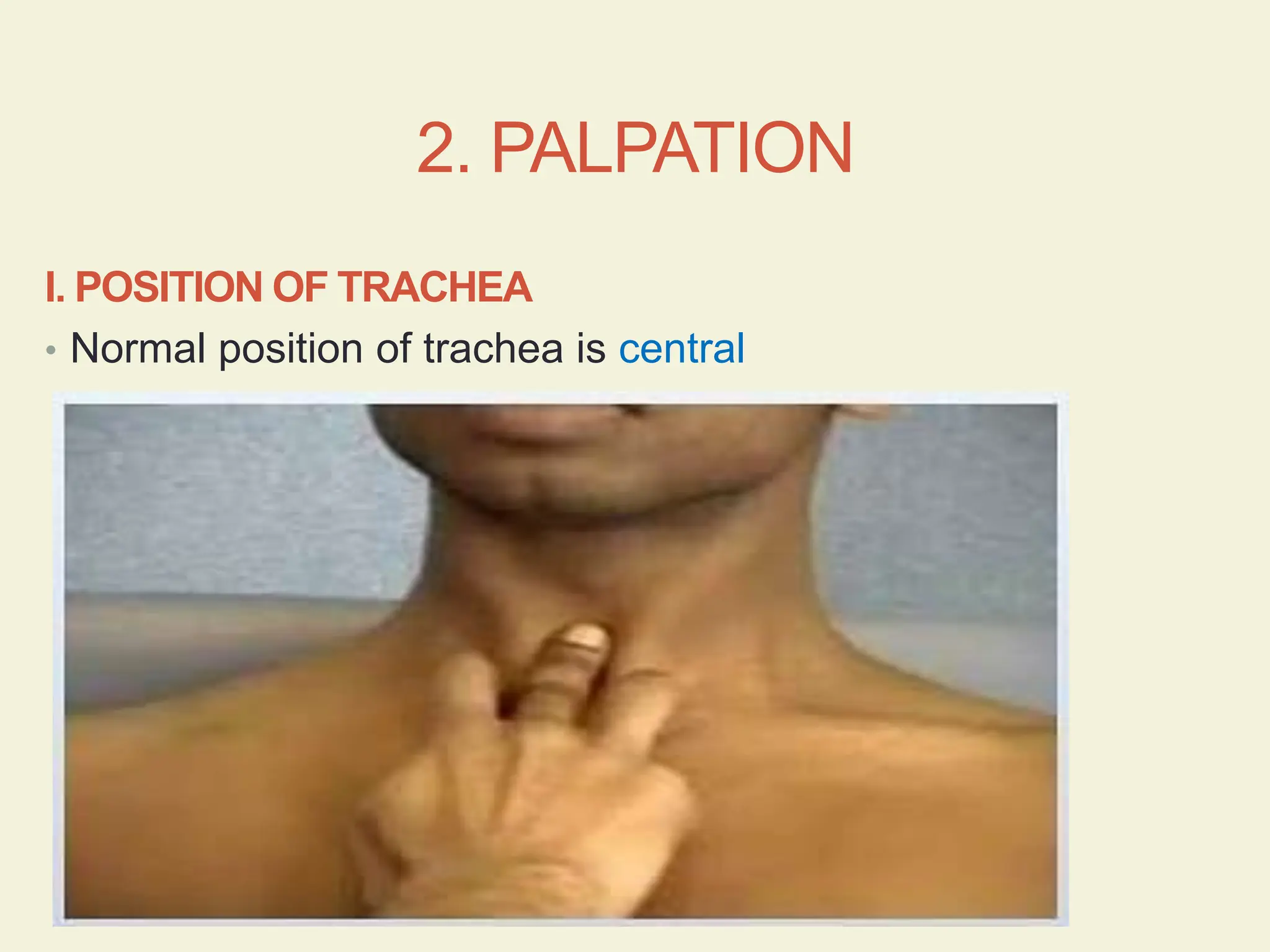
RESPIRATORY SYSTEM
EXAMINATION
DR AXELLE SAVERETTIAR
Position of subject
The respiratory system examination is ideally done with the
subject comfortably resting on the bed, sitting at an angle
of 45˚ and supported by pillows.
RESPIRATORY SYSTEM
EXAMINATION
1.Inspection 2. Palpation 3. Percussion 4. Auscultation
1.INSPECTION I. SHAPE OF THE CHEST
II. MOVEMENTS OF THE CHEST
a. Rate of respiration
b. Depth of respiration
c. Rhythm of respiration
d. Expansion of chest
e. Type of respiration
1. INSPECTION
I. SHAPE OF THE CHEST
Bilaterally symmetrical
and elliptical in cross
section.
Anteroposterior diameter
is less than the transverse
diameter in a ratio of 5:7.
Abnormal forms of chest
A. PIGEON CHEST
Chest is elliptical with
prominent sternum.
Seen in rickets.
Abnormal forms of chest
B. SCOLIOSIS
It is the lateral bending
of the vertebral column.
Prominence of front of
the chest on one side
with flattening on the
other side.
Abnormal forms of chest
C. KYPHOSIS
It is the forward bending
of the vertebral column
Shortening of the chest
with prominent sternum.
Abnormal forms of chest
D. BARREL SHAPE CHEST
AP diameter of chest
increases.
It is seen in:
Emphysema
Bronchial asthma
Fibrosis
Collapse of lungs
1. INSPECTION
II. MOVEMENTS OF THE CHEST
a. Rate of respiration
• Normal rate is 12 – 16 breaths per minute
Increased
respiratory rate
(tachypnea)
• Exercise
• Fever
• Anemia
Decreased
respiratory rate
(bradypnea)
• Brain damage
II. MOVEMENTS OF THE CHEST
1. INSPECTION
1. INSPECTION
II. MOVEMENTS OF THE
CHEST
b. Depth of respiration
• Deep breathing – brain
damage
• Shallow breathing –
bronchial asthma
1. INSPECTION
II. MOVEMENTS OF THE CHEST
c. Rhythm of respiration
• The normal respiration has regular rhythm with inspiration
longer than expiration.
1. INSPECTION
II. MOVEMENTS OF THE CHEST
d. Expansion of chest
• Normally both sides of the chest wall move symmetrically
(equal on both sides)
Diminished or asymmetrical movements:
• Consolidation
• Collapse
• Fibrosis
• Pleural effusion
• Hydro pneumothorax
1. INSPECTION
II. MOVEMENTS OF THE CHEST
e. Type of respiration
• Normal – Abdominal
2. PALPATION I. POSTION OF TRACHEA
II. POSITION OF APEX BEAT
III. EXPANSION OF CHEST
IV. VOCAL FREMITUS
2. PALPATION
I. POSITION OF TRACHEA
• Normal position of trachea is central
2. PALPATION
Trachea pulled towards the
affected side
• Collapse
• Fibrosis of lung
Trachea pushed away from
affected side
• Pleural effusion
• Pneumothorax
2. PALPATION
II. POSITION OFAPEX BEAT
Normal position - 9 cm from midline in the left 5th intercostal
space.
2. PALPATION
• II. POSITION OFAPEX BEAT
• Cardiac impulse refers to movements occurring due to the
impact of the heart against the chest wall during the
systole.
• The apex beat is the lowest and outermost point of
definite cardiac impulse, where it is seen / felt most
forcibly (point of maximum impulse).
2. PALPATION
Absent apex
beat
• Obesity
• If lying behind a
rib
• Dextrocardia
Prominent apex
beat
• Anxiety
• After exercise
Displaced apex
beat
• Enlargement of
left ventricle
• Pleural or
pulmonary
diseases
• Deformities of
the chest wall
or the thoracic
vertebrae –
scoliosis
2. PALPATION
III. EXPANSION OF THE CHEST
Two methods:
1. Using a measuring tape
• Normal chest expansion is 5 – 8 cm.
2. Grasping the chest
2. PALPATION
III. EXPANSION OF THE CHEST
Using a measuring tape
• Chest expansion can be measured using a measuring tape around the
chest at the level of the nipples.
Grasping the chest
• Place finger tips of both hands on either side of the lower rib cage.
• Tips of the thumbs meet in the midline in the front but not touch the
chest.
• Ask subject to take deep breath so that the distance between the
thumbs will increase and it will indicate degree of expansion.
• If one thumb remains closer to midline this means that there is
decreased expansion of that side.
EXAMINATION OF CHEST
RIGHT LEFT
ANTERIOR Supra - clavicular Supra - clavicular
Supra - mammary Supra - mammary
Mammary Mammary
Infra - mammary Infra - mammary
Axillary
Infra - axillary
POSTERIOR Supra – scapular Supra – scapular
Inter – scapular
Infra – scapular Infra – scapular
EXAMINATION OF CHEST
2. PALPATION
IV. VOCAL FREMITUS
• Vocal: pertaining to voice
• Fremitus: vibratory sensation perceived by palpation
• Sound vibrations from the larynx pass down the bronchi
and cause the lungs and the chest wall to vibrate.
2. PALPATION
IV. VOCAL FREMITUS
• The vibration may be detected by palpation by ulnar
border of hand placed flat on the chest.
• The subject is then asked to repeat 1 1 1 or 9 9 9 or Ram
Ram Ram
• The vibrations are perceived by the examining hand.
• Compare right and left side
2. PALPATION
IV. VOCAL FREMITUS
2. PALPATION
Consolidation Bronchial
obstruction
Pleural effusion
Pneumothorax
Fibrosis
Collapse
IV. VOCAL FREMITUS
3. PERCUSSION
• Normal percussion note of the lungs is resonant
3. PERCUSSION
Lower limit of left lung:
Note changes from
resonant to tympanic
due to presence of
stomach
Lower limit of right lung:
• 6th rib – Mammary line
• 8th rib – Mid axillary
line
• 10th rib – Scapular line
3. PERCUSSION
Pneumothorax Thickening of pleura
Pleural effusion
4. AUSCULTATION I. BREATH SOUNDS
a. Vesicular
b. Bronchial
II. VOCAL RESONANCE
III.ADDED SOUNDS
a. Pleural rub
b. Rhonchi or wheeze
c. Crepitations or crackles
d. Stridor
4. AUSCULTATION
I. BREATH SOUNDS
Two main types of breath sounds can normally be
heard over the chest:
• Vesicular
• Bronchial
4. AUSCULTATION
I. BREATH SOUNDS
4. AUSCULTATION
I. BREATH SOUNDS
a. Vesicular breath sound
• It is characterized by active inspiration due to passage of
air into bronchi and alveoli followed without a pause by
passive expiration due to elastic recoil of the alveoli which
occurs maximally in the early phases giving an apparent
impression of short expiration.
4. AUSCULTATION
I. BREATH SOUNDS
4. AUSCULTATION
I. BREATH SOUNDS
b. Bronchial breath sound
• It is characterized by active inspiration due to passage of
air into the bronchi. The alveolar phase is absent
(because of consolidation in alveoli) and hence expiration
is also active occupying the same duration time as
inspiration.
VESICULAR BREATH
SOUNDS
BRONCHIAL BREATH
SOUNDS
Origin In larger airways In larger airways
When the lung between the
airways and chest wall is airless
as in consolidation, fibrosis and
collapse of lungs
Character Low pitch rustling sound High frequency and harsh
Inspiration
and
Expiration
 No gap between end of
inspiration and beginning of
expiration
 Inspiration is twice as long
as expiration
 Gap between end of
inspiration and beginning of
expiration
 Inspiration and expiration are
of same duration
Sites Heard all over healthy lungs
(chest)
Heard over trachea
Heard in patients with
consolidation, fibrosis and
collapse of lungs
4. AUSCULTATION
II. VOCAL RESONANCE
• It is the sound of the voice heard on auscultation of the
chest.
• When subject repeats 1 1 1 or 9 9 9, the ear perceives
not only the distinct syllables but a resonant sound.
4. AUSCULTATION
Bronchophony -
consolidation
Egophony - above the
level of pleural effusion
Whispering pectoriloquy
- cavity of lungs and
consolidation
Pleural effusion
Pneumothorax
4. AUSCULTATION
III.ADDED SOUNDS
a. Pleural rub
• It occurs due to inflammation of pleura and it gives rise to
a characteristic friction rub.
b. Rhonchi or wheeze
• They are continuous musical sounds associated with
airway narrowing and particularly heard during expiration
• Seen in bronchitis and bronchial asthma
4. AUSCULTATION
III.ADDED SOUNDS
c. Crepitations or crackles
• They are crackling sounds produced by sudden change in
pressure related to sudden opening of previously closed
airways.
• It is heard both during expiration and inspiration.
• Seen in pneumonia.
d. Stridor
• It is a loud inspiratory sound heard over the airways due
to obstruction to the respiratory tract mainly the larynx and
trachea.
498485741-Respiratory-System-Examination.pptx

498485741-Respiratory-System-Examination.pptx

  • 1.
  • 2.
    Position of subject Therespiratory system examination is ideally done with the subject comfortably resting on the bed, sitting at an angle of 45˚ and supported by pillows.
  • 3.
    RESPIRATORY SYSTEM EXAMINATION 1.Inspection 2.Palpation 3. Percussion 4. Auscultation
  • 4.
    1.INSPECTION I. SHAPEOF THE CHEST II. MOVEMENTS OF THE CHEST a. Rate of respiration b. Depth of respiration c. Rhythm of respiration d. Expansion of chest e. Type of respiration
  • 5.
    1. INSPECTION I. SHAPEOF THE CHEST Bilaterally symmetrical and elliptical in cross section. Anteroposterior diameter is less than the transverse diameter in a ratio of 5:7.
  • 6.
    Abnormal forms ofchest A. PIGEON CHEST Chest is elliptical with prominent sternum. Seen in rickets.
  • 8.
    Abnormal forms ofchest B. SCOLIOSIS It is the lateral bending of the vertebral column. Prominence of front of the chest on one side with flattening on the other side.
  • 9.
    Abnormal forms ofchest C. KYPHOSIS It is the forward bending of the vertebral column Shortening of the chest with prominent sternum.
  • 10.
    Abnormal forms ofchest D. BARREL SHAPE CHEST AP diameter of chest increases. It is seen in: Emphysema Bronchial asthma Fibrosis Collapse of lungs
  • 11.
    1. INSPECTION II. MOVEMENTSOF THE CHEST a. Rate of respiration • Normal rate is 12 – 16 breaths per minute
  • 12.
    Increased respiratory rate (tachypnea) • Exercise •Fever • Anemia Decreased respiratory rate (bradypnea) • Brain damage II. MOVEMENTS OF THE CHEST 1. INSPECTION
  • 13.
    1. INSPECTION II. MOVEMENTSOF THE CHEST b. Depth of respiration • Deep breathing – brain damage • Shallow breathing – bronchial asthma
  • 14.
    1. INSPECTION II. MOVEMENTSOF THE CHEST c. Rhythm of respiration • The normal respiration has regular rhythm with inspiration longer than expiration.
  • 15.
    1. INSPECTION II. MOVEMENTSOF THE CHEST d. Expansion of chest • Normally both sides of the chest wall move symmetrically (equal on both sides) Diminished or asymmetrical movements: • Consolidation • Collapse • Fibrosis • Pleural effusion • Hydro pneumothorax
  • 16.
    1. INSPECTION II. MOVEMENTSOF THE CHEST e. Type of respiration • Normal – Abdominal
  • 17.
    2. PALPATION I.POSTION OF TRACHEA II. POSITION OF APEX BEAT III. EXPANSION OF CHEST IV. VOCAL FREMITUS
  • 18.
    2. PALPATION I. POSITIONOF TRACHEA • Normal position of trachea is central
  • 19.
    2. PALPATION Trachea pulledtowards the affected side • Collapse • Fibrosis of lung Trachea pushed away from affected side • Pleural effusion • Pneumothorax
  • 20.
    2. PALPATION II. POSITIONOFAPEX BEAT Normal position - 9 cm from midline in the left 5th intercostal space.
  • 21.
    2. PALPATION • II.POSITION OFAPEX BEAT • Cardiac impulse refers to movements occurring due to the impact of the heart against the chest wall during the systole. • The apex beat is the lowest and outermost point of definite cardiac impulse, where it is seen / felt most forcibly (point of maximum impulse).
  • 22.
    2. PALPATION Absent apex beat •Obesity • If lying behind a rib • Dextrocardia Prominent apex beat • Anxiety • After exercise Displaced apex beat • Enlargement of left ventricle • Pleural or pulmonary diseases • Deformities of the chest wall or the thoracic vertebrae – scoliosis
  • 24.
    2. PALPATION III. EXPANSIONOF THE CHEST Two methods: 1. Using a measuring tape • Normal chest expansion is 5 – 8 cm. 2. Grasping the chest
  • 25.
    2. PALPATION III. EXPANSIONOF THE CHEST Using a measuring tape • Chest expansion can be measured using a measuring tape around the chest at the level of the nipples. Grasping the chest • Place finger tips of both hands on either side of the lower rib cage. • Tips of the thumbs meet in the midline in the front but not touch the chest. • Ask subject to take deep breath so that the distance between the thumbs will increase and it will indicate degree of expansion. • If one thumb remains closer to midline this means that there is decreased expansion of that side.
  • 26.
    EXAMINATION OF CHEST RIGHTLEFT ANTERIOR Supra - clavicular Supra - clavicular Supra - mammary Supra - mammary Mammary Mammary Infra - mammary Infra - mammary Axillary Infra - axillary POSTERIOR Supra – scapular Supra – scapular Inter – scapular Infra – scapular Infra – scapular
  • 27.
  • 28.
    2. PALPATION IV. VOCALFREMITUS • Vocal: pertaining to voice • Fremitus: vibratory sensation perceived by palpation • Sound vibrations from the larynx pass down the bronchi and cause the lungs and the chest wall to vibrate.
  • 29.
    2. PALPATION IV. VOCALFREMITUS • The vibration may be detected by palpation by ulnar border of hand placed flat on the chest. • The subject is then asked to repeat 1 1 1 or 9 9 9 or Ram Ram Ram • The vibrations are perceived by the examining hand. • Compare right and left side
  • 30.
  • 31.
    2. PALPATION Consolidation Bronchial obstruction Pleuraleffusion Pneumothorax Fibrosis Collapse IV. VOCAL FREMITUS
  • 32.
    3. PERCUSSION • Normalpercussion note of the lungs is resonant
  • 33.
    3. PERCUSSION Lower limitof left lung: Note changes from resonant to tympanic due to presence of stomach Lower limit of right lung: • 6th rib – Mammary line • 8th rib – Mid axillary line • 10th rib – Scapular line
  • 34.
    3. PERCUSSION Pneumothorax Thickeningof pleura Pleural effusion
  • 35.
    4. AUSCULTATION I.BREATH SOUNDS a. Vesicular b. Bronchial II. VOCAL RESONANCE III.ADDED SOUNDS a. Pleural rub b. Rhonchi or wheeze c. Crepitations or crackles d. Stridor
  • 36.
    4. AUSCULTATION I. BREATHSOUNDS Two main types of breath sounds can normally be heard over the chest: • Vesicular • Bronchial
  • 37.
  • 38.
    4. AUSCULTATION I. BREATHSOUNDS a. Vesicular breath sound • It is characterized by active inspiration due to passage of air into bronchi and alveoli followed without a pause by passive expiration due to elastic recoil of the alveoli which occurs maximally in the early phases giving an apparent impression of short expiration.
  • 39.
  • 40.
    4. AUSCULTATION I. BREATHSOUNDS b. Bronchial breath sound • It is characterized by active inspiration due to passage of air into the bronchi. The alveolar phase is absent (because of consolidation in alveoli) and hence expiration is also active occupying the same duration time as inspiration.
  • 41.
    VESICULAR BREATH SOUNDS BRONCHIAL BREATH SOUNDS OriginIn larger airways In larger airways When the lung between the airways and chest wall is airless as in consolidation, fibrosis and collapse of lungs Character Low pitch rustling sound High frequency and harsh Inspiration and Expiration  No gap between end of inspiration and beginning of expiration  Inspiration is twice as long as expiration  Gap between end of inspiration and beginning of expiration  Inspiration and expiration are of same duration Sites Heard all over healthy lungs (chest) Heard over trachea Heard in patients with consolidation, fibrosis and collapse of lungs
  • 42.
    4. AUSCULTATION II. VOCALRESONANCE • It is the sound of the voice heard on auscultation of the chest. • When subject repeats 1 1 1 or 9 9 9, the ear perceives not only the distinct syllables but a resonant sound.
  • 43.
    4. AUSCULTATION Bronchophony - consolidation Egophony- above the level of pleural effusion Whispering pectoriloquy - cavity of lungs and consolidation Pleural effusion Pneumothorax
  • 44.
    4. AUSCULTATION III.ADDED SOUNDS a.Pleural rub • It occurs due to inflammation of pleura and it gives rise to a characteristic friction rub. b. Rhonchi or wheeze • They are continuous musical sounds associated with airway narrowing and particularly heard during expiration • Seen in bronchitis and bronchial asthma
  • 45.
    4. AUSCULTATION III.ADDED SOUNDS c.Crepitations or crackles • They are crackling sounds produced by sudden change in pressure related to sudden opening of previously closed airways. • It is heard both during expiration and inspiration. • Seen in pneumonia. d. Stridor • It is a loud inspiratory sound heard over the airways due to obstruction to the respiratory tract mainly the larynx and trachea.