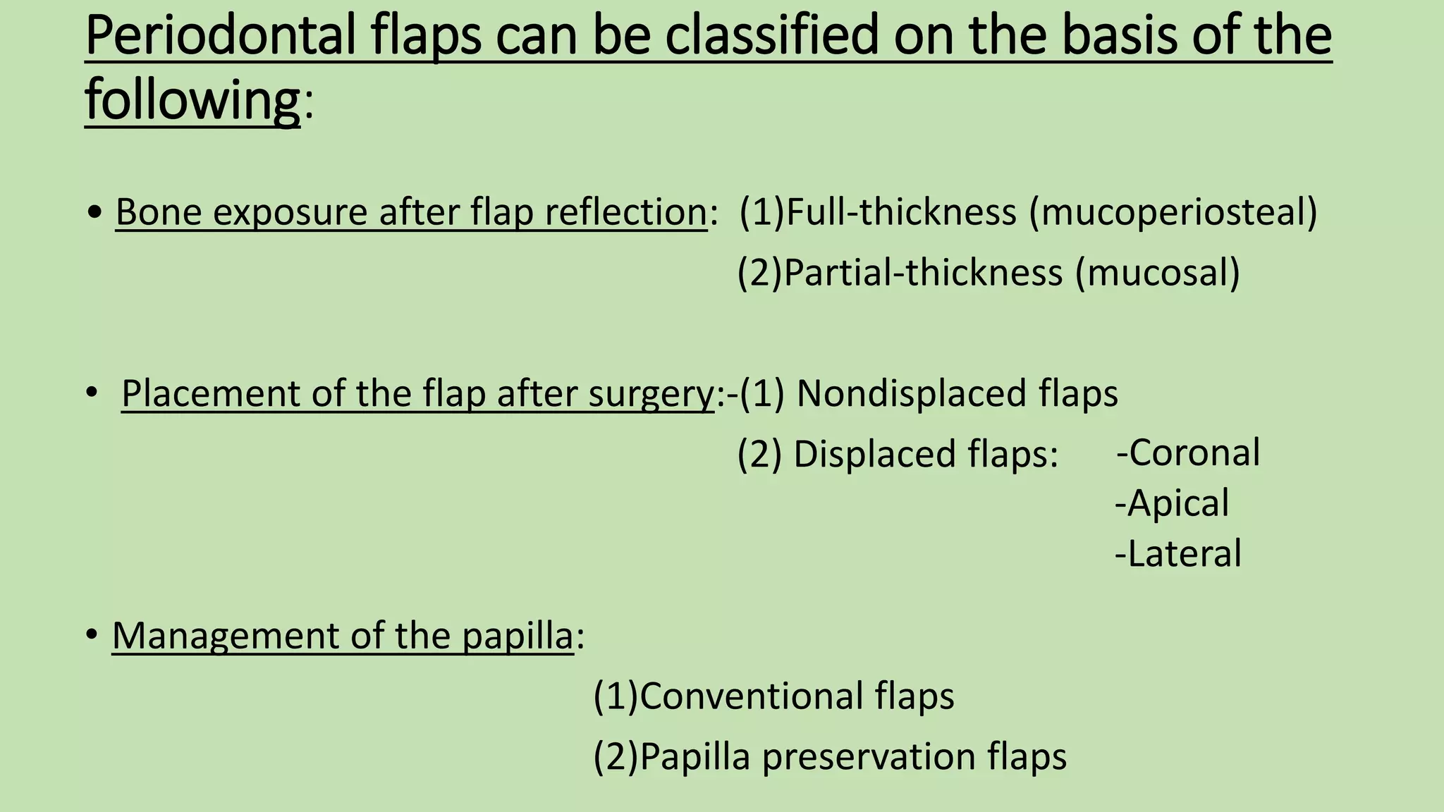
The Flap Technique for Pocket
Therapy
PRESENTED BY: Dr. MANOJ M. PARADHI
1st YEAR PG
Dept. Of Periodontics And Oral Implantology
Introduction
• Periodontal flap is defined as ‘the section of gingiva and/or
mucosa surgically elevated from the underlying tissues to
provide visibility and accessibility to the bone and root
surfaces’.
• The flap also allows gingiva to be displaced to a different
location in patients with mucogingival involvement.
OBJECTIVES
1. Increase accessibility to root deposits for scaling and root
planning
2. Eliminate or reduce pocket depth via resection of the pocket wall
3. Gain access for osseous resective surgery.
Periodontal flaps can be classified on the basis of the
following:
• Bone exposure after flap reflection: (1)Full-thickness (mucoperiosteal)
(2)Partial-thickness (mucosal)
• Placement of the flap after surgery:-(1) Nondisplaced flaps
(2) Displaced flaps:
• Management of the papilla:
(1)Conventional flaps
(2)Papilla preservation flaps
-Coronal
-Apical
-Lateral
Technique for Access and Pocket Depth
Reduction or Elimination:
(1)Modified Widman flap
(2)Undisplaced flap
(3)Apically displaced flap
Incisions
• All three flap techniques
use of the basic incisions:
1. Internal bevel incision
/Reverse bevel incision
2. Crevicular incision
3. Interdental incision
Internal bevel incisions for the different types of flaps
MODIFIED WIDMAN FLAP
• Morris (1965) called it the “unrepositioned mucoperiosteal flap.”
Ramfjord and Nissle (1974) called “modified Widman flap”.
• It is used whenever reattachment with minimal gingival recession is
desired.
o INDICATIONS :
• Moderately deep pockets
• Moderate furcation involvement
• Patient with a high caries rate and root sensitivity problem
• Step 1: The initial incision is an internal bevel incision to the alveolar crest
starting 0.5 mm to 1 mm away from the gingival margin .
• Step 2: The gingiva is reflected with a periosteal elevator.
• Step 3: A crevicular incision is made from the bottom of the pocket to the
bone in such a way that it circumscribes the triangular wedge of tissue
that contains the pocket lining.
Steps outline
• Step 4: Third incision is made in the interdental spaces coronal to the bone
with a curette or an interproximal knife, and the gingival collar is removed.
• Step 5: Tissue tags and granulation tissue are removed with a curette.
• Step 6: Adapt the facial and lingual interproximal tissue adjacent to each
other in such a way that no interproximal bone remains exposed at the time
of suturing.
• Step 7: Continuous, independent sling sutures are placed in both the facial
and palatal areas and covered with a periodontal surgical pack.
Advantages of Modified Widman Flap:
•Obtaining close adaptation of soft tissues to root surfaces.
• Less exposure of root surfaces therefore it offers esthetic
advantage in the anterior segments ( Ramfjord and
Nissle,1974).
• Complete removal of pocket epithelium.
• Esthetically superior to gingivectomy.
UNDISPLACED FLAP
• Pocket elimination procedure using internal bevel incision.
Also called as INTERNAL BEVEL GINGIVECTOMY.
• Pocket wall is eliminated with first incision.
• Elimination of ‘dead space’ as the flap margin is place over
bonecrest postoperatively
• However, sufficient attached gingiva is a pre-requisite
• Usually used for pocket elimination of palatal pockets
Steps outline
• Step 1: The pockets are measured with the periodontal probe, and a
bleeding point is produced on the outer surface of the gingiva to
mark the pocket bottom.
• Step 2: The initial or internal bevel incision is made after scalloping
the bleeding marks on the gingiva.
• Step 3: The second or crevicular incision is made from the bottom of
the pocket to the bone to detach the connective tissue from the
bone.
• Step 4: The flap is reflected with a periosteal elevator from the
internal bevel incision
• Step 5: The third or interdental incision is made with an
interdental knife to separate the connective tissue from the
bone.
• Step 6: The triangular wedge of tissue created by the three
incisions is removed with a curette.
• Step 7: The area is debrided to remove all tissue tags and
granulation tissue with the use of sharp curettes.
• Step 8: A continuous sling suture is used to secure the facial
and lingual or palatal flaps. The area is covered with a
periodontal pack.
APICALLY DISPLACED FLAP
• This flap technique used for:
- Pocket eradication
- Widening the zone of attached gingiva
- Crown lengthening procedures
• Apically displaced flap can be:
- Full-thickness (mucoperiosteal) flap
- Split-thickness (mucosal) flap
OBJECTIVES
• Apical displacement of entire mucogingival unit to
eliminate the pockets while retaining the attached
gingiva.
• Surgical access for osseous surgery, treatment of
infrabony pockets and root planing.
Steps outline
• Step 1: An internal bevel incision is made . To preserve as much of the
keratinized and attached gingiva as possible, it should be no more
than about 1 mm from the crest of the gingiva and directed to the
crest of the bone.
• Step 2: Crevicular incisions are made, and this is followed by the
initial elevation of the flap. Interdental incisions are then performed,
and the wedge of tissue that contains the pocket wall is removed.
• Step 3: Vertical incisions are made extending beyond the
mucogingival junction.
• Step 4: After the removal of all granulation tissue, scaling and root
planing, and osseous surgery if needed, the flap is displaced apically.
• Step 5:
Full-thickness flap was created, a sling suture around the tooth prevents
the flap from sliding to a position more apical than what is desired, and the
periodontal dressing can avoid its movement in a coronal direction.
Partial-thickness flap is sutured to the periosteum with the use of a direct
loop suture or a combination of loop and anchor suture. A dry foil is placed
over the flap before it is covered with the dressing to prevent the
introduction of pack under the flap
Advantages
• Reduction of probing depth,
• Preserving or increasing the presurgical zone of gingiva,
• Facilitation of healing, accessibility to bone, roots, furcations,
subgingival caries, and other anatomical aberrations,
• Controlling the tissue placement.
Disadvantages
• Sacrifice of crestal alveolar process and supporting bone
• Extensive exposure of root surfaces.
CONCLUSION
• Periodontal flap procedures for pocket therapy include flaps that
are created only for access to root surfaces and bone margins, flaps
for the precise processes of osseous surgery, and flaps for
periodontal regeneration.
• All of these approaches have specific flap designs and step-by-step
elements, and all of them include calculus removal and root
planing as part of the essential treatment protocol.

The flap technique for pocket therapy

  • 1.
    The Flap Techniquefor Pocket Therapy PRESENTED BY: Dr. MANOJ M. PARADHI 1st YEAR PG Dept. Of Periodontics And Oral Implantology
  • 2.
    Introduction • Periodontal flapis defined as ‘the section of gingiva and/or mucosa surgically elevated from the underlying tissues to provide visibility and accessibility to the bone and root surfaces’. • The flap also allows gingiva to be displaced to a different location in patients with mucogingival involvement.
  • 3.
    OBJECTIVES 1. Increase accessibilityto root deposits for scaling and root planning 2. Eliminate or reduce pocket depth via resection of the pocket wall 3. Gain access for osseous resective surgery.
  • 4.
    Periodontal flaps canbe classified on the basis of the following: • Bone exposure after flap reflection: (1)Full-thickness (mucoperiosteal) (2)Partial-thickness (mucosal) • Placement of the flap after surgery:-(1) Nondisplaced flaps (2) Displaced flaps: • Management of the papilla: (1)Conventional flaps (2)Papilla preservation flaps -Coronal -Apical -Lateral
  • 5.
    Technique for Accessand Pocket Depth Reduction or Elimination: (1)Modified Widman flap (2)Undisplaced flap (3)Apically displaced flap
  • 6.
    Incisions • All threeflap techniques use of the basic incisions: 1. Internal bevel incision /Reverse bevel incision 2. Crevicular incision 3. Interdental incision
  • 7.
    Internal bevel incisionsfor the different types of flaps
  • 8.
    MODIFIED WIDMAN FLAP •Morris (1965) called it the “unrepositioned mucoperiosteal flap.” Ramfjord and Nissle (1974) called “modified Widman flap”. • It is used whenever reattachment with minimal gingival recession is desired. o INDICATIONS : • Moderately deep pockets • Moderate furcation involvement • Patient with a high caries rate and root sensitivity problem
  • 9.
    • Step 1:The initial incision is an internal bevel incision to the alveolar crest starting 0.5 mm to 1 mm away from the gingival margin . • Step 2: The gingiva is reflected with a periosteal elevator. • Step 3: A crevicular incision is made from the bottom of the pocket to the bone in such a way that it circumscribes the triangular wedge of tissue that contains the pocket lining. Steps outline
  • 10.
    • Step 4:Third incision is made in the interdental spaces coronal to the bone with a curette or an interproximal knife, and the gingival collar is removed. • Step 5: Tissue tags and granulation tissue are removed with a curette. • Step 6: Adapt the facial and lingual interproximal tissue adjacent to each other in such a way that no interproximal bone remains exposed at the time of suturing. • Step 7: Continuous, independent sling sutures are placed in both the facial and palatal areas and covered with a periodontal surgical pack.
  • 15.
    Advantages of ModifiedWidman Flap: •Obtaining close adaptation of soft tissues to root surfaces. • Less exposure of root surfaces therefore it offers esthetic advantage in the anterior segments ( Ramfjord and Nissle,1974). • Complete removal of pocket epithelium. • Esthetically superior to gingivectomy.
  • 16.
    UNDISPLACED FLAP • Pocketelimination procedure using internal bevel incision. Also called as INTERNAL BEVEL GINGIVECTOMY. • Pocket wall is eliminated with first incision. • Elimination of ‘dead space’ as the flap margin is place over bonecrest postoperatively • However, sufficient attached gingiva is a pre-requisite • Usually used for pocket elimination of palatal pockets
  • 17.
    Steps outline • Step1: The pockets are measured with the periodontal probe, and a bleeding point is produced on the outer surface of the gingiva to mark the pocket bottom. • Step 2: The initial or internal bevel incision is made after scalloping the bleeding marks on the gingiva. • Step 3: The second or crevicular incision is made from the bottom of the pocket to the bone to detach the connective tissue from the bone.
  • 18.
    • Step 4:The flap is reflected with a periosteal elevator from the internal bevel incision • Step 5: The third or interdental incision is made with an interdental knife to separate the connective tissue from the bone. • Step 6: The triangular wedge of tissue created by the three incisions is removed with a curette. • Step 7: The area is debrided to remove all tissue tags and granulation tissue with the use of sharp curettes. • Step 8: A continuous sling suture is used to secure the facial and lingual or palatal flaps. The area is covered with a periodontal pack.
  • 22.
    APICALLY DISPLACED FLAP •This flap technique used for: - Pocket eradication - Widening the zone of attached gingiva - Crown lengthening procedures • Apically displaced flap can be: - Full-thickness (mucoperiosteal) flap - Split-thickness (mucosal) flap
  • 23.
    OBJECTIVES • Apical displacementof entire mucogingival unit to eliminate the pockets while retaining the attached gingiva. • Surgical access for osseous surgery, treatment of infrabony pockets and root planing.
  • 24.
    Steps outline • Step1: An internal bevel incision is made . To preserve as much of the keratinized and attached gingiva as possible, it should be no more than about 1 mm from the crest of the gingiva and directed to the crest of the bone. • Step 2: Crevicular incisions are made, and this is followed by the initial elevation of the flap. Interdental incisions are then performed, and the wedge of tissue that contains the pocket wall is removed. • Step 3: Vertical incisions are made extending beyond the mucogingival junction.
  • 25.
    • Step 4:After the removal of all granulation tissue, scaling and root planing, and osseous surgery if needed, the flap is displaced apically. • Step 5: Full-thickness flap was created, a sling suture around the tooth prevents the flap from sliding to a position more apical than what is desired, and the periodontal dressing can avoid its movement in a coronal direction. Partial-thickness flap is sutured to the periosteum with the use of a direct loop suture or a combination of loop and anchor suture. A dry foil is placed over the flap before it is covered with the dressing to prevent the introduction of pack under the flap
  • 30.
    Advantages • Reduction ofprobing depth, • Preserving or increasing the presurgical zone of gingiva, • Facilitation of healing, accessibility to bone, roots, furcations, subgingival caries, and other anatomical aberrations, • Controlling the tissue placement. Disadvantages • Sacrifice of crestal alveolar process and supporting bone • Extensive exposure of root surfaces.
  • 31.
    CONCLUSION • Periodontal flapprocedures for pocket therapy include flaps that are created only for access to root surfaces and bone margins, flaps for the precise processes of osseous surgery, and flaps for periodontal regeneration. • All of these approaches have specific flap designs and step-by-step elements, and all of them include calculus removal and root planing as part of the essential treatment protocol.