SlideShare a Scribd company logo
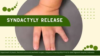
Department of plastic, Reconstructive and aesthestic surgery. Udayana University/RSUP Prof Dr IGNG Ngoerah Hospital, Indonesia
• Syndactyly is one of the most common
congenital hand anomalies treated by pediatric
plastic surgeons.
• Numerous surgical methods have been
described, many of which involve the use of
local flaps to reconstruct the commissure and
full-thickness skin grafts for coverage of raw
areas.
• Recently, reconstructive techniques without the
use of skin grafts have been devised, which work
well for certain indications. Special
considerations are described for complete,
complex, and syndromic syndactylies
Syndactyly is a congenital anomaly in which there is
fusion of adjacent digits due to failure of separation of
developing phalanges during organogenesis.
Normally, webbing between the digits regresses
during 6 to 8 weeks of gestation in a distal to
proximal direction.
Dependent on apoptosis of some portions of the apical
ectodermal ridge, and is mediated by cytokines such as bone
morphogenetic proteins, transforming growth factor-β,
fibroblast growth factors, and retinoic acid.
Syndactyly occurs in approximately 1 in every 2,000 to
3,000 births.
Syndactyly is bilateral in half of the patients and may be
symmetrical or asymmetrical.
Males are affected twice as frequently as females.
It can be inherited autosomal dominantly with variable
expression or reduced penetrance, and can also occur
sporadically
Environmental factors associated with syndactyly
include maternal smoking, lower nutritional status,
lower socioeconomic status, and increased meat and
egg consumption during pregnancy
The most commonly involved interspace in isolated
syndactyly is between the middle and ring finger,
followed by the interspace between the ring and little
finger
when the fingers are fused along the full length of the
digits including the nailfold
Complete Syndactyly
Incomplete Syndactyly when the nailfold is not involved
When fingers are connected by only skin and soft
tissue, and “complex syndactyly” when osseous or
cartilaginous unions are present between adjacent
digits
Simple Syndactyly
Complicated syndactyly is useful to describe abnormalities that extend
beyond simple side-to-side fusion of digits, such as
accessory phalanges or abnormal tendons, muscles,
and nerves interposed within the fused interspaces.
when osseous or cartilaginous unions are present
between adjacent digits
Complex syndactyly
Incomplete Syndactyly Describe abnormalities that extend beyond simple
side-to-side fusion of digits, such as accessory
phalanges or abnormal tendons, muscles, and
nerves interposed within the fused interspaces
Syndactyly associated with syndromes is often
classified as complicated syndactyly; examples include
central polysyndactyly and typical cleft hand
Syndactyly may occur in isolation or as a common
feature of at least 28 syndromes.
• Physical examination is performed that includes
the entire affected upper limb, contralateral
hand, chest wall, and feet to search for additional
anatomical differences.
• Radiographs of the hand are taken to confirm the
skeletal deformities and detect any concealed
extra digits or articular deformities.
• In the case of complex syndactyly, magnetic
resonance imaging or ultrasound can determine
flexor tendon and vascular anatomy.
• Surgery is indicated for nearly all cases of
syndactyly, as the potential for improved
functionality outweighs the risks of the procedure
• a mild, incomplete syndactyly that does not impair
function, medical conditions that preclude surgery,
or complex syndactyly thst risk further functional
impairment
• The goal of surgery is to create a
normalwebspace and improve the appearance of
the involved fingers.
• Soft tissue deficits usually require skin grafting,
although many techniques have been described
to avoid skin grafting.
• Smallerdeficits may alternatively be allowed to
heal by secondary intention.
• We generally perform surgery around 12 months
of age.
• Surgery before 12 months of age is generally
associated with a higher incidence of scar
contracture
• If the defect is bilateral, procedures should be
performed on both hands simultaneously in non
ambulatory children younger than 12 to 14
months
• If both sides of a finger are involved, multiple surgical procedures are required.
• All releases should be performed before school age when fine motor skills of the hand are
essential, and preferably before 24 months
• Operating on both sides of the finger at one time
puts the patient at risk for neurovascular
compromise. In addition, the bordering digits are
typically operated on first given the greater
degree of size discrepancies and the potential for
tripod pinch following release.
• The most common reconstructive design for the
commissure, and that used at our institution,
involves a rectangular, proximally-based dorsal
flap.
• Flap design is heavily dependent on training and
surgeon preference, and can be easily modified
to attempt a more anatomical webspace
reconstruction.
• In cases where construction of a commissure flap
is not needed, such as very mild syndactylies or
mild constriction of the first webspace, a z-plasty
can be employed to simplylengthen the soft
tissue and increase range of motion
• The incision location and pattern must also be
determined, though most surgeons prefer a
zigzag pattern.
• Cronin’s technique of matched zigzag incisions on
the flexor and dorsal surfaces was developed
after his observation that all postoperative
contractures arose from straight incisions on the
flexor surface
• An alternativemethod uses straight-line
midlateral incisions, which are closed with skin
grafts, splinted to prevent contracture, and then
medially excised
Skin grafting has been used to resurface these rawareas for the last century,
and its introduction greatly decreased the contracture rate that resulted
from large raw surfaces left to heal secondarily
Table-21Syndactyly release with skin grafting
Issues with skin grafts include the need for postoperative immobilization, contraction
leading toweb creep and flexion contractures, hyperpigmentation, hair growth, and
donorsite morbidity
Table-1 Syndactyly release with skin grafting
• Grafts are frequently obtained from the groin,
which allows full-thickness harvest with minimal
morbidity.
• Grafts from this region should be taken lateral to
the femoral artery to avoid hair growth at
puberty.
• Grafts from the groin tend to be darker than the
normal hand skin, however, and this may become
an issue as the child becomes older.
• Other possible donor sites are the distal wrist
crease or medial upper arm
• Grafts from the dorsal metacarpal region leave a
visible scar on the hand
• Foreskin has been used, but tends to take poorly
and pigments.
• Patients must be consented before skin grafting,
as the scar at the donor site might hypertrophy.
• Surgery is performed under general
anesthesia with a tourniquet and loupe
magnification.
• The hand and ASIS area are both
prepped and draped.
• The rectangular flap is designed,
extending from the metacarpal heads to
two-thirds the length of the proximal
phalanx.
• The longitudinal edges of the flap can be
curved to fit the roundness of the finger.
Figure 4 (A) The dorsal rectangular flap for
reconstruction of the commissure extends from
the metacarpal heads to two-thirds the length of
the proximal phalanx. A zigzag incision is used to
separate the digits.
• . The dorsal zigzag incision begins at the
apex of the flap, and then extends to the
midline of the proximal interphalangeal
(PIP) joint of the adjacent finger.
• It traverses to the midline of the
neighboring middle phalanx, and back
across to the midline of the distal
interphalangeal joint.
• The palmar zigzag is designed in the
opposite manner so they are mirror
images, with a proximal midline vertical
incision extending from the proximal
zigzag to the level of the desired
webspace
(Figure 4 B) The palmar zigzag incision is a mirror
image of the dorsal incision, with a proximal
midline incision extending to the neo-webspace. A
T incision will be added at the proximal end of the
straight incision to allow insetting of the flaps.
• The proximal palmar incision will have a
small T incision added at the end to
allow insetting of the flap. The T incision
can be adjusted during surgery to allow a
better fit between the flaps
• Once this is done, the hand is
exsanguinated with a Martin bandage
and the tourniquet elevated to 250 mm
Hg pressure
(Figure 4 B) The palmar zigzag incision is a mirror
image of the dorsal incision, with a proximal
midline incision extending to the neo-webspace. A
T incision will be added at the proximal end of the
straight incision to allow insetting of the flaps.
• The proximal palmar incision will have a small T incision
added at the end to allow insetting of the flap. The T incision
can be adjusted during surgery to allow a better fit between
the flaps
• Once this is done, the hand is exsanguinated with a Martin
bandage and the tourniquet elevated to 250 mm Hg pressure
• The dorsal triangular flaps are raised first with
sharp dissection, while controlling bleeding
points with bipolar cautery.
• Attention is then turned to the volar flaps, which
are raised, and neurovascular bundles are
identified. Next, the separation of digits occurs in
the distal to proximal direction.
• During the separation, neurovascular bundles
and venous plexuses on the dorsum of digits and
within the flaps are preserved.
• The proximal dissection is limited by the
bifurcation of the digital artery, which
thereby limits the location of the new
webspace.
• Each digit must have one viable digital
artery.
• The digital artery can be ligated as long
as the other side of the digit has not
been operated on, or the other artery is
known to be intact
• It is thus essential to keep proper
documentation of the digital vasculature
during all surgeries for future reference.
(Fig. 5 )Dorsal and palmar interdigitating zigzag flaps
will resurface the lateral aspects of the digits. After
triangular flaps are lifted, the dorsal rectangular flap
is raised and all flaps judiciously defatted.
• Instead of ligating the artery, a vein graft can be
used to extend the artery and allow a more proximal
webspace, though thiswould rarely be necessary.
• If the digital nerve bifurcates distal to the webspace,
microdissection is used to separate it.
• Attention is then returned to the dorsal side to
elevate the rectangular flap, which is partially
defatted
(Fig. 5 )Dorsal and palmar interdigitating
zigzag flaps will resurface the lateral aspects
of the digits. After triangular flaps are lifted,
the dorsal rectangular flap is raised and all
flaps judiciously defatted.
• On the volar side, a vertical incision ismade
proximally and extended to overcorrect the
webspace.
• A small T incision is then made at the proximal
palmar incision to bring up the flaps on the sides.
Wounds are then irrigated and bleeding
controlled.
• All flaps in the finger are defatted carefully, and
then tacked down in place at the tips with 4–0
chromic suture to ensure they fit properly. If
necessary, the T incision can be lengthened to
allow the flaps to fit better.
• The tourniquet is then let down,
bleeding controlled, and the remaining
flaps sutured in place with interrupted
4–0 chromic sutures.
• To cover any remaining skin defects, an
appropriately sized graft is marked over
the ASIS.
• The full-thickness skin graft is elevated
using the knife and hemostasis achieved.
(Figure 6 (A) Grafts from anterior to the anterior
superior iliac spine covering skin deficits at the
proximal aspect of the
lateral fingers.
• The donor site is closed by advancing
the skin and then closing with
interrupted 4–0 Vicryl (Xeroform, Inc.)
and running subcuticular 4–0 Monocryl
(Ethicon, Inc.) sutures.
• Steri-Strips (3M, Inc.) are placed over the
closure.
• The graft is then defatted and trimmed,
and sutured in place over the skin
defects with interrupted 4–0 chromic
sutures.
Figure 6. (B) (B) The dorsal rectangular flap is
advanced to reconstruct the commissure.
Surgical methods using grafts need both compression and
immobilization to allow for graft take.
Grafts and incisions are covered with one layer of Xeroform
gauze followed by cotton in thewebspace,with the fingers in
abduction to avoid kinking of commissuralflaps.
In young children, the compressed dressing is reinforced with
an above-the-elbow plaster castwith the elbowflexed at 90
degrees to prevent the cast from slipping off the arm.
• Graft-free reconstruction improves overall skin match and decreases
operative time.
• However, not all cases are amenable to primary closure.
Table-2 Syndactyly release without skin grafting
• Simple syndactylies do not extend beyond the
PIP joint may allow for adequate separation of
the digits and deepening of the webspace
without the need for a graft.
• In the complex syndactyly (extend more distally),
local tissue becomes too scarce for adequate
circumferential coverage, regardless of flap
design.
• Primary closure without grafting can be achieved
using two major principles: importation of excess
dorsal tissue and/or extensive defatting of the
involved digits.
• No single technique has been identified as a gold
standard for surgical correction.
• However, the most common techniques involve a
pedicled dorsal metacarpal artery flap that
receives numerous small contributory vessels
• The common disadvantage of a graft-free
syndactyly release is a more conspicuous scar on
the dorsum of the hand.
• Defatting, rather than flap design, has been
suggested as the more important determinant of
whether primary closure can be accomplished.
• Fat tissue is judiciously dissected fromthe
commissure to the distal end of fusion and must
be removal laterally along the syndactylized
digits to the middorsal line.
• However, excess fat removal can lead to
complications such as neurovascular injury, poor
venous drainage, and a withered finger
appearance.
• Inadequate defatting can lead to tight sutures
and ischemia.
• After defatting, if the skin is still unable to be
closed primarily without tension, small areas
(preferably < 2 mmin size) may be left open to
heal secondarily.
• In general, very young patients, from 3 to 6
months old, exhibit the most digital fat and are
the most amenable to defatting.
• Older patients and syndromic patients have less
digital fat and are more likely to require grafting
or healing by secondary intention
• Simple syndactyly release usually successfully improves hand appearance and creates independent digits
that are freely mobile , On the contrary, complex syndactyly release is often associated with loss of mobility
due to the higher risk of contracture and scarring and also has a higher reoperation rate.
• Short-term complications including infection, graft or flap maceration, and graft failure are frequently related to the child’s
activity or inadequate immobilization. Causes of graft failure, which are more common in infants, include hematoma, seroma, and
loss of postoperative bandages
• Web creep is defined as the distal migration of the web- space with growth and is due to scar contracture (►Fig. 8). Incidence ranges in studies from 7.5 to
60% and is more common when the patient is under 18 months old at the time of surgery. Factors that increase incidence of web creep include complex
syndactyly, use of split-thickness skin graft, secondary intention healing after graft loss, poor flap design with longitudinal scars at base of finger
• Short-term complications including infection, graft or flap maceration, and graft failure are frequently related to the child’s activity or
inadequate immobilization. Causes of graft failure, which are more common in infants, include hematoma, seroma, and loss of postoperative
bandages
• Web creep is defined as the distal migration of the web- space with growth and is due to scar contracture (►Fig. 8). Incidence ranges in studies from 7.5 to 60% and is more
common when the patient is under 18 months old at the time of surgery. Factors that increase incidence of web creep include complex syndactyly, use of split-thickness skin
graft, secondary intention healing after graft loss, poor flap design with longitudinal scars at base of finger
• Web creep can be prevented with full- thickness skin grafts, early release of border digits, and
overcompensating the webspace deepening by recessing the webspace more proximally.
• The release of complex syndactyly can result in joint instability from insufficient collateral ligaments, and can be
corrected with arthrodesis as a revision procedure at skeletal maturity.
• Syndactyly is a common congenital hand deformity that
can manifest in an isolated or syndromic form.
• Patients can present with simple syndactyly, which
occurs when the web space moves distally and there is a
fusion of the soft tissue and skin but not of the nail bed,
or with complex syndactyly, where there is bone
involvement in addition to skin and soft tissue
involvement.
• There should be several separate surgeries if one digit is
involved in multiple fusions, as performing both releases
in one surgery can lead to neurovascular compromise.
SYNDACTYLY RELEASE..pptx

More Related Content

What's hot

Congenital anomalies of upper limb
Congenital anomalies of upper limbCongenital anomalies of upper limb
Congenital anomalies of upper limb
Victor Effiom
 
G04 compartment syndrome
G04 compartment syndromeG04 compartment syndrome
G04 compartment syndrome
Claudiu Cucu
 
Syndactyly Hand
Syndactyly Hand Syndactyly Hand
Syndactyly Hand
Dr Kushal Shah
 
Thoracic outlet syndrome.
Thoracic outlet syndrome.Thoracic outlet syndrome.
Thoracic outlet syndrome.
SethiNet presentations
 
Congenital anamolies upper limb - Dr.KK
Congenital anamolies upper  limb - Dr.KKCongenital anamolies upper  limb - Dr.KK
Congenital anamolies upper limb - Dr.KK
Queen Mary Hospital
 
Complications of Fractures
Complications of Fractures Complications of Fractures
Complications of Fractures
Siddhartha Sinha
 
Limb salvage vs amputation final
Limb salvage vs amputation finalLimb salvage vs amputation final
Limb salvage vs amputation final
Sagar Savsani
 
Nerve injuries of Upper Limb
Nerve injuries of Upper LimbNerve injuries of Upper Limb
Nerve injuries of Upper Limb
Pradeep Singh Narwat
 
Supraspinatus tear - medial information
Supraspinatus tear - medial information Supraspinatus tear - medial information
Supraspinatus tear - medial information
martinshaji
 
Back Pain in Children.ppt
Back Pain in Children.pptBack Pain in Children.ppt
Back Pain in Children.ppt
Shama
 
Extensor tendon injuries
Extensor tendon injuriesExtensor tendon injuries
Extensor tendon injuries
mhmad farooq
 
Inflamatory arthritis
Inflamatory arthritisInflamatory arthritis
Inflamatory arthritis
drangelosmith
 
Various ankle injuries and it’s management
Various ankle injuries and it’s managementVarious ankle injuries and it’s management
Various ankle injuries and it’s management
Amol Gaikwad
 
Developmental disorders(MUSCULOSKELETAL)
Developmental disorders(MUSCULOSKELETAL)Developmental disorders(MUSCULOSKELETAL)
Developmental disorders(MUSCULOSKELETAL)
Rajani Cartor
 
Congenital hand anomalies
Congenital hand anomaliesCongenital hand anomalies
Congenital hand anomalies
Subhakanta Mohapatra
 
Injuries around the shoulder
Injuries around the shoulderInjuries around the shoulder
Injuries around the shoulder
Siddhartha Sinha
 
Syndactyly
SyndactylySyndactyly
Syndactyly
drJaishreeRai
 
Spondylolisthesis
SpondylolisthesisSpondylolisthesis
Spondylolisthesis
ratish mishra
 
Compartment syndromes
Compartment syndromesCompartment syndromes
Compartment syndromes
mithilesh216
 
Flaps Basics and Important Leg Flaps and Trauma to Leg management
Flaps Basics and Important Leg Flaps and Trauma to Leg managementFlaps Basics and Important Leg Flaps and Trauma to Leg management
Flaps Basics and Important Leg Flaps and Trauma to Leg management
Dr. Hardik Dodia
 

What's hot (20)

Congenital anomalies of upper limb
Congenital anomalies of upper limbCongenital anomalies of upper limb
Congenital anomalies of upper limb
 
G04 compartment syndrome
G04 compartment syndromeG04 compartment syndrome
G04 compartment syndrome
 
Syndactyly Hand
Syndactyly Hand Syndactyly Hand
Syndactyly Hand
 
Thoracic outlet syndrome.
Thoracic outlet syndrome.Thoracic outlet syndrome.
Thoracic outlet syndrome.
 
Congenital anamolies upper limb - Dr.KK
Congenital anamolies upper  limb - Dr.KKCongenital anamolies upper  limb - Dr.KK
Congenital anamolies upper limb - Dr.KK
 
Complications of Fractures
Complications of Fractures Complications of Fractures
Complications of Fractures
 
Limb salvage vs amputation final
Limb salvage vs amputation finalLimb salvage vs amputation final
Limb salvage vs amputation final
 
Nerve injuries of Upper Limb
Nerve injuries of Upper LimbNerve injuries of Upper Limb
Nerve injuries of Upper Limb
 
Supraspinatus tear - medial information
Supraspinatus tear - medial information Supraspinatus tear - medial information
Supraspinatus tear - medial information
 
Back Pain in Children.ppt
Back Pain in Children.pptBack Pain in Children.ppt
Back Pain in Children.ppt
 
Extensor tendon injuries
Extensor tendon injuriesExtensor tendon injuries
Extensor tendon injuries
 
Inflamatory arthritis
Inflamatory arthritisInflamatory arthritis
Inflamatory arthritis
 
Various ankle injuries and it’s management
Various ankle injuries and it’s managementVarious ankle injuries and it’s management
Various ankle injuries and it’s management
 
Developmental disorders(MUSCULOSKELETAL)
Developmental disorders(MUSCULOSKELETAL)Developmental disorders(MUSCULOSKELETAL)
Developmental disorders(MUSCULOSKELETAL)
 
Congenital hand anomalies
Congenital hand anomaliesCongenital hand anomalies
Congenital hand anomalies
 
Injuries around the shoulder
Injuries around the shoulderInjuries around the shoulder
Injuries around the shoulder
 
Syndactyly
SyndactylySyndactyly
Syndactyly
 
Spondylolisthesis
SpondylolisthesisSpondylolisthesis
Spondylolisthesis
 
Compartment syndromes
Compartment syndromesCompartment syndromes
Compartment syndromes
 
Flaps Basics and Important Leg Flaps and Trauma to Leg management
Flaps Basics and Important Leg Flaps and Trauma to Leg managementFlaps Basics and Important Leg Flaps and Trauma to Leg management
Flaps Basics and Important Leg Flaps and Trauma to Leg management
 

Similar to SYNDACTYLY RELEASE..pptx

Soft tissue managment
Soft tissue managmentSoft tissue managment
Soft tissue managment
Muhammed Omar
 
Flexor and extensor tendon injury
Flexor and extensor tendon injuryFlexor and extensor tendon injury
Flexor and extensor tendon injury
Dr. Anurag Mittal
 
Gingiva.pptx
Gingiva.pptxGingiva.pptx
Gingiva.pptx
Meghna490298
 
Hand anatomy 1
Hand anatomy 1 Hand anatomy 1
Hand anatomy 1
Mahmoud Gouda
 
Non microsurgical coverage of hand
Non microsurgical coverage of handNon microsurgical coverage of hand
Non microsurgical coverage of hand
Punith Vasanthan
 
Recent advances in minimal access surgery.pptx
Recent advances in minimal access surgery.pptxRecent advances in minimal access surgery.pptx
Recent advances in minimal access surgery.pptx
Manoj H.V
 
Syndactyly.pptx
 Syndactyly.pptx Syndactyly.pptx
Syndactyly.pptx
RenuSingla3
 
Congenital hand diseases: managment
Congenital hand diseases: managmentCongenital hand diseases: managment
Congenital hand diseases: managment
Khaldoon Alaghbari
 
Periodontal flap
Periodontal flapPeriodontal flap
Periodontal flap
akshay shete
 
Finger tip injuries & management
Finger tip injuries & managementFinger tip injuries & management
Finger tip injuries & management
Rajesh Gantayat
 
Flap techniques for pocket therapy
Flap techniques for pocket therapy  Flap techniques for pocket therapy
Flap techniques for pocket therapy
Dr. Archana Balakrishnan
 
Gingivectomy
GingivectomyGingivectomy
Gingivectomy
pragy mallik
 
Hypermobility and ankylosis
Hypermobility and ankylosisHypermobility and ankylosis
Hypermobility and ankylosis
Hanan Shanab
 
Hand injuries (compiled by Dr. Sanjib Kumar Das)
Hand injuries (compiled by Dr. Sanjib Kumar Das)Hand injuries (compiled by Dr. Sanjib Kumar Das)
Hand injuries (compiled by Dr. Sanjib Kumar Das)
Dr. Sanjib Kumar Das
 
Infections of hand
Infections of handInfections of hand
Infections of hand
Mirant Dave
 
Acute Extensor tendon injuries diagnosis and management.pptx
Acute Extensor tendon injuries diagnosis and management.pptxAcute Extensor tendon injuries diagnosis and management.pptx
Acute Extensor tendon injuries diagnosis and management.pptx
Rohie3
 
Mucogingival surgery
Mucogingival surgeryMucogingival surgery
Mucogingival surgery
Athraa Ali
 
Abdominal Incisions and sutures
Abdominal Incisions and sutures Abdominal Incisions and sutures
Abdominal Incisions and sutures
AlaaZeineh
 
Pollicization
PollicizationPollicization
Surgicalincisions 150519180458-lva1-app6892
Surgicalincisions 150519180458-lva1-app6892Surgicalincisions 150519180458-lva1-app6892
Surgicalincisions 150519180458-lva1-app6892
Mahar852
 

Similar to SYNDACTYLY RELEASE..pptx (20)

Soft tissue managment
Soft tissue managmentSoft tissue managment
Soft tissue managment
 
Flexor and extensor tendon injury
Flexor and extensor tendon injuryFlexor and extensor tendon injury
Flexor and extensor tendon injury
 
Gingiva.pptx
Gingiva.pptxGingiva.pptx
Gingiva.pptx
 
Hand anatomy 1
Hand anatomy 1 Hand anatomy 1
Hand anatomy 1
 
Non microsurgical coverage of hand
Non microsurgical coverage of handNon microsurgical coverage of hand
Non microsurgical coverage of hand
 
Recent advances in minimal access surgery.pptx
Recent advances in minimal access surgery.pptxRecent advances in minimal access surgery.pptx
Recent advances in minimal access surgery.pptx
 
Syndactyly.pptx
 Syndactyly.pptx Syndactyly.pptx
Syndactyly.pptx
 
Congenital hand diseases: managment
Congenital hand diseases: managmentCongenital hand diseases: managment
Congenital hand diseases: managment
 
Periodontal flap
Periodontal flapPeriodontal flap
Periodontal flap
 
Finger tip injuries & management
Finger tip injuries & managementFinger tip injuries & management
Finger tip injuries & management
 
Flap techniques for pocket therapy
Flap techniques for pocket therapy  Flap techniques for pocket therapy
Flap techniques for pocket therapy
 
Gingivectomy
GingivectomyGingivectomy
Gingivectomy
 
Hypermobility and ankylosis
Hypermobility and ankylosisHypermobility and ankylosis
Hypermobility and ankylosis
 
Hand injuries (compiled by Dr. Sanjib Kumar Das)
Hand injuries (compiled by Dr. Sanjib Kumar Das)Hand injuries (compiled by Dr. Sanjib Kumar Das)
Hand injuries (compiled by Dr. Sanjib Kumar Das)
 
Infections of hand
Infections of handInfections of hand
Infections of hand
 
Acute Extensor tendon injuries diagnosis and management.pptx
Acute Extensor tendon injuries diagnosis and management.pptxAcute Extensor tendon injuries diagnosis and management.pptx
Acute Extensor tendon injuries diagnosis and management.pptx
 
Mucogingival surgery
Mucogingival surgeryMucogingival surgery
Mucogingival surgery
 
Abdominal Incisions and sutures
Abdominal Incisions and sutures Abdominal Incisions and sutures
Abdominal Incisions and sutures
 
Pollicization
PollicizationPollicization
Pollicization
 
Surgicalincisions 150519180458-lva1-app6892
Surgicalincisions 150519180458-lva1-app6892Surgicalincisions 150519180458-lva1-app6892
Surgicalincisions 150519180458-lva1-app6892
 

Recently uploaded

Histololgy of Female Reproductive System.pptx
Histololgy of Female Reproductive System.pptxHistololgy of Female Reproductive System.pptx
Histololgy of Female Reproductive System.pptx
AyeshaZaid1
 
Vestibulocochlear Nerve by Dr. Rabia Inam Gandapore.pptx
Vestibulocochlear Nerve by Dr. Rabia Inam Gandapore.pptxVestibulocochlear Nerve by Dr. Rabia Inam Gandapore.pptx
Vestibulocochlear Nerve by Dr. Rabia Inam Gandapore.pptx
Dr. Rabia Inam Gandapore
 
Complementary feeding in infant IAP PROTOCOLS
Complementary feeding in infant IAP PROTOCOLSComplementary feeding in infant IAP PROTOCOLS
Complementary feeding in infant IAP PROTOCOLS
chiranthgowda16
 
K CỔ TỬ CUNG.pdf tự ghi chép, chữ hơi xấu
K CỔ TỬ CUNG.pdf tự ghi chép, chữ hơi xấuK CỔ TỬ CUNG.pdf tự ghi chép, chữ hơi xấu
K CỔ TỬ CUNG.pdf tự ghi chép, chữ hơi xấu
HongBiThi1
 
CLEAR ALIGNER THERAPY IN ORTHODONTICS .pptx
CLEAR ALIGNER THERAPY IN ORTHODONTICS .pptxCLEAR ALIGNER THERAPY IN ORTHODONTICS .pptx
CLEAR ALIGNER THERAPY IN ORTHODONTICS .pptx
Government Dental College & Hospital Srinagar
 
Adhd Medication Shortage Uk - trinexpharmacy.com
Adhd Medication Shortage Uk - trinexpharmacy.comAdhd Medication Shortage Uk - trinexpharmacy.com
Adhd Medication Shortage Uk - trinexpharmacy.com
reignlana06
 
Ear and its clinical correlations By Dr. Rabia Inam Gandapore.pptx
Ear and its clinical correlations By Dr. Rabia Inam Gandapore.pptxEar and its clinical correlations By Dr. Rabia Inam Gandapore.pptx
Ear and its clinical correlations By Dr. Rabia Inam Gandapore.pptx
Dr. Rabia Inam Gandapore
 
Cervical Disc Arthroplasty ORSI 2024.pptx
Cervical Disc Arthroplasty ORSI 2024.pptxCervical Disc Arthroplasty ORSI 2024.pptx
Cervical Disc Arthroplasty ORSI 2024.pptx
LEFLOT Jean-Louis
 
10 Benefits an EPCR Software should Bring to EMS Organizations
10 Benefits an EPCR Software should Bring to EMS Organizations   10 Benefits an EPCR Software should Bring to EMS Organizations
10 Benefits an EPCR Software should Bring to EMS Organizations
Traumasoft LLC
 
The Nervous and Chemical Regulation of Respiration
The Nervous and Chemical Regulation of RespirationThe Nervous and Chemical Regulation of Respiration
The Nervous and Chemical Regulation of Respiration
MedicoseAcademics
 
Promoting Wellbeing - Applied Social Psychology - Psychology SuperNotes
Promoting Wellbeing - Applied Social Psychology - Psychology SuperNotesPromoting Wellbeing - Applied Social Psychology - Psychology SuperNotes
Promoting Wellbeing - Applied Social Psychology - Psychology SuperNotes
PsychoTech Services
 
MERCURY GROUP.BHMS.MATERIA MEDICA.HOMOEOPATHY
MERCURY GROUP.BHMS.MATERIA MEDICA.HOMOEOPATHYMERCURY GROUP.BHMS.MATERIA MEDICA.HOMOEOPATHY
MERCURY GROUP.BHMS.MATERIA MEDICA.HOMOEOPATHY
DRPREETHIJAMESP
 
share - Lions, tigers, AI and health misinformation, oh my!.pptx
share - Lions, tigers, AI and health misinformation, oh my!.pptxshare - Lions, tigers, AI and health misinformation, oh my!.pptx
share - Lions, tigers, AI and health misinformation, oh my!.pptx
Tina Purnat
 
Physical demands in sports - WCSPT Oslo 2024
Physical demands in sports - WCSPT Oslo 2024Physical demands in sports - WCSPT Oslo 2024
Physical demands in sports - WCSPT Oslo 2024
Torstein Dalen-Lorentsen
 
Clinic ^%[+27633867063*Abortion Pills For Sale In Tembisa Central
Clinic ^%[+27633867063*Abortion Pills For Sale In Tembisa CentralClinic ^%[+27633867063*Abortion Pills For Sale In Tembisa Central
Clinic ^%[+27633867063*Abortion Pills For Sale In Tembisa Central
19various
 
Post-Menstrual Smell- When to Suspect Vaginitis.pptx
Post-Menstrual Smell- When to Suspect Vaginitis.pptxPost-Menstrual Smell- When to Suspect Vaginitis.pptx
Post-Menstrual Smell- When to Suspect Vaginitis.pptx
FFragrant
 
Efficacy of Avartana Sneha in Ayurveda
Efficacy of Avartana Sneha in AyurvedaEfficacy of Avartana Sneha in Ayurveda
Efficacy of Avartana Sneha in Ayurveda
Dr. Jyothirmai Paindla
 
CBL Seminar 2024_Preliminary Program.pdf
CBL Seminar 2024_Preliminary Program.pdfCBL Seminar 2024_Preliminary Program.pdf
CBL Seminar 2024_Preliminary Program.pdf
suvadeepdas911
 
Does Over-Masturbation Contribute to Chronic Prostatitis.pptx
Does Over-Masturbation Contribute to Chronic Prostatitis.pptxDoes Over-Masturbation Contribute to Chronic Prostatitis.pptx
Does Over-Masturbation Contribute to Chronic Prostatitis.pptx
walterHu5
 
Demystifying Fallopian Tube Blockage- Grading the Differences and Implication...
Demystifying Fallopian Tube Blockage- Grading the Differences and Implication...Demystifying Fallopian Tube Blockage- Grading the Differences and Implication...
Demystifying Fallopian Tube Blockage- Grading the Differences and Implication...
FFragrant
 

Recently uploaded (20)

Histololgy of Female Reproductive System.pptx
Histololgy of Female Reproductive System.pptxHistololgy of Female Reproductive System.pptx
Histololgy of Female Reproductive System.pptx
 
Vestibulocochlear Nerve by Dr. Rabia Inam Gandapore.pptx
Vestibulocochlear Nerve by Dr. Rabia Inam Gandapore.pptxVestibulocochlear Nerve by Dr. Rabia Inam Gandapore.pptx
Vestibulocochlear Nerve by Dr. Rabia Inam Gandapore.pptx
 
Complementary feeding in infant IAP PROTOCOLS
Complementary feeding in infant IAP PROTOCOLSComplementary feeding in infant IAP PROTOCOLS
Complementary feeding in infant IAP PROTOCOLS
 
K CỔ TỬ CUNG.pdf tự ghi chép, chữ hơi xấu
K CỔ TỬ CUNG.pdf tự ghi chép, chữ hơi xấuK CỔ TỬ CUNG.pdf tự ghi chép, chữ hơi xấu
K CỔ TỬ CUNG.pdf tự ghi chép, chữ hơi xấu
 
CLEAR ALIGNER THERAPY IN ORTHODONTICS .pptx
CLEAR ALIGNER THERAPY IN ORTHODONTICS .pptxCLEAR ALIGNER THERAPY IN ORTHODONTICS .pptx
CLEAR ALIGNER THERAPY IN ORTHODONTICS .pptx
 
Adhd Medication Shortage Uk - trinexpharmacy.com
Adhd Medication Shortage Uk - trinexpharmacy.comAdhd Medication Shortage Uk - trinexpharmacy.com
Adhd Medication Shortage Uk - trinexpharmacy.com
 
Ear and its clinical correlations By Dr. Rabia Inam Gandapore.pptx
Ear and its clinical correlations By Dr. Rabia Inam Gandapore.pptxEar and its clinical correlations By Dr. Rabia Inam Gandapore.pptx
Ear and its clinical correlations By Dr. Rabia Inam Gandapore.pptx
 
Cervical Disc Arthroplasty ORSI 2024.pptx
Cervical Disc Arthroplasty ORSI 2024.pptxCervical Disc Arthroplasty ORSI 2024.pptx
Cervical Disc Arthroplasty ORSI 2024.pptx
 
10 Benefits an EPCR Software should Bring to EMS Organizations
10 Benefits an EPCR Software should Bring to EMS Organizations   10 Benefits an EPCR Software should Bring to EMS Organizations
10 Benefits an EPCR Software should Bring to EMS Organizations
 
The Nervous and Chemical Regulation of Respiration
The Nervous and Chemical Regulation of RespirationThe Nervous and Chemical Regulation of Respiration
The Nervous and Chemical Regulation of Respiration
 
Promoting Wellbeing - Applied Social Psychology - Psychology SuperNotes
Promoting Wellbeing - Applied Social Psychology - Psychology SuperNotesPromoting Wellbeing - Applied Social Psychology - Psychology SuperNotes
Promoting Wellbeing - Applied Social Psychology - Psychology SuperNotes
 
MERCURY GROUP.BHMS.MATERIA MEDICA.HOMOEOPATHY
MERCURY GROUP.BHMS.MATERIA MEDICA.HOMOEOPATHYMERCURY GROUP.BHMS.MATERIA MEDICA.HOMOEOPATHY
MERCURY GROUP.BHMS.MATERIA MEDICA.HOMOEOPATHY
 
share - Lions, tigers, AI and health misinformation, oh my!.pptx
share - Lions, tigers, AI and health misinformation, oh my!.pptxshare - Lions, tigers, AI and health misinformation, oh my!.pptx
share - Lions, tigers, AI and health misinformation, oh my!.pptx
 
Physical demands in sports - WCSPT Oslo 2024
Physical demands in sports - WCSPT Oslo 2024Physical demands in sports - WCSPT Oslo 2024
Physical demands in sports - WCSPT Oslo 2024
 
Clinic ^%[+27633867063*Abortion Pills For Sale In Tembisa Central
Clinic ^%[+27633867063*Abortion Pills For Sale In Tembisa CentralClinic ^%[+27633867063*Abortion Pills For Sale In Tembisa Central
Clinic ^%[+27633867063*Abortion Pills For Sale In Tembisa Central
 
Post-Menstrual Smell- When to Suspect Vaginitis.pptx
Post-Menstrual Smell- When to Suspect Vaginitis.pptxPost-Menstrual Smell- When to Suspect Vaginitis.pptx
Post-Menstrual Smell- When to Suspect Vaginitis.pptx
 
Efficacy of Avartana Sneha in Ayurveda
Efficacy of Avartana Sneha in AyurvedaEfficacy of Avartana Sneha in Ayurveda
Efficacy of Avartana Sneha in Ayurveda
 
CBL Seminar 2024_Preliminary Program.pdf
CBL Seminar 2024_Preliminary Program.pdfCBL Seminar 2024_Preliminary Program.pdf
CBL Seminar 2024_Preliminary Program.pdf
 
Does Over-Masturbation Contribute to Chronic Prostatitis.pptx
Does Over-Masturbation Contribute to Chronic Prostatitis.pptxDoes Over-Masturbation Contribute to Chronic Prostatitis.pptx
Does Over-Masturbation Contribute to Chronic Prostatitis.pptx
 
Demystifying Fallopian Tube Blockage- Grading the Differences and Implication...
Demystifying Fallopian Tube Blockage- Grading the Differences and Implication...Demystifying Fallopian Tube Blockage- Grading the Differences and Implication...
Demystifying Fallopian Tube Blockage- Grading the Differences and Implication...
 

SYNDACTYLY RELEASE..pptx

  • 1. Department of plastic, Reconstructive and aesthestic surgery. Udayana University/RSUP Prof Dr IGNG Ngoerah Hospital, Indonesia
  • 2. • Syndactyly is one of the most common congenital hand anomalies treated by pediatric plastic surgeons. • Numerous surgical methods have been described, many of which involve the use of local flaps to reconstruct the commissure and full-thickness skin grafts for coverage of raw areas. • Recently, reconstructive techniques without the use of skin grafts have been devised, which work well for certain indications. Special considerations are described for complete, complex, and syndromic syndactylies
  • 3. Syndactyly is a congenital anomaly in which there is fusion of adjacent digits due to failure of separation of developing phalanges during organogenesis. Normally, webbing between the digits regresses during 6 to 8 weeks of gestation in a distal to proximal direction. Dependent on apoptosis of some portions of the apical ectodermal ridge, and is mediated by cytokines such as bone morphogenetic proteins, transforming growth factor-β, fibroblast growth factors, and retinoic acid.
  • 4. Syndactyly occurs in approximately 1 in every 2,000 to 3,000 births. Syndactyly is bilateral in half of the patients and may be symmetrical or asymmetrical. Males are affected twice as frequently as females.
  • 5. It can be inherited autosomal dominantly with variable expression or reduced penetrance, and can also occur sporadically Environmental factors associated with syndactyly include maternal smoking, lower nutritional status, lower socioeconomic status, and increased meat and egg consumption during pregnancy The most commonly involved interspace in isolated syndactyly is between the middle and ring finger, followed by the interspace between the ring and little finger
  • 6. when the fingers are fused along the full length of the digits including the nailfold Complete Syndactyly Incomplete Syndactyly when the nailfold is not involved
  • 7. When fingers are connected by only skin and soft tissue, and “complex syndactyly” when osseous or cartilaginous unions are present between adjacent digits Simple Syndactyly Complicated syndactyly is useful to describe abnormalities that extend beyond simple side-to-side fusion of digits, such as accessory phalanges or abnormal tendons, muscles, and nerves interposed within the fused interspaces.
  • 8. when osseous or cartilaginous unions are present between adjacent digits Complex syndactyly Incomplete Syndactyly Describe abnormalities that extend beyond simple side-to-side fusion of digits, such as accessory phalanges or abnormal tendons, muscles, and nerves interposed within the fused interspaces
  • 9. Syndactyly associated with syndromes is often classified as complicated syndactyly; examples include central polysyndactyly and typical cleft hand Syndactyly may occur in isolation or as a common feature of at least 28 syndromes.
  • 10. • Physical examination is performed that includes the entire affected upper limb, contralateral hand, chest wall, and feet to search for additional anatomical differences. • Radiographs of the hand are taken to confirm the skeletal deformities and detect any concealed extra digits or articular deformities. • In the case of complex syndactyly, magnetic resonance imaging or ultrasound can determine flexor tendon and vascular anatomy.
  • 11.
  • 12. • Surgery is indicated for nearly all cases of syndactyly, as the potential for improved functionality outweighs the risks of the procedure • a mild, incomplete syndactyly that does not impair function, medical conditions that preclude surgery, or complex syndactyly thst risk further functional impairment
  • 13. • The goal of surgery is to create a normalwebspace and improve the appearance of the involved fingers. • Soft tissue deficits usually require skin grafting, although many techniques have been described to avoid skin grafting. • Smallerdeficits may alternatively be allowed to heal by secondary intention.
  • 14. • We generally perform surgery around 12 months of age. • Surgery before 12 months of age is generally associated with a higher incidence of scar contracture • If the defect is bilateral, procedures should be performed on both hands simultaneously in non ambulatory children younger than 12 to 14 months • If both sides of a finger are involved, multiple surgical procedures are required. • All releases should be performed before school age when fine motor skills of the hand are essential, and preferably before 24 months
  • 15. • Operating on both sides of the finger at one time puts the patient at risk for neurovascular compromise. In addition, the bordering digits are typically operated on first given the greater degree of size discrepancies and the potential for tripod pinch following release.
  • 16. • The most common reconstructive design for the commissure, and that used at our institution, involves a rectangular, proximally-based dorsal flap. • Flap design is heavily dependent on training and surgeon preference, and can be easily modified to attempt a more anatomical webspace reconstruction. • In cases where construction of a commissure flap is not needed, such as very mild syndactylies or mild constriction of the first webspace, a z-plasty can be employed to simplylengthen the soft tissue and increase range of motion
  • 17. • The incision location and pattern must also be determined, though most surgeons prefer a zigzag pattern. • Cronin’s technique of matched zigzag incisions on the flexor and dorsal surfaces was developed after his observation that all postoperative contractures arose from straight incisions on the flexor surface • An alternativemethod uses straight-line midlateral incisions, which are closed with skin grafts, splinted to prevent contracture, and then medially excised
  • 18. Skin grafting has been used to resurface these rawareas for the last century, and its introduction greatly decreased the contracture rate that resulted from large raw surfaces left to heal secondarily Table-21Syndactyly release with skin grafting
  • 19. Issues with skin grafts include the need for postoperative immobilization, contraction leading toweb creep and flexion contractures, hyperpigmentation, hair growth, and donorsite morbidity Table-1 Syndactyly release with skin grafting
  • 20. • Grafts are frequently obtained from the groin, which allows full-thickness harvest with minimal morbidity. • Grafts from this region should be taken lateral to the femoral artery to avoid hair growth at puberty. • Grafts from the groin tend to be darker than the normal hand skin, however, and this may become an issue as the child becomes older. • Other possible donor sites are the distal wrist crease or medial upper arm
  • 21. • Grafts from the dorsal metacarpal region leave a visible scar on the hand • Foreskin has been used, but tends to take poorly and pigments. • Patients must be consented before skin grafting, as the scar at the donor site might hypertrophy.
  • 22. • Surgery is performed under general anesthesia with a tourniquet and loupe magnification. • The hand and ASIS area are both prepped and draped. • The rectangular flap is designed, extending from the metacarpal heads to two-thirds the length of the proximal phalanx. • The longitudinal edges of the flap can be curved to fit the roundness of the finger. Figure 4 (A) The dorsal rectangular flap for reconstruction of the commissure extends from the metacarpal heads to two-thirds the length of the proximal phalanx. A zigzag incision is used to separate the digits.
  • 23. • . The dorsal zigzag incision begins at the apex of the flap, and then extends to the midline of the proximal interphalangeal (PIP) joint of the adjacent finger. • It traverses to the midline of the neighboring middle phalanx, and back across to the midline of the distal interphalangeal joint. • The palmar zigzag is designed in the opposite manner so they are mirror images, with a proximal midline vertical incision extending from the proximal zigzag to the level of the desired webspace (Figure 4 B) The palmar zigzag incision is a mirror image of the dorsal incision, with a proximal midline incision extending to the neo-webspace. A T incision will be added at the proximal end of the straight incision to allow insetting of the flaps.
  • 24. • The proximal palmar incision will have a small T incision added at the end to allow insetting of the flap. The T incision can be adjusted during surgery to allow a better fit between the flaps • Once this is done, the hand is exsanguinated with a Martin bandage and the tourniquet elevated to 250 mm Hg pressure (Figure 4 B) The palmar zigzag incision is a mirror image of the dorsal incision, with a proximal midline incision extending to the neo-webspace. A T incision will be added at the proximal end of the straight incision to allow insetting of the flaps.
  • 25. • The proximal palmar incision will have a small T incision added at the end to allow insetting of the flap. The T incision can be adjusted during surgery to allow a better fit between the flaps • Once this is done, the hand is exsanguinated with a Martin bandage and the tourniquet elevated to 250 mm Hg pressure
  • 26. • The dorsal triangular flaps are raised first with sharp dissection, while controlling bleeding points with bipolar cautery. • Attention is then turned to the volar flaps, which are raised, and neurovascular bundles are identified. Next, the separation of digits occurs in the distal to proximal direction. • During the separation, neurovascular bundles and venous plexuses on the dorsum of digits and within the flaps are preserved.
  • 27. • The proximal dissection is limited by the bifurcation of the digital artery, which thereby limits the location of the new webspace. • Each digit must have one viable digital artery. • The digital artery can be ligated as long as the other side of the digit has not been operated on, or the other artery is known to be intact • It is thus essential to keep proper documentation of the digital vasculature during all surgeries for future reference. (Fig. 5 )Dorsal and palmar interdigitating zigzag flaps will resurface the lateral aspects of the digits. After triangular flaps are lifted, the dorsal rectangular flap is raised and all flaps judiciously defatted.
  • 28. • Instead of ligating the artery, a vein graft can be used to extend the artery and allow a more proximal webspace, though thiswould rarely be necessary. • If the digital nerve bifurcates distal to the webspace, microdissection is used to separate it. • Attention is then returned to the dorsal side to elevate the rectangular flap, which is partially defatted (Fig. 5 )Dorsal and palmar interdigitating zigzag flaps will resurface the lateral aspects of the digits. After triangular flaps are lifted, the dorsal rectangular flap is raised and all flaps judiciously defatted.
  • 29. • On the volar side, a vertical incision ismade proximally and extended to overcorrect the webspace. • A small T incision is then made at the proximal palmar incision to bring up the flaps on the sides. Wounds are then irrigated and bleeding controlled. • All flaps in the finger are defatted carefully, and then tacked down in place at the tips with 4–0 chromic suture to ensure they fit properly. If necessary, the T incision can be lengthened to allow the flaps to fit better.
  • 30. • The tourniquet is then let down, bleeding controlled, and the remaining flaps sutured in place with interrupted 4–0 chromic sutures. • To cover any remaining skin defects, an appropriately sized graft is marked over the ASIS. • The full-thickness skin graft is elevated using the knife and hemostasis achieved. (Figure 6 (A) Grafts from anterior to the anterior superior iliac spine covering skin deficits at the proximal aspect of the lateral fingers.
  • 31. • The donor site is closed by advancing the skin and then closing with interrupted 4–0 Vicryl (Xeroform, Inc.) and running subcuticular 4–0 Monocryl (Ethicon, Inc.) sutures. • Steri-Strips (3M, Inc.) are placed over the closure. • The graft is then defatted and trimmed, and sutured in place over the skin defects with interrupted 4–0 chromic sutures. Figure 6. (B) (B) The dorsal rectangular flap is advanced to reconstruct the commissure.
  • 32. Surgical methods using grafts need both compression and immobilization to allow for graft take. Grafts and incisions are covered with one layer of Xeroform gauze followed by cotton in thewebspace,with the fingers in abduction to avoid kinking of commissuralflaps. In young children, the compressed dressing is reinforced with an above-the-elbow plaster castwith the elbowflexed at 90 degrees to prevent the cast from slipping off the arm.
  • 33. • Graft-free reconstruction improves overall skin match and decreases operative time. • However, not all cases are amenable to primary closure. Table-2 Syndactyly release without skin grafting
  • 34. Table-2 Syndactyly release without skin grafting
  • 35. Table-2 Syndactyly release without skin grafting
  • 36. • Simple syndactylies do not extend beyond the PIP joint may allow for adequate separation of the digits and deepening of the webspace without the need for a graft. • In the complex syndactyly (extend more distally), local tissue becomes too scarce for adequate circumferential coverage, regardless of flap design. • Primary closure without grafting can be achieved using two major principles: importation of excess dorsal tissue and/or extensive defatting of the involved digits.
  • 37. • No single technique has been identified as a gold standard for surgical correction. • However, the most common techniques involve a pedicled dorsal metacarpal artery flap that receives numerous small contributory vessels • The common disadvantage of a graft-free syndactyly release is a more conspicuous scar on the dorsum of the hand.
  • 38. • Defatting, rather than flap design, has been suggested as the more important determinant of whether primary closure can be accomplished. • Fat tissue is judiciously dissected fromthe commissure to the distal end of fusion and must be removal laterally along the syndactylized digits to the middorsal line. • However, excess fat removal can lead to complications such as neurovascular injury, poor venous drainage, and a withered finger appearance.
  • 39. • Inadequate defatting can lead to tight sutures and ischemia. • After defatting, if the skin is still unable to be closed primarily without tension, small areas (preferably < 2 mmin size) may be left open to heal secondarily. • In general, very young patients, from 3 to 6 months old, exhibit the most digital fat and are the most amenable to defatting. • Older patients and syndromic patients have less digital fat and are more likely to require grafting or healing by secondary intention
  • 40. • Simple syndactyly release usually successfully improves hand appearance and creates independent digits that are freely mobile , On the contrary, complex syndactyly release is often associated with loss of mobility due to the higher risk of contracture and scarring and also has a higher reoperation rate.
  • 41. • Short-term complications including infection, graft or flap maceration, and graft failure are frequently related to the child’s activity or inadequate immobilization. Causes of graft failure, which are more common in infants, include hematoma, seroma, and loss of postoperative bandages • Web creep is defined as the distal migration of the web- space with growth and is due to scar contracture (►Fig. 8). Incidence ranges in studies from 7.5 to 60% and is more common when the patient is under 18 months old at the time of surgery. Factors that increase incidence of web creep include complex syndactyly, use of split-thickness skin graft, secondary intention healing after graft loss, poor flap design with longitudinal scars at base of finger
  • 42. • Short-term complications including infection, graft or flap maceration, and graft failure are frequently related to the child’s activity or inadequate immobilization. Causes of graft failure, which are more common in infants, include hematoma, seroma, and loss of postoperative bandages • Web creep is defined as the distal migration of the web- space with growth and is due to scar contracture (►Fig. 8). Incidence ranges in studies from 7.5 to 60% and is more common when the patient is under 18 months old at the time of surgery. Factors that increase incidence of web creep include complex syndactyly, use of split-thickness skin graft, secondary intention healing after graft loss, poor flap design with longitudinal scars at base of finger
  • 43. • Web creep can be prevented with full- thickness skin grafts, early release of border digits, and overcompensating the webspace deepening by recessing the webspace more proximally. • The release of complex syndactyly can result in joint instability from insufficient collateral ligaments, and can be corrected with arthrodesis as a revision procedure at skeletal maturity.
  • 44. • Syndactyly is a common congenital hand deformity that can manifest in an isolated or syndromic form. • Patients can present with simple syndactyly, which occurs when the web space moves distally and there is a fusion of the soft tissue and skin but not of the nail bed, or with complex syndactyly, where there is bone involvement in addition to skin and soft tissue involvement. • There should be several separate surgeries if one digit is involved in multiple fusions, as performing both releases in one surgery can lead to neurovascular compromise.