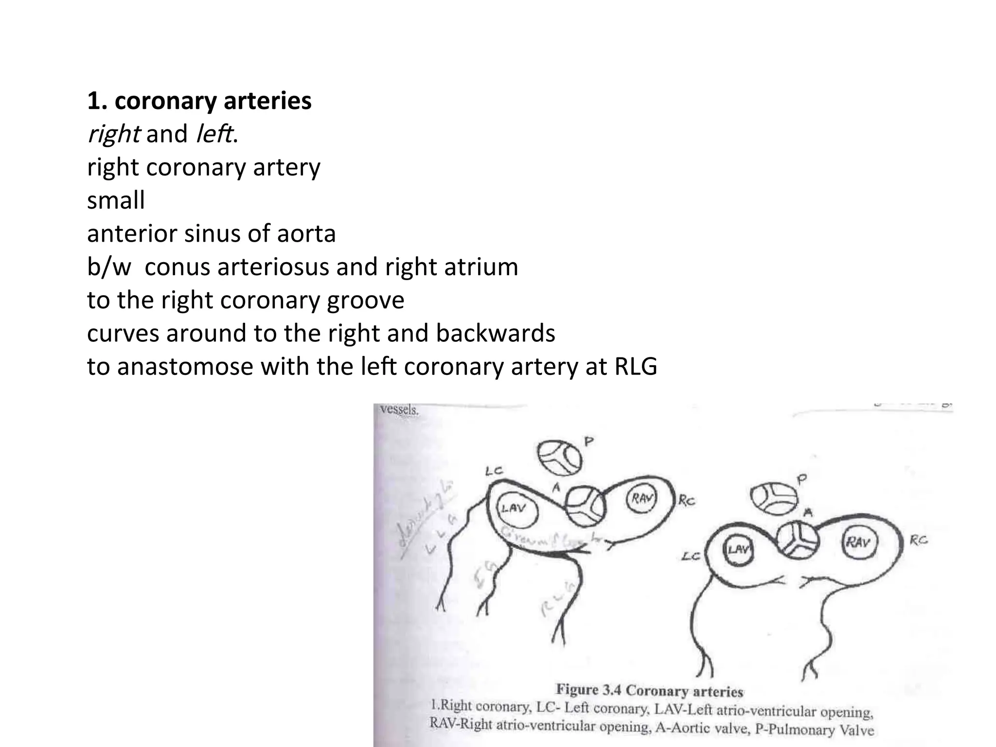
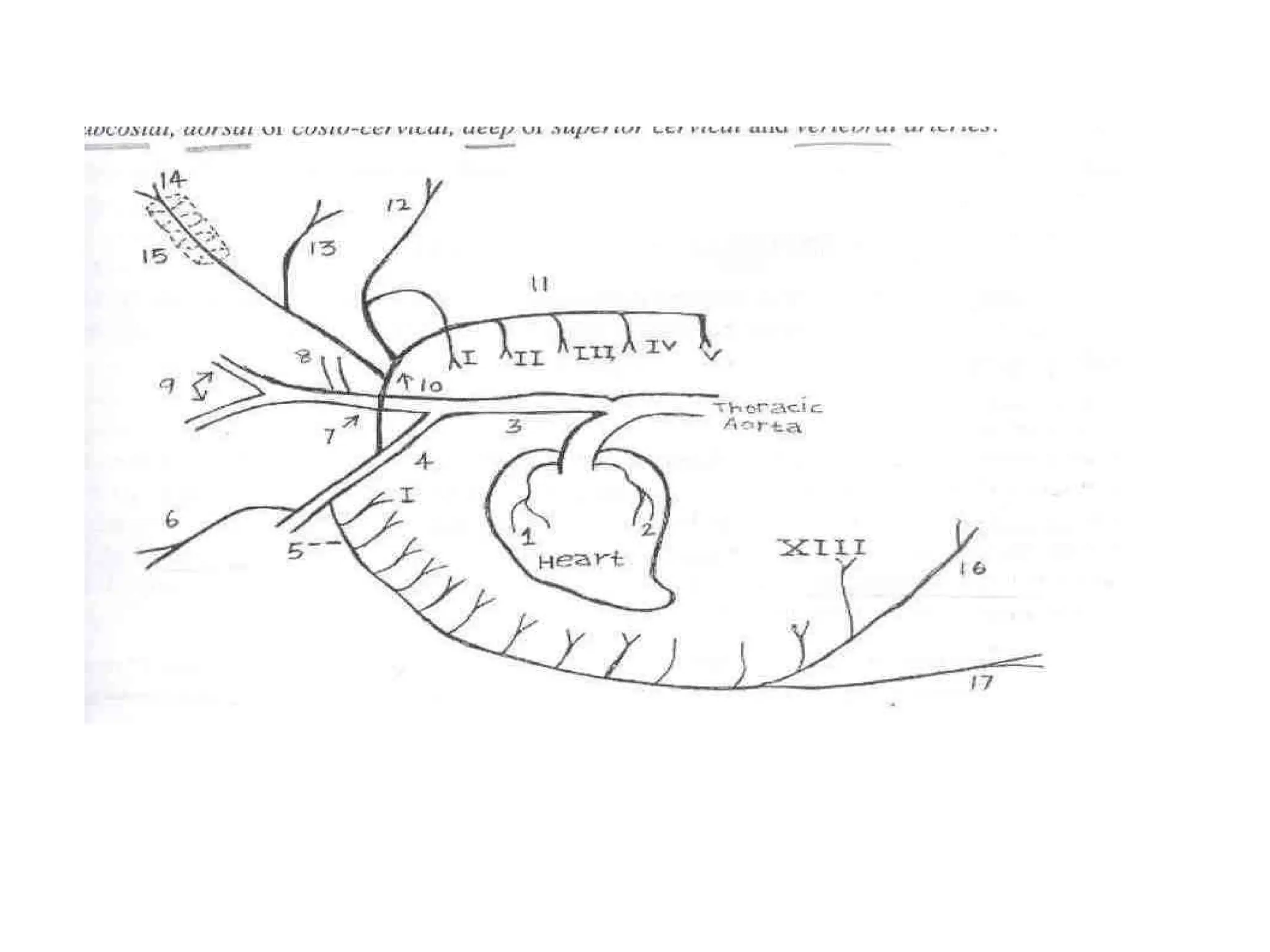
1. coronary arteries
rightand left.
right coronary artery
small
anterior sinus of aorta
b/w conus arteriosus and right atrium
to the right coronary groove
curves around to the right and backwards
to anastomose with the left coronary artery at RLG
3.
The left coronaryartery
larger
left posterior sinus of aorta
emerges behind the origin of the pulmonary artery
gives off in its course dorsal and ventral branches to wall of the aorta
divides into descending branch AND circumflex branch
descending branch - left longitudinal groove
circumflex branch - coronary groove, gives off a branch in the intermediate groove
curves to right and descends into the right longitudinal groove
Venous blood from myocardium - right and left ascending coronary veins
Accompany descending branch of left coronary artery
Join to GREAT CARDIAC VEIN
GCV + VHA open into CORONARY SINUS
4.
2. COMMON BRACHIOCEPHALICTRUNK (CBCT)
Convexity of the aortic arch
10 to 13 cm
anterior mediastinum
left side - vagus and cardiac nerves
right side - anterior vena cava
opposite to 2ND
ICS brachiocephalic and left brachial arteries
brachiocephalic - beneath the trachea
Opposite first rib - right brachial artery
continued as bicarotid trunk
right and left common carotid arteries.
6.
3. BRONCHIAL ARTERY
Visceralunpaired
nutrient artery of the lungs
posterior face of the aorta
left face of the oesophagus
right and left bronchial arteries
right is larger
reaches the root of the lung
passes on dorsal face of bronchi
4. OESOPHAGEAL ARTERY
Visceral unpaired
posterior face of thoracic aorta
supply the thoracic part of the oesophagus
7.
5. INTERCOSTAL ARTERIES
Parietalpaired
13 pairs
first intercostal arises from dorsal artery
second to fifth pair from sub costal artery
rest from thoracic aorta
dorsal face of the thoracic aorta
pass upwards across the bodies of the dorsal vertebrae
cross the vena hemiazygos on the left
reach upper part of ICS - gives off branches to the pleura and vertebra
divides into a dorsospinal and an intercostal branch
dorsospinal branch divides into spinal and muscular branches
intercostal branch reaches the posterior border of the rib
runs down in company with the nerve and vein
perforating intercostal arteries - serratus thoracis and abdominal muscles and skin
terminal branches anastomose with ascending branches of
the internal thoracic and musculophrenic arteries
8.
6. PHRENIC ARTERIES
Parietalunpaired
two or three branches
arise either directly from thoracic aorta / coeliac artery /
left ruminal artery / intercostal
supply the crura of diaphragm
9.
BRACHIAL ARTERIES
left brachialartery - branch of CBCT
right brachial artery - branch of BCA
Each brachial artery - towards the thoracic inlet
medial face of the first rib
winding round the anterior border of the first rib
below insertion of scalenus ventralis
reach the axilla
brachial plexus
included in the loop formed by musculocutaneous and median nerve
turns distally on the medial surface of the arm
below its medial condyle - median artery
in front and in contact is median nerve
10.
1. INTRATHORACIC BRANCHESOF BRACHIAL ARTERY
(1) A common trunk
dorsal face of brachial artery close to its origin
divides into subcostal, dorsal or costo-cervical, deep or superior cervical and
vertebral arteries
subcostal artery
ventral aspect of the bodies of the thoracic vertebrae
second to the fifth intercostal arteries
dorsal artery (costo cervical)
small
leaves thoracic cavity in front of first costotransverse articulation
first intercostal artery
then passes deeply to supply complexus, splenius, serratus cervicis, rhomboideus
and trapezius.
12.
deep cervical artery(superior cervical)
superior face of the vertebral artery
level of the transverse process of the seventh cervical vertebra
passes on deep face of complexus
supplies all the muscles of the lateral cervical group, ligamentum nuchae and skin
vertebral artery
large
emerges between scalenus and longus colli,
under transverse process of the seventh cervical vertebra detaches superior cervical
artery
enters foreman transversarium of the sixth cervical vertebra
In its course gives branches to the intertransversales colli, spinal cord and its meninges
b/w the second and the third cervical vertebrae detaches a thick muscular branch
enters the spinal canal
run in floor of this canal
divide in the ring of atlas into medial and lateral
medial branch or cerebrospinal artery - joins the rete mirabile cerebri
lateral branch - a twig to the rete mirabile,
emerges out through intervertebral foramen of atlas to supply the muscles
13.
2. INTERNAL THORACICARTERY
large vessel
ventral face of brachial artery opposite the first rib
terminates into musculophrenic and anterior abdominal arteries
Collateral Branches:
At each ICS - dorsal and ventral branches
dorsal branches anastomoses with the intercostal arteries of the aorta
ventral branches - transversus thoracis, pleura, pericardium
perforating intercostal arteries anastomose with branches of external thoracic artery
In the young animals - some branches to the thymus
musculophrenic - groove between the 8th and 9th costal cartilages
diaphragm, intercostal muscle and transversus abdominis
anterior abdominal artery pass b/w the 9th
costal cartilage and xiphoid cartilage
deep face of rectus abdominis
anastomoses with branches of posterior abdominal artery
14.
3. INFERIOR CERVICALARTERY
dorsal face of brachial artery just before it winds round the first rib
downwards and forward
deep face of brachiocephalicus
supplies posterior cervical lymph glands, brachiocephalicus, omotransversarius,
prescapular lymph glands and supraspinatus