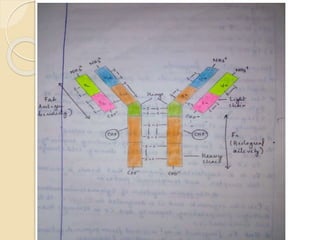
Structure
of
Antibody
Sijo.A
B.sc.Botany &
Biotechnology
Mar ivanios college
Tvm, Kerala
What is Antibody?
 Antibody(Ab) or immunoglobulin(Ig) is the large Y
shaped glycoprotein produced by the body’s immune
system when it detects harmful substances are called
antigens.
 They are synthesized by B lymphocytes and secreted
by plasma cells.
 Depending on the electrophoretic migration, 3 types of
globulins are present in the blood, namely α, β and γ
 So antibodies are gamma (γ) globulin.
Structure of Antibody
 The structure of antibody was discovered by Rodney.R.porter
and Gerald Edelman in 1962.
 All antibodies have a common basic structure.
 Each antibody molecule has 4 polypeptide chains.
 Two small called light chain(L) and two longer called heavy
chain(H). Hence an antibody represented as H2L2.
 Each polypeptide chain contains an (NH3+) amino terminal
and carboxy (COO-) terminal.
 They are linked by disulfide bonds.
 L-chains have a molecular weight of 20-25kd.
 The central region of heavy chain is called hinge region and it
is rich in proline.
 Proline prevents the peptide chains from assuming an alpha-
heix conformation.
Variable & constant region
 L & H chains are further divided into variable &
constant region.
 These regions are composed of 3D folded repeating
segments are called domains. Each domain contains
110 amino acids.
 The variable region concern with antigen binding and
constant region concern with various biological
functions like complement activation and binding to cell
surface receptors.
 The light chain consists of one variable(vL) & one
constant region(cL).
 The heavy chain consists of one variable (vH) & three
constant (cH) regions.
Hypervariability region or
CDR
 The variable region of both L and H contains 3 variable
loops for binding antigens. These antigen binding site is
called CDR (Complementary Determining Region).
 The CDR determines the specificity & affinity of antigen.
 Immunoglobulins are classified on the
basis of amino acid sequence in the constant region of
light chain. They are kappa(k) and lamda(Λ).
 Immunoglobulins are again classified on the basis of
aminoacid sequence in the constant region of heavy
chain. They are:
i) Ig G - γ ii) Ig A - α
iii) Ig M - μ iv) Ig E - ε
v) Ig D - δ
Immunoglobulin fragments
 Immunoglobulin fragments are produced by proteolytic digestion.
 The papain (proteolytic enzyme of papaya) digestion of Ig G
molecules produced by 3 fragments. Out of which 2 fragments
are identical and it has antigen binding capacity. So it is termed
as Fab.
 The antigen binding part of antibody is called Fab.
 The third fragment has no Ag binding capacity and observed that
it crystalize during cold storage. So it termed as Fc.
 Fc is a crystalisable fragment that binds to various cell receptors
and complement proteins.
 The pepsin digestion of Ig G molecules
produced 2 Fab like segments & it is designated as F(ab)’2. it
has antigen precipitating capacity & it is stronger than one
combination.
 But Fc fragment is not recovered from pepsin digestion, because
it digested into multiple fragments.

 THANK YOU…

structureofantibody-170624101143.pdf

  • 1.
  • 2.
    What is Antibody? Antibody(Ab) or immunoglobulin(Ig) is the large Y shaped glycoprotein produced by the body’s immune system when it detects harmful substances are called antigens.  They are synthesized by B lymphocytes and secreted by plasma cells.  Depending on the electrophoretic migration, 3 types of globulins are present in the blood, namely α, β and γ  So antibodies are gamma (γ) globulin.
  • 3.
    Structure of Antibody The structure of antibody was discovered by Rodney.R.porter and Gerald Edelman in 1962.  All antibodies have a common basic structure.  Each antibody molecule has 4 polypeptide chains.  Two small called light chain(L) and two longer called heavy chain(H). Hence an antibody represented as H2L2.  Each polypeptide chain contains an (NH3+) amino terminal and carboxy (COO-) terminal.  They are linked by disulfide bonds.  L-chains have a molecular weight of 20-25kd.  The central region of heavy chain is called hinge region and it is rich in proline.  Proline prevents the peptide chains from assuming an alpha- heix conformation.
  • 4.
    Variable & constantregion  L & H chains are further divided into variable & constant region.  These regions are composed of 3D folded repeating segments are called domains. Each domain contains 110 amino acids.  The variable region concern with antigen binding and constant region concern with various biological functions like complement activation and binding to cell surface receptors.  The light chain consists of one variable(vL) & one constant region(cL).  The heavy chain consists of one variable (vH) & three constant (cH) regions.
  • 5.
    Hypervariability region or CDR The variable region of both L and H contains 3 variable loops for binding antigens. These antigen binding site is called CDR (Complementary Determining Region).  The CDR determines the specificity & affinity of antigen.  Immunoglobulins are classified on the basis of amino acid sequence in the constant region of light chain. They are kappa(k) and lamda(Λ).  Immunoglobulins are again classified on the basis of aminoacid sequence in the constant region of heavy chain. They are: i) Ig G - γ ii) Ig A - α iii) Ig M - μ iv) Ig E - ε v) Ig D - δ
  • 6.
    Immunoglobulin fragments  Immunoglobulinfragments are produced by proteolytic digestion.  The papain (proteolytic enzyme of papaya) digestion of Ig G molecules produced by 3 fragments. Out of which 2 fragments are identical and it has antigen binding capacity. So it is termed as Fab.  The antigen binding part of antibody is called Fab.  The third fragment has no Ag binding capacity and observed that it crystalize during cold storage. So it termed as Fc.  Fc is a crystalisable fragment that binds to various cell receptors and complement proteins.  The pepsin digestion of Ig G molecules produced 2 Fab like segments & it is designated as F(ab)’2. it has antigen precipitating capacity & it is stronger than one combination.  But Fc fragment is not recovered from pepsin digestion, because it digested into multiple fragments. 
  • 8.