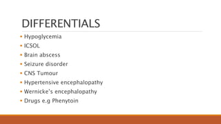
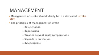
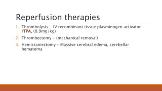
Management of Acute
Stroke
BY
DR. IBRAHIM ADAMU
DEPT. OF INTERNAL MEDICINE
STATE SPECIALIST HOSPITAL, GOMBE
31ST JANUARY, 2023
OUTLINE
 Introduction
 Epidemiology
 Classification
 Risk factors
 Pathophysiology
 Clinical presentation
 Management
 Conclusion
 References
INTRODUCTION
 Cerebrovascular diseases include some of the most common and
devastating disorders
 Major cause of adult disability
 The word “stroke” was first introduced into medicine in 1689 by
William Cole in a ‘physico-medical essay concerning apoplexies’
Apoplexy, from the Greek word meaning “to struck down with
violence," first appeared in Hippocratic Circa 400 BC writings
which describe this phenomenon.
DEFINITION
Stroke
◦ ‘Rapidly developing focal (or global) disturbance of cerebral
function, lasting 24 hours or longer, or leading to death, with no
apparent cause other than of vascular origin’ (WHO 1970)
Transient Ischemic attack (TIA)
◦ ‘Episodes of temporary and focal dysfunction of vascular origin,
which are variable in duration, commonly lasting from 2 to 15
minutes, but occasionally lasting as long as a day (24 hours)
(WHO 1975)
DEFINITION
Stroke : Clinical syndrome of rapidly evolving focal disturbance of
cerebral function, with no apparent cause other than of vascular
origin with an objective neuroimaging evidence of infarcton
irrespective of duration of symptoms.
TIA : a transient episode of neurological dysfunction caused by focal
brain, spinal cord, or retinal ischemia without objective evidence of
acute infarction
The risk of developing a stroke after a hemispheric TIA can be as high as 20% within the first month, with
the greatest risk within the first 48 hours.
EPIDEMIOLOGY
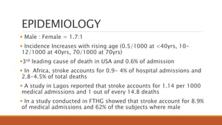
 Common neurological emergency associated with morbidity and
mortality
 Stroke is the 4th most common neurological disorder after
headache, epilepsy and neuropathy
 Someone suffers a stroke every 53 seconds and someone dies
from stroke every 3.3 minutes
 Second leading cause of preventable deaths in adults worldwide.
 15 million cases annually [WHO]
- 5 million deaths
- 5 million left with disability
- 5 million recover
EPIDEMIOLOGY
 Male : Female = 1.7:1
 Incidence Increases with rising age (0.5/1000 at <40yrs, 10-
12/1000 at 40yrs, 70/1000 at 70yrs)
3rd leading cause of death in USA and 0.6% of admission
 In Africa, stroke accounts for 0.9- 4% of hospital admissions and
2.8-4.5% of total deaths
 A study in Lagos reported that stroke accounts for 1.14 per 1000
medical admissions and 1 out of every 14.8 deaths
 In a study conducted in FTHG showed that stroke account for 8.9%
of medical admissions and 62% of the subjects where male
EPIDEMIOLOGY
 Among stroke survivors
- 30% require assistance with activities of daily living
- 20% require assistance with ambulation and
- 16% institutional care.
CLASSIFICATION
Broadly classified into pathologic types
1. Infarctive/Ischemic stroke (80%)
i) Thrombotic (50%)
ii) Embolic (30%)
iii) Small vessel stroke( lacunar)
iv) Hypoperfusion
2. Haemorrhagic Stroke (20%)
i) Intra-cerebral haemorrhage (ICH)-15%
ii) Subarachnoid haemorrhage (SAH)-5%
CLASSIFICATION OF STROKE
CLASSIFICATION
 Anterior Circulation stroke
◦ Total (TACS)
◦ Partial (PACS)
 Posterior Circulation Stroke (POCS)
(OxfordshireCommunityclassification)
RISK FACTORS
Non-modifiable
◦ Age
◦ Gender
◦ Race Black
◦ Prior stroke
◦ Family History
◦ Migraine with aura
RISK FACTORS
Modifiable risk factors
High blood pressure
Cigarette smoking
Transient ischemic attacks
Heart disease
Diabetes mellitus
Hypercoagulopathy
Systemic illnesses e.g CKD, SCDx
Alcohol consumption
Obesity
Sedentary life style
Homocystenaemia
Antiphospholipid syndrome
Drugs e.g OCP, HRT, cocaine, and
steroids
Infectons e.g HIV, CMV, Herpes
simplex
PATHOPHYSIOLOGY
Infarctive Stroke
 Cerebral auto-regulation is lost and cerebral blood flow will
depend on blood pressure
 There is attempt at mobilization of collateral circulation following
occlusion of cerebral blood vessel:
 Inner core of infarct (umbra) has CBF 10 mls/100g/min, this area
is energy depleted with disruption membrane ion transport and
mitochondrial failure leading to release of proteolytic enzymes and
ultimately liquefactive necrosis
 The surrounding core area
(penumbra) has CBF 10-
20mls/100g/min
 The penumbra tissue looses
electrical activity but can be
salvaged if blood flow is
restored during the
therapeutic window period
(3-6hrs)
 Reduction in BP or
dehydration will thus worsen
ischemia
PATHOPHYSIOLOGY
PATHOPHYSIOLOGY
Hemorrhagic stroke
 Chronic hypertension with
charcot-bouchard aneurysms and
berry/saccular aneurysms,
congenital AV malformations,
amyloid angiopathy,
anticoagulant therapy and drugs
all results to rupture of cerebral
vessels
 Bleeding into the surface of the
brain is commonly due to
aneurysm while bleeding into the
tissue of the brain is commonly
caused by HTN
PATHOPHYSIOLOGY
Explosive entry of blood into the brain parenchyma with structural
disruption of neuronal activity by
• Compression of neurons and vessels leading to additional ischemic
damage
• Vasospasm from direct neurotoxicity of blood
• Raised ICP from Cerebral oedema
• Large hemorrhages can cause trans-tentorial coning and rapid death due
to severely elevated intracranial pressure
Predilection sites for bleed includes putaminal (35%), lobar (25%), thalamic (20%), cerebellar (8%),
pontine (7%)
Hemorrhagic
 - Longstanding hypertension with poor compliance
 - Associated with emotional excitement/activity
 - Headache & vomiting
 - Alteration in level of consciousness
 - Seizures
CLINICAL PRESENTATION
Thrombotic
- History of TIA
- Stroke is progressive
- Usually happens in the early morning hours
CLINICAL PRESENTATION
Embolic
- Abrupt with no warning
- Patients with known heart disease like VHDx, AF, IHD etc.
- Maximal deficit at onset
- Rapid recovery
Stroke Syndromes
1. MCA Stroke Syndrome
- Dominant hemisphere
- Non-dominant hemisphere
2. ACA Stroke Syndrome
3. PCA Stroke Syndrome
CLINICAL PRESENTATION
General signs
 Fever
 Hypertension
 Elevated blood sugar
 Precordium- murmurs, cardiac arrhythmias
 Altered level of consciousness or coma
 Neck- carotid bruit, nuchal rigidity
 Eye- retinal hemorrhages
Anisocoria (pupillary dilatation/constriction)
PHYSICAL EXAMINATION
Physical is directed toward 5 major areas:
(1) assessing the airway, breathing, and circulation (ABCs)
(2) defining the severity of the patient's neurologic deficits (level of
consciousness, visual function, motor function, sensation and
neglect, cerebellar function, and language)
(3) identifying potential causes of the stroke
(4) identifying potential stroke mimics
(5) identifying comorbid conditions
DIAGNOSIS
 History
 Exam
 Imaging
◦ CT Scan
◦ MRI
◦ CT/MR Angiography
CT SCANS
 CT is highly sensitive for the diagnosis of haemorrhage in the
acute setting
 Early CT Scan is valuable to make diagnosis and to exclude stroke
mimics
MRI
 MRI is more sensitive than CT for the diagnosis of stroke but
changes are not imminent in the early acute stage
 Although new generation CT scanners may identify subtle
indicators of infarction within six hours of stroke onset in a
significant number of patients
Ancillary investigation
1. Lipid profile
2. Glucose
3. Urea/electrolytes/creatinine
4. Urinalysis
5. ECG /Echocardiography
6. Chest X-ray
7. FBC & Clotting profile
Others: Carotid USS, Homocysteine levels, Cardiac troponins,
Genotype, Viral screen
DIFFERENTIALS
 Hypoglycemia
 ICSOL
 Brain abscess
 Seizure disorder
 CNS Tumour
 Hypertensive encephalopathy
 Wernicke’s encephalopathy
 Drugs e.g Phenytoin
MANAGEMENT
 Management of stroke should ideally be in a dedicated ‘stroke
unit’
 The principles of management of stroke
- Resuscitation
- Reperfusion
- Treat or prevent acute complications
- Secondary prevention
- Rehabilitation
Reperfusion therapies
1. Thrombolysis - IV recombinant tissue plasminogen activator -
rTPA, (0.9mg/kg)
2. Thrombectomy - (mechanical removal)
3. Hemicraniectomy – Massive cerebral edema, cerebellar
hematoma
Treatment of acute complications
Neurological
•Raised ICP
•Seizure
•Hydrocephalus
Non-neurological
CHEST: Aspiration pneumonia, P.E
ENDOCRINE: SIADH, Hyperglycemia, Hypoglycemia
CVS: Arrhythmias, Hypertension
GIT: Constipation
Treatment of acute complications
 Hypertension: Target is <220/120mmHg (MAP <145) in ischemic
stroke and <160/90mmHg (MAP 135) in hemorrhagic stroke.
Presence of end organ damage may require urgent BP reduction
 Studies have shown that use of Aspirin as 300mg in the first 24
hrs improves morbidity. It is given for 2 weeks then tapered
 The use of anti-oxidant has no place now in management of
stroke. Infact Vit-E may worsen hemorrhagic stroke
 The IVF of choice in stroke is N/S
Secondary prevention
 Prevent late complications e.g depression, decubitus ulcer, joint
contractures
 Blood pressure control
◦ Diuretics +/- ACE inhibitors or ARBs
 Diabetes management
 Lipid management
 Smoking cessation
 Alcohol moderation
 Weight reduction
 Anti-platelet agents/Anti-coagulants
Rehabilitation
 The aim is to restore function
- Physiotherapy
- Speech therapy
- Occupational therapy
COMPLICATIONS
 Acute (< 7 days) e.g Cerebral edema, Aspiration pneumonitis,
Hypoglycemia
 Subacute (2wk – 3mo) e.g DVT, UTI, Chest infections, Sores,
Malnutrition
 Chronic ( > 3mo) e.g Contractures, Depression, Paraplegia
PROGNOSIS
The following are associated with poor prognosis
◦ Increased patient age
◦ Raised temperature
◦ Hyperglycemia
◦ Increased blood pressure
◦ Increased stroke severity
◦ Access to specialist care
◦ Availability of stroke facilities
Chances of mortality decreases significantly after the first week
CONCLUSION
 Stroke is a common neurological emergency and a major cause of
adult disability and mortality world-wide
 Despite newer drugs and advances in medical intensive care
technology, the mortality and long-term morbidity rates is still
significantly high
 Incidence varies among different parts of the world and increases
with age
 Risk factors can be modifiable and non-modifiable, with
hypertension and diabetes as one of the most recognized risk
factors especially among the elderly
 Outcome depends on type, time of presentation, facilities available
and presence of co-morbid states
 In poor countries, the problem is compounded by increasing level
of poverty, ignorance and poor drug compliance etc.
REFERENCES
 Lecture notes on cerebrovascular disorders by Dr. Fadimatu Kabir
delivered on 24th February, 2022
 Robert M. Kliegman, Bonita F. Stanton, Joseph W. St. Geme III,
Nina F. Schor, & Richard E. Behrman ‘Update on management of
Stroke’ The New England Journal of Medicine. Article No.
10.1056/NEJMoa223456
 Professor Parveen Kumar, Dr. Micheal Clark MD Textbook of
Clinical Medicine 8th edition
 CT Patterns of Stoke in Adults Patients at FTHG by Dr Yunusa
Dahiru Mohammed
 Website: http://www.emedicine/medscape.com/stroke50763.
Accessed on Friday, 27th March 2023, 3:36pm
Stroke .pptx

Stroke .pptx

  • 1.
    Management of Acute Stroke BY DR.IBRAHIM ADAMU DEPT. OF INTERNAL MEDICINE STATE SPECIALIST HOSPITAL, GOMBE 31ST JANUARY, 2023
  • 2.
    OUTLINE  Introduction  Epidemiology Classification  Risk factors  Pathophysiology  Clinical presentation  Management  Conclusion  References
  • 3.
    INTRODUCTION  Cerebrovascular diseasesinclude some of the most common and devastating disorders  Major cause of adult disability  The word “stroke” was first introduced into medicine in 1689 by William Cole in a ‘physico-medical essay concerning apoplexies’ Apoplexy, from the Greek word meaning “to struck down with violence," first appeared in Hippocratic Circa 400 BC writings which describe this phenomenon.
  • 4.
    DEFINITION Stroke ◦ ‘Rapidly developingfocal (or global) disturbance of cerebral function, lasting 24 hours or longer, or leading to death, with no apparent cause other than of vascular origin’ (WHO 1970) Transient Ischemic attack (TIA) ◦ ‘Episodes of temporary and focal dysfunction of vascular origin, which are variable in duration, commonly lasting from 2 to 15 minutes, but occasionally lasting as long as a day (24 hours) (WHO 1975)
  • 5.
    DEFINITION Stroke : Clinicalsyndrome of rapidly evolving focal disturbance of cerebral function, with no apparent cause other than of vascular origin with an objective neuroimaging evidence of infarcton irrespective of duration of symptoms. TIA : a transient episode of neurological dysfunction caused by focal brain, spinal cord, or retinal ischemia without objective evidence of acute infarction The risk of developing a stroke after a hemispheric TIA can be as high as 20% within the first month, with the greatest risk within the first 48 hours.
  • 6.
    EPIDEMIOLOGY  Common neurologicalemergency associated with morbidity and mortality  Stroke is the 4th most common neurological disorder after headache, epilepsy and neuropathy  Someone suffers a stroke every 53 seconds and someone dies from stroke every 3.3 minutes  Second leading cause of preventable deaths in adults worldwide.  15 million cases annually [WHO] - 5 million deaths - 5 million left with disability - 5 million recover
  • 7.
    EPIDEMIOLOGY  Male :Female = 1.7:1  Incidence Increases with rising age (0.5/1000 at <40yrs, 10- 12/1000 at 40yrs, 70/1000 at 70yrs) 3rd leading cause of death in USA and 0.6% of admission  In Africa, stroke accounts for 0.9- 4% of hospital admissions and 2.8-4.5% of total deaths  A study in Lagos reported that stroke accounts for 1.14 per 1000 medical admissions and 1 out of every 14.8 deaths  In a study conducted in FTHG showed that stroke account for 8.9% of medical admissions and 62% of the subjects where male
  • 8.
    EPIDEMIOLOGY  Among strokesurvivors - 30% require assistance with activities of daily living - 20% require assistance with ambulation and - 16% institutional care.
  • 9.
    CLASSIFICATION Broadly classified intopathologic types 1. Infarctive/Ischemic stroke (80%) i) Thrombotic (50%) ii) Embolic (30%) iii) Small vessel stroke( lacunar) iv) Hypoperfusion 2. Haemorrhagic Stroke (20%) i) Intra-cerebral haemorrhage (ICH)-15% ii) Subarachnoid haemorrhage (SAH)-5%
  • 10.
  • 11.
    CLASSIFICATION  Anterior Circulationstroke ◦ Total (TACS) ◦ Partial (PACS)  Posterior Circulation Stroke (POCS) (OxfordshireCommunityclassification)
  • 12.
    RISK FACTORS Non-modifiable ◦ Age ◦Gender ◦ Race Black ◦ Prior stroke ◦ Family History ◦ Migraine with aura
  • 13.
    RISK FACTORS Modifiable riskfactors High blood pressure Cigarette smoking Transient ischemic attacks Heart disease Diabetes mellitus Hypercoagulopathy Systemic illnesses e.g CKD, SCDx Alcohol consumption Obesity Sedentary life style Homocystenaemia Antiphospholipid syndrome Drugs e.g OCP, HRT, cocaine, and steroids Infectons e.g HIV, CMV, Herpes simplex
  • 14.
    PATHOPHYSIOLOGY Infarctive Stroke  Cerebralauto-regulation is lost and cerebral blood flow will depend on blood pressure  There is attempt at mobilization of collateral circulation following occlusion of cerebral blood vessel:  Inner core of infarct (umbra) has CBF 10 mls/100g/min, this area is energy depleted with disruption membrane ion transport and mitochondrial failure leading to release of proteolytic enzymes and ultimately liquefactive necrosis
  • 15.
     The surroundingcore area (penumbra) has CBF 10- 20mls/100g/min  The penumbra tissue looses electrical activity but can be salvaged if blood flow is restored during the therapeutic window period (3-6hrs)  Reduction in BP or dehydration will thus worsen ischemia PATHOPHYSIOLOGY
  • 16.
    PATHOPHYSIOLOGY Hemorrhagic stroke  Chronichypertension with charcot-bouchard aneurysms and berry/saccular aneurysms, congenital AV malformations, amyloid angiopathy, anticoagulant therapy and drugs all results to rupture of cerebral vessels  Bleeding into the surface of the brain is commonly due to aneurysm while bleeding into the tissue of the brain is commonly caused by HTN
  • 17.
    PATHOPHYSIOLOGY Explosive entry ofblood into the brain parenchyma with structural disruption of neuronal activity by • Compression of neurons and vessels leading to additional ischemic damage • Vasospasm from direct neurotoxicity of blood • Raised ICP from Cerebral oedema • Large hemorrhages can cause trans-tentorial coning and rapid death due to severely elevated intracranial pressure Predilection sites for bleed includes putaminal (35%), lobar (25%), thalamic (20%), cerebellar (8%), pontine (7%)
  • 18.
    Hemorrhagic  - Longstandinghypertension with poor compliance  - Associated with emotional excitement/activity  - Headache & vomiting  - Alteration in level of consciousness  - Seizures CLINICAL PRESENTATION
  • 19.
    CLINICAL PRESENTATION Thrombotic - Historyof TIA - Stroke is progressive - Usually happens in the early morning hours
  • 20.
    CLINICAL PRESENTATION Embolic - Abruptwith no warning - Patients with known heart disease like VHDx, AF, IHD etc. - Maximal deficit at onset - Rapid recovery
  • 21.
    Stroke Syndromes 1. MCAStroke Syndrome - Dominant hemisphere - Non-dominant hemisphere 2. ACA Stroke Syndrome 3. PCA Stroke Syndrome
  • 22.
    CLINICAL PRESENTATION General signs Fever  Hypertension  Elevated blood sugar  Precordium- murmurs, cardiac arrhythmias  Altered level of consciousness or coma  Neck- carotid bruit, nuchal rigidity  Eye- retinal hemorrhages Anisocoria (pupillary dilatation/constriction)
  • 23.
    PHYSICAL EXAMINATION Physical isdirected toward 5 major areas: (1) assessing the airway, breathing, and circulation (ABCs) (2) defining the severity of the patient's neurologic deficits (level of consciousness, visual function, motor function, sensation and neglect, cerebellar function, and language) (3) identifying potential causes of the stroke (4) identifying potential stroke mimics (5) identifying comorbid conditions
  • 24.
    DIAGNOSIS  History  Exam Imaging ◦ CT Scan ◦ MRI ◦ CT/MR Angiography
  • 25.
    CT SCANS  CTis highly sensitive for the diagnosis of haemorrhage in the acute setting  Early CT Scan is valuable to make diagnosis and to exclude stroke mimics
  • 26.
    MRI  MRI ismore sensitive than CT for the diagnosis of stroke but changes are not imminent in the early acute stage  Although new generation CT scanners may identify subtle indicators of infarction within six hours of stroke onset in a significant number of patients
  • 27.
    Ancillary investigation 1. Lipidprofile 2. Glucose 3. Urea/electrolytes/creatinine 4. Urinalysis 5. ECG /Echocardiography 6. Chest X-ray 7. FBC & Clotting profile Others: Carotid USS, Homocysteine levels, Cardiac troponins, Genotype, Viral screen
  • 28.
    DIFFERENTIALS  Hypoglycemia  ICSOL Brain abscess  Seizure disorder  CNS Tumour  Hypertensive encephalopathy  Wernicke’s encephalopathy  Drugs e.g Phenytoin
  • 29.
    MANAGEMENT  Management ofstroke should ideally be in a dedicated ‘stroke unit’  The principles of management of stroke - Resuscitation - Reperfusion - Treat or prevent acute complications - Secondary prevention - Rehabilitation
  • 30.
    Reperfusion therapies 1. Thrombolysis- IV recombinant tissue plasminogen activator - rTPA, (0.9mg/kg) 2. Thrombectomy - (mechanical removal) 3. Hemicraniectomy – Massive cerebral edema, cerebellar hematoma
  • 31.
    Treatment of acutecomplications Neurological •Raised ICP •Seizure •Hydrocephalus Non-neurological CHEST: Aspiration pneumonia, P.E ENDOCRINE: SIADH, Hyperglycemia, Hypoglycemia CVS: Arrhythmias, Hypertension GIT: Constipation
  • 32.
    Treatment of acutecomplications  Hypertension: Target is <220/120mmHg (MAP <145) in ischemic stroke and <160/90mmHg (MAP 135) in hemorrhagic stroke. Presence of end organ damage may require urgent BP reduction  Studies have shown that use of Aspirin as 300mg in the first 24 hrs improves morbidity. It is given for 2 weeks then tapered  The use of anti-oxidant has no place now in management of stroke. Infact Vit-E may worsen hemorrhagic stroke  The IVF of choice in stroke is N/S
  • 33.
    Secondary prevention  Preventlate complications e.g depression, decubitus ulcer, joint contractures  Blood pressure control ◦ Diuretics +/- ACE inhibitors or ARBs  Diabetes management  Lipid management  Smoking cessation  Alcohol moderation  Weight reduction  Anti-platelet agents/Anti-coagulants
  • 34.
    Rehabilitation  The aimis to restore function - Physiotherapy - Speech therapy - Occupational therapy
  • 35.
    COMPLICATIONS  Acute (<7 days) e.g Cerebral edema, Aspiration pneumonitis, Hypoglycemia  Subacute (2wk – 3mo) e.g DVT, UTI, Chest infections, Sores, Malnutrition  Chronic ( > 3mo) e.g Contractures, Depression, Paraplegia
  • 36.
    PROGNOSIS The following areassociated with poor prognosis ◦ Increased patient age ◦ Raised temperature ◦ Hyperglycemia ◦ Increased blood pressure ◦ Increased stroke severity ◦ Access to specialist care ◦ Availability of stroke facilities Chances of mortality decreases significantly after the first week
  • 37.
    CONCLUSION  Stroke isa common neurological emergency and a major cause of adult disability and mortality world-wide  Despite newer drugs and advances in medical intensive care technology, the mortality and long-term morbidity rates is still significantly high  Incidence varies among different parts of the world and increases with age  Risk factors can be modifiable and non-modifiable, with hypertension and diabetes as one of the most recognized risk factors especially among the elderly  Outcome depends on type, time of presentation, facilities available and presence of co-morbid states  In poor countries, the problem is compounded by increasing level of poverty, ignorance and poor drug compliance etc.
  • 38.
    REFERENCES  Lecture noteson cerebrovascular disorders by Dr. Fadimatu Kabir delivered on 24th February, 2022  Robert M. Kliegman, Bonita F. Stanton, Joseph W. St. Geme III, Nina F. Schor, & Richard E. Behrman ‘Update on management of Stroke’ The New England Journal of Medicine. Article No. 10.1056/NEJMoa223456  Professor Parveen Kumar, Dr. Micheal Clark MD Textbook of Clinical Medicine 8th edition  CT Patterns of Stoke in Adults Patients at FTHG by Dr Yunusa Dahiru Mohammed  Website: http://www.emedicine/medscape.com/stroke50763. Accessed on Friday, 27th March 2023, 3:36pm

Editor's Notes

  • #5 24 hrs time based: obsolete, confusing; misleading as permanent injury may occur sooner
  • #10 Emboli may either be of cardiac or arterial origin. Cardiac sources include Atrial fibrillation, Recent myocardial infarction Prosthetic valves
  • #13 Age >65 yrs Gender M > F Race Black > asians> whites
  • #26 Hyperdense middle cerebral artery and midline deviation due to tissue swelling is the tale tale sign of ischemic stroke Hypodense with ventricular extension for hemorrhagic
  • #31 Criteria for thrombolysis: 3 hrs from onset ICH excluded by imaging SBP < 185; DBP < 110 mm Hg Platelets > 100,000 Pt not on anticoagulants No recent surgery or GI bleeding No seizures at onset
  • #32 Management of raised ICP: Mannitol and laxis, Nurse in 30, Mechanical thrombectomy, Hemicraniectomy, Hyperventillation, Selective head cooling, Dexamethasone