Download as PDF, PPTX


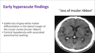


The document discusses types of strokes, focusing on ischemic (85%) and hemorrhagic (15%), and outlines the role of non-contrast CT scans as the first imaging modality for acute ischemic stroke evaluation. It details the stages of stroke progression, findings in various time frames, and emphasizes the importance of CT in excluding hemorrhage and diagnosing strokes. Additionally, the document describes increased intracranial pressure, its symptoms, and potential treatments.


