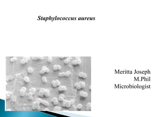
Staphylococcus aureus
•Download as PPTX, PDF•
0 likes•177 views

Breif discussion about the organism and food through which the outbreaks have occured. It is also added with Bacteriological Analytical Methods (BAM) for the isolation and enumeration of the organism from the food sample.

Recommended
More Related Content
What's hot
What's hot (20)
Similar to Staphylococcus aureus
Similar to Staphylococcus aureus (20)
Recently uploaded
Recently uploaded (20)
Staphylococcus aureus
- 2. First described by Scottish surgeon Sir. Alexander Ogston Causes pyogenic infection in human Greek: staphyle, bunch of grapes; coccus, a grain or berry Gram positive coccus Spherical to ovoid cells of 1 micrometer diameter. Catalase positive, Oxidase negative and glucose fermentation
- 3. Mesophile with a growth temperature range between 7 and 48ºC and an optimum at 37ºC Enterotoxin is produced at an optimum of 35-40ºC. Production of enterotoxin is adversely affected by anaerobic conditions. Growth occurs at pH 6 -7 with minimum and maximum limits of 4.0 and 9.8 – 10. Readily grows in media containing 5 – 7% NaCl and some capable of growth in 20%. It can grow down to an water activity of 0.83
- 5. First outbreak of illness through cheese in Michigan. Barber (1914) demonstrated staphylococci were able to cause poisoning in milk from cow with staphylococcal mastitis. Dack (1930) showed staphylococcal food poisoning by a filterable enterotoxin. The infections are relatively mild and short lived type of infections. Enterotoxin toxin producing strains include S.aureus, S.intermedius and S.hycius
- 6. Characterized by short incubation period 2 – 4h. Nausea, vomiting, stomach cramps, retching and prostration are the predominant symptoms, diarrhoea is also often. Severe cases dehydration, marked pallor and collapse may require treatment by intravenous infusion. S.aureus produces 11 enterotoxins (SEA to SEJ). There are three variants of SEC. When illness is of short incubation period, it is due to ingestion of a pre-formed toxin in the food. Frequently implicated in outbreaks of food, toxins A and D
- 8. Naturally occur in poultry and other raw meat as skin microflora. Isolated from raw milk and milk products as enterotoxin can survive pasteurization Contamination by food handlers. Contaminated from infected skin lesions or by coughing and sneezing. Poultry products and cold, cooked meats. Salted meats such as ham and corned beef, Canned foods, hard cheeses, cold sweets, custards and cream-filled bakery products
- 10. Media and Reagents Baird-Parker medium Trypticase (tryptic) soy agar (TSA) Brain heart infusion (BHI) broth Coagulase plasma (rabbit) with EDTA Toluidine blue-DNA agar Lysostaphin (Schwartz-Mann, Mountain View Ave., Orangeburg, NY 10962) Tryptone yeast extract agar Paraffin oil, sterile 0.02 M phosphate-saline buffer , containing 1% NaCl Catalase test
- 11. Serial diution of the sample to be analysed Aseptically transfer 1 ml sample suspension to 3 plates of Baird-Parker agar, distributing 1 ml of inoculum equitably to 3 plates. Select plates containing 20-200 colonies, unless only plates at lower dilutions (>200 colonies) have colonies with typical appearance of S. aureus. Count and record colonies. When plates of the lowest dilution contain <20 colonies, these may be used. Plates containing >200 colonies have colonies with the typical appearance of S. aureus and typical colonies do not appear at higher dilutions, use these plates for the enumeration of S. aureus, but do not count nontypical colonies.
- 12. COLONY MORPHOLOGY Colonies of S. aureus are circular, smooth, convex, moist, 2-3 mm in diameter on uncrowded plates, gray to jet-black, frequently with light-colored (off-white) margin, surrounded by opaque zone and frequently with an outer clear zone; colonies have buttery to gummy consistency when touched with inoculating needle.
- 13. Select > 1 colony of each type counted and test for coagulase production. Add number of colonies on triplicate plates represented by colonies giving positive coagulase test and multiply by the sample dilution factor. Report this number as number of S. aureus/g of food tested.
- 14. Transfer suspect S. aureus colonies into small tubes containing 0.2-0.3 ml BHI broth and emulsify thoroughly Inoculate agar slant of suitable maintenance medium, e.g., TSA, with loopful of BHI suspension. Incubate BHI culture suspension and slants 18-24 h at 35-37°C. Add 0.5 ml reconstituted coagulase plasma with EDTA (B-4, above) to the BHI culture and mix thoroughly. Incubate at 35-37°C and examine periodically over 6 h period for clot formation
- 15. Only firm and complete clot that stays in place when tube is tilted or inverted is considered positive for S. aureus. Test known positive and negative cultures simultaneously with suspect cultures of unknown coagulase activity. Stain all suspect cultures with Gram reagent and observe microscopically. A latex agglutination test (AUREUS TESTTM, Trisum Corp., Taipei, Taiwan) may be substituted for the coagulase test if a more rapid procedure is desired.
- 18. Catalase test :
- 19. Inoculate tube of carbohydrate fermentation medium containing glucose (0.5%). Immediately inoculate each tube heavily with wire loop. Make certain inoculum reaches bottom of tube. Cover surface of agar with layer of sterile paraffin oil at least 25 mm thick. Incubate 5 days at 35-37°C. Acid is produced anaerobically if indicator changes to yellow throughout tube, indicating presence of S. aureus. Run controls simultaneously (positive and negative cultures and medium controls).
- 21. Repeat 2, above, using mannitol as carbohydrate in medium. S. aureus is usually positive but some strains are negative. Run controls simultaneously. Anaerobic utilization of mannitol.
- 22. Transfer isolated colony from agar plate with inoculating loop to 0.2 ml phosphate-saline buffer, and emulsify. Transfer half of suspended cells to another tube (13 × 100 mm) and mix with 0.1 ml phosphate-saline buffer as control. Add 0.1 ml lysostaphin (dissolved in 0.02 M phosphate- saline buffer containing 1% NaCl) to original tube for concentration of 25 µg lysostaphin/ml. Incubate both tubes at 35-37°C for not more than 2 h. If turbidity clears in test mixture, test is considered positive. If clearing has not occurred in 2 h, test is negative. S. aureus is generally positive. Lysostaphin sensitivity.