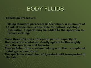
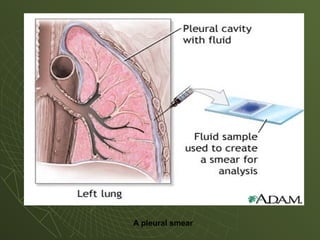
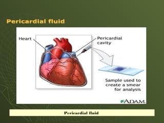
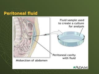
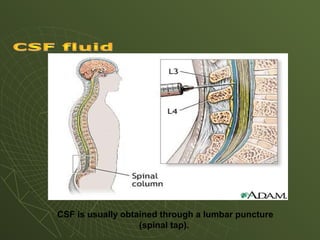
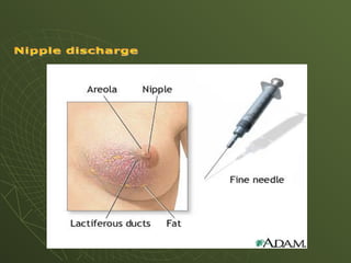
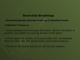
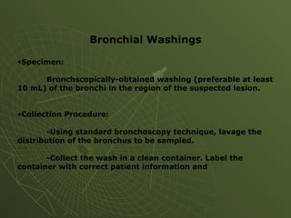
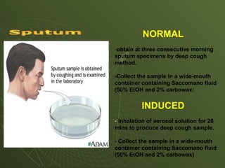
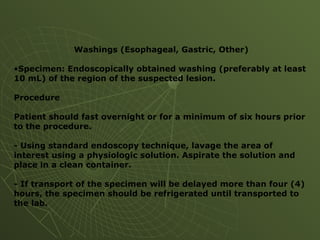
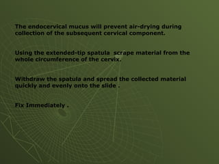
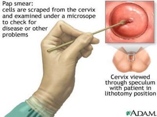
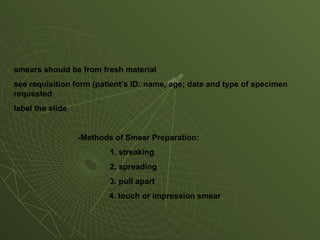
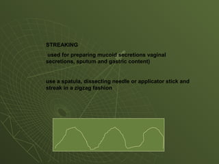
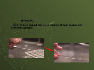
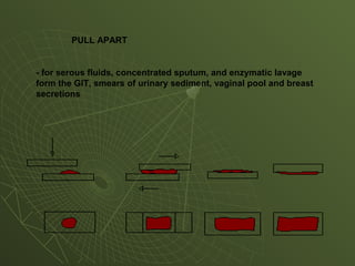
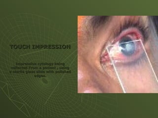
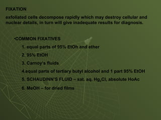
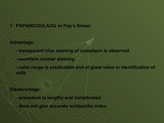
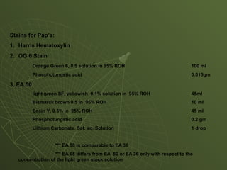
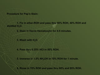
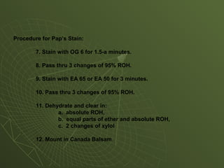
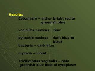
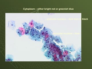
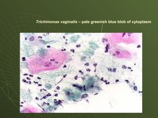
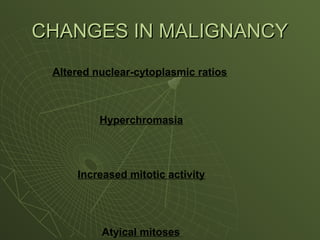
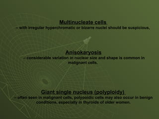
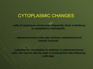
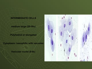
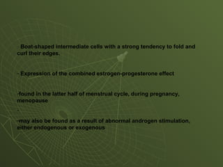
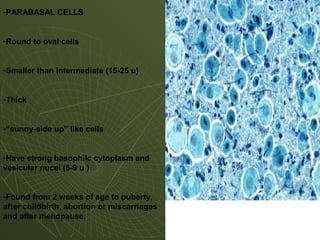
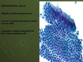
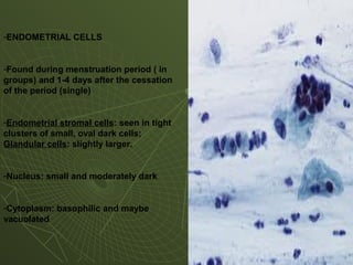
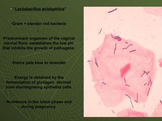
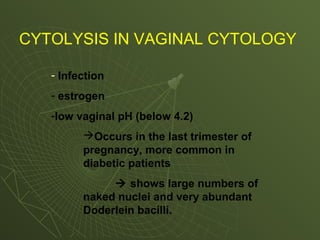
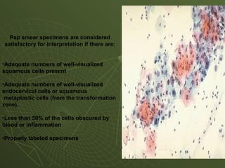
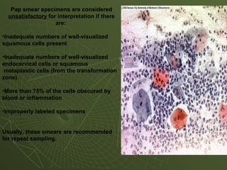
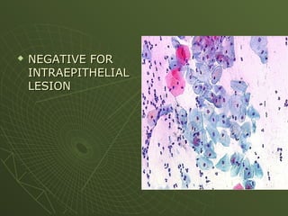
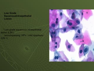
2. a branch of Cytology which deals wit the microscopic study of cells that
have been desquamated from the epithelial surfaces.
recommended for :
Detection of malignant cells or precancerous lesions in the body
Detection of asymptomatic or precancerous cervical lesions in women
Assessment of female hormonal status in case of sterility and endocrine
disorders
Determination of genetic (phenotypic) sex
Detection of the presence of infectious microorganisms
4. • Collection Procedure:
- Using standard paracentesis technique. A minimum of
10 mL of specimen is desirable for optimal cytologic
evaluation. Heparin may be added to the specimen to
reduce clotting.
- Place three (3) units of heparin per mL capacity of
the collection container. Gently agitate to thoroughly
mix the specimen and heparin.
- Always Submit the specimen along with the completed
cytology request form.
- The specimen should be refrigerated until transported to
the lab.
BODY FLUIDS
BODY FLUIDS
8. CSF is usually obtained through a lumbar puncture
(spinal tap).
9.
10. • Gently strip the sub-areolar area and nipple with the thumb and
forefinger.
• Place the slide upon the nipple and draw it quickly across the
nipple. ***If 1 drop is obtained, get another slide and do the pull-
apart technique.
• Immediately immersed the slide into a bottle of 95% isopropyl
ROH or use spray fixative.
NIPPLE DISCHARGE
NIPPLE DISCHARGE
12. - Bronchoscopically-directed brushing of identified lesion.
•Collection Procedure:
- Using standard bronchoscopy technique, identify the lesion in
question and obtain a brushing sample of the lesion.
- Gently apply the sample on to glass slide and immediately
immerse slide into 3% glacial acetic acid alcohol fixative.
- The brush tip should also be cut off into the solution.
Bronchial Brushings
13. Bronchial Washings
•Specimen:
Bronchscopically-obtained washing (preferable at least
10 mL) of the bronchi in the region of the suspected lesion.
•Collection Procedure:
-Using standard bronchoscopy technique, lavage the
distribution of the bronchus to be sampled.
-Collect the wash in a clean container. Label the
container with correct patient information and
14. NORMAL
-obtain at three consecutive morning
sputum specimens by deep cough
method.
-Collect the sample in a wide-mouth
container containing Saccomano fluid
(50% EtOH and 2% carbowax)
INDUCED
- Inhalation of aerosol solution for 20
mins to produce deep cough sample.
- Collect the sample in a wide-mouth
container containing Saccomano fluid
(50% EtOH and 2% carbowax)
15.
16. Washings (Esophageal, Gastric, Other)
•Specimen: Endoscopically obtained washing (preferably at least
10 mL) of the region of the suspected lesion.
Procedure
Patient should fast overnight or for a minimum of six hours prior
to the procedure.
- Using standard endoscopy technique, lavage the area of
interest using a physiologic solution. Aspirate the solution and
place in a clean container.
- If transport of the specimen will be delayed more than four (4)
hours, the specimen should be refrigerated until transported to
the lab.
17. The endocervical mucus will prevent air-drying during
collection of the subsequent cervical component.
Using the extended-tip spatula scrape material from the
whole circumference of the cervix.
Withdraw the spatula and spread the collected material
quickly and evenly onto the slide .
Fix Immediately .
18.
19. smears should be from fresh material
see requisition form (patient’s ID: name, age; date and type of specimen
requested
label the slide
-Methods of Smear Preparation:
1. streaking
2. spreading
3. pull apart
4. touch or impression smear
20. STREAKING
used for preparing mucoid secretions vaginal
secretions, sputum and gastric content)
use a spatula, dissecting needle or applicator stick and
streak in a zigzag fashion
21. SPREADING
- used for thick mucoid secretions (smears of fresh sputum and
bronchial aspirates)
22. PULL APART
- for serous fluids, concentrated sputum, and enzymatic lavage
form the GIT, smears of urinary sediment, vaginal pool and breast
secretions
23. TOUCH IMPRESSION
TOUCH IMPRESSION
Impression cytology being
collected From a patient , using
a sterile glass slide with polished
edges.
edges.
24. FIXATION
exfoliated cells decompose rapidly which may destroy cellular and
nuclear details, in turn will give inadequate results for diagnosis.
•COMMON FIXATIVES
1. equal parts of 95% EtOh and ether
2. 95% EtOH
3. Carnoy’s fluids
4.equal parts of tertiary butyl alcohol and 1 part 95% EtOH
5. SCHAUDINN’S FLUID – sat. aq. Hg2Cl, absolute HoAc
6. MeOH – for dried films
25. 1. PAPANICOULAOU or Pap’s Smear
Advantage:
- transparent blue staining of cytoplasm is observed
- excellent nuclear staining
- color range is predictable and of great value in identification of
cells
Disadvantage:
- procedure is lengthy and complicated
- does not give accurate acidophilic index
26. Stains for Pap’s:
1. Harris Hematoxylin
2. OG 6 Stain
Orange Green 6, 0.5 solution in 95% ROH 100 ml
Phosphotungstic acid 0.015gm
3. EA 50
light green SF, yellowish 0.1% solution in 95% ROH 45ml
Bismarck brown 0.5 in 95% ROH 10 ml
Eosin Y, 0.5% in 95% ROH 45 ml
Phosphotungstic acid 0.2 gm
Lithium Carbonate. Sat. aq. Solution 1 drop
*** EA 50 is comparable to EA 36
*** EA 65 differs from EA 50 or EA 36 only with respect to the
concentration of the light green stock solution
27. Procedure for Pap’s Stain:
1. Fix in ether-ROH and pass thru 80% ROH, 40% ROH and
distilled H2O.
2. Stain in Harris Hematoxylin for 4-5 minutes.
3. Wash with H2O.
4. Pass thru 0.25% HCl in 50% ROH.
5. Immerse in 1.5% NH4OH in 70% ROH for 1 minute.
6. Rinse in 70% ROH and pass thru 80% and 95% ROH.
28. Procedure for Pap’s Stain:
7. Stain with OG 6 for 1.5-a minutes.
8. Pass thru 3 changes of 95% ROH.
9. Stain with EA 65 or EA 50 for 3 minutes.
10. Pass thru 3 changes of 95% ROH.
11. Dehydrate and clear in:
a. absolute ROH,
b. equal parts of ether and absolute ROH,
c. 2 changes of xylol
12. Mount in Canada Balsam.
29. Results:
Cytoplasm – either bright red or
greenish blue
vesicular nucleus – blue
pyknotic nucleus – dark blue to
black
bacteria – dark blue
mycelia – violet
Trichimonas vaginalis – pale
greenish blue blob of cytoplasm
30. Cytoplasm – either bright red or greenish blue
vesicular nucleus – blue
pyknotic nucleus – dark blue to black
33. Multinucleate cells
– with irregular hyperchromatic or bizarre nuclei should be suspicious,
Anisokaryosis
– considerable variation in nuclear size and shape is common in
malignant cells.
Giant single nucleus (polyploidy)
– often seen in malignant cells, polypoidic cells may also occur in benign
conditions, especially in thyroids of older women.
34. CYTOPLASMIC CHANGES
- cells of squamous carcinomas frequently show a tendency
to cytoplasmic eosinophila.
- adenocarcinoma cells may enclose, endometrial and
colonic cancers)
- cytoplasmic vacuolation is common in adenocarcinoma
cells, but may be also be seen in endometrial cells following
cutterage.
35. In general:
- malignant cells show reduced cohesiveness, possibly related to a
defect of the intercellular “zippers” i.e., desmosomes.
-Cancer cells are larger than their normal counterparts and
frequently show bizarre and grotesque shape.
Occasionally:
- exfoliated cells from epithelial tumors assume a greatly
elongated, fibrocyte-like appearance.
36. CELL PATTERN
•Examine for the small groups or clusters of cells
-Oblivious patterns
e.g. acini in adenocacinoma arising in glandular tissues
-Rosettes in neuroblastomas and ependymoma
-Stratification in squamous epithelial growths
-Whorls in mesotheliomas
If there are no patterns focus up and down the on cell
clumps
-In strips epithelium, irregular stratification of anisokaryotic,
hyperchromatic cells are helpful in diagnosing carcinoma of
cervix and bronchus.
37. OTHER CRITERIA
1. In bronchial secretion smear, abundant lymphocytes
are common in presence of malignancy, but may also
be found in certain inflammatory conditions and in
leukaemia.
2. The presence of old blood and blood pigments is a
minor indirect clue, but has many other etiologies.
38. VAGINAL CYTOLOGY
Vaginal cytology is a type of endocrine assay. Tracking changes in
the morphology of desquamated vaginal epithelial cells provides a
convenient means of assaying changes in estrogen levels.
39. •Vaginal smears may be taken regularly and often.
•Hormonal changes are best mirrored in the upper third of
the vagina.
•They can also be taken from the lateral walls because their
more accessible and less likely to be contaminated by
cellular debris or discharge.
42. Anucleate cells are abnormal which may be derived from:
1. smear contamination by the cells from the vulva
2. epidermization of the vagina or cervix resulting from
prolapse
3. leukoplakia of the cervix
4. ruptured membranes in pregnant women
5. marked hyper-estrinism
43. INTERMEDIATE CELLS
-
medium large (20-30u)
-
Polyhedral or elongated
-
Cytoplasm: basophilic with vacuoles
-
Vesicular nuclei (6-9u)
44. - Boat-shaped intermediate cells with a strong tendency to fold and
curl their edges.
- Expression of the combined estrogen-progesterone effect
-found in the latter half of menstrual cycle, during pregnancy,
menopause
-
-may also be found as a result of abnormal androgen stimulation,
either endogenous or exogenous
45. -PARABASAL CELLS
-Round to oval cells
-Smaller than intermediate (15-25 u)
-Thick
-“sunny-side up” like cells
-Have strong basophilc cytoplasm and
vesicular nucei (6-9 u )
-Found from 2 weeks of age to puberty,
after childbirth, abortion or miscarriages
and after menopause.
46. -ENDOCERVICAL CELLS
-Slightly cylindrical appearance
-occurs in groups and strips of three
or more cells
-cytoplasm: deeply basophilic the
that of the parabasal cells
47. -ENDOMETRIAL CELLS
-Found during menstruation period ( in
groups) and 1-4 days after the cessation
of the period (single)
-Endometrial stromal cells: seen in tight
clusters of small, oval dark cells;
Glandular cells: slightly larger.
-Nucleus: small and moderately dark
-Cytoplasm: basophilic and maybe
vacuolated
48. - “Lactobacillus acidophilus”
-Gram + slender rod bacteria
-
-Predominant organism of the vaginal
normal flora: establishes the low pH
that inhibits the growth of pathogens
-Stains pale blue to lavender
-Energy is obtained by the
fermentation of glycogen derived
from disintegrating epithelial cells
-Numerous in the luteal phase and
during pregnancy
49. CYTOLYSIS IN VAGINAL CYTOLOGY
- Infection
- estrogen
-low vaginal pH (below 4.2)
Occurs in the last trimester of
pregnancy, more common in
diabetic patients
shows large numbers of
naked nuclei and very abundant
Doderlein bacilli.
50. The BETHESDA SYSTEM
Specimen Adequacy
Satisfactory
Limited
Unsatisfactory
General Categorization:
Negative for Intraepithelial lesion or malignant cell
Epithelial cell abnormality
Descriptive Diagnosis:
Atypical squamous cells of unknown significance
Low grade squamous intraepithelial lesion
High grade squamous intraepithelial lesion
Squamous Cell Carcinoma
Glandular cell abnormality
Atypical glandular cells
Adenocarcinoma
Others
51. Pap smear specimens are considered
satisfactory for interpretation if there are:
•Adequate numbers of well-visualized
squamous cells present
•Adequate numbers of well-visualized
endocervical cells or squamous
metaplastic cells (from the transformation
zone).
•Less than 50% of the cells obscured by
blood or inflammation
•Properly labeled specimens
Specimen Satisfactory
52. Pap smear specimens are considered
unsatisfactory for interpretation if there
are:
•Inadequate numbers of well-visualized
squamous cells present
•Inadequate numbers of well-visualized
endocervical cells or squamous
metaplastic cells (from the transformation
zone).
•More than 75% of the cells obscured by
blood or inflammation
•Improperly labeled specimens
Usually, these smears are recommended
for repeat sampling.
53. Specimens subjected to rejection
1. Specimen is submitted without a requisition.
2. Specimen is not labeled with the patient name.
3. The patient name (or other identifying information) on the
specimen and requisition do not correspond.
4. The specimen is labeled appropriately but the requisition is not
labeled.
5. The specimen slide(s) is (are) irreparably broken.
6. Specimen is submitted from an unauthorized source.
57. •High grade squamous intraepithelial
lesion (HSIL)
(encompassing: moderate and severe
dysplasia, CIS, CIN 2
and CIN 3)
•- with features suspicious for invasion (if
invasion
is suspected)
Severe Dysplasia or CIS