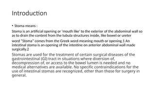
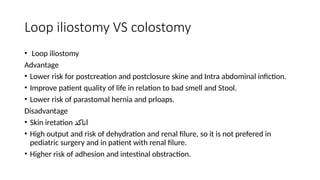
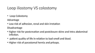
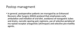
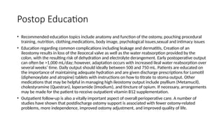
The document discusses various aspects of stomas, including their definitions, types, surgical indications, and management principles, particularly in relation to bowel trauma. It outlines different stoma types such as end stomas, loop stomas, and colostomies, as well as their advantages and disadvantages in clinical settings. Additionally, it emphasizes postoperative care, education for patients, and complications associated with ostomy procedures.


![For an end stoma (see the images below), the bowel is divided, and the proximal end is brought
through the abdominal wall. The distal nonfunctioning limb can be brought out through the same
abdominal wall opening as the end stoma (ie, double-barrel stoma), it can be brought out through a
separate incision (ie, mucous fistula), or it can be closed and left in the peritoneal cavity (ie,
Hartmann procedure).
• A loop stoma is created by maturing a segment of bowel over a rod or tube without completely
dividing the bowel. Loop stomas provide excellent decompression and have the advantage of
simple closure without the need for a separate laparotomy in most cases. However, loop stomas are
not completely diverting, because proximal contents can spill over into the distal limb. Therefore,
they should be used with caution in patients in whom stool in the distal bowel may be problematic.
• A decompressing stoma, or blowhole, is created in patients in unstable condition by opening the
antimesenteric border of bowel without mobilizing the entire loop of bowel.
• Stomas can also be formed in association with an anastomosis for proximal or distal venting or
irrigation (ie, Bishop-Koop [12]
and Santulli stomas; see the image below). These stomas were initially
designed for the treatment of infants with meconium ileus but have been adapted for many other
purposes.](https://image.slidesharecdn.com/stomapresentation-241125150001-da5e2dee/85/Stoma-in-abdomen-types-and-procedure-complication-Presentation-3-320.jpg)

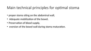

![• The stoma is distant from the incision, through the midportion of
the rectus muscle away from skin folds (eg, groin, flank), bony
prominences (eg, rib cage, iliac spine), and umbilicus (see the image
below).
• Stoma location in infants and neonates (see the image below)
follows these same principles whenever possible; however, the small
size of the abdominal wall in infants and the short mesentery of the
bowel chosen for the stoma often limit the options. For temporary
stomas in infants, the bowel can be brought out directly through or
adjacent to the umbilicus (see the images below). [13]
This site is
easier for appliance placement and results in a cosmetically superior
scar when the stoma is ultimately closed.](https://image.slidesharecdn.com/stomapresentation-241125150001-da5e2dee/85/Stoma-in-abdomen-types-and-procedure-complication-Presentation-11-320.jpg)


![Basics of Stoma and Management care[1].pptx](https://cdn.slidesharecdn.com/ss_thumbnails/basicsofstomaandmanagement1-250417022629-8799f789-thumbnail.jpg?width=640&height=640&fit=bounds)


![ONFH[AVN HIP] -TRIPLE REGIME -A NOVAL SURGICAL CONCEPT .pptx](https://cdn.slidesharecdn.com/ss_thumbnails/onfhavnhip2026koaconcalicutdrgokuldevdrmashraf-260210064517-213ec005-thumbnail.jpg?width=640&height=640&fit=bounds)


