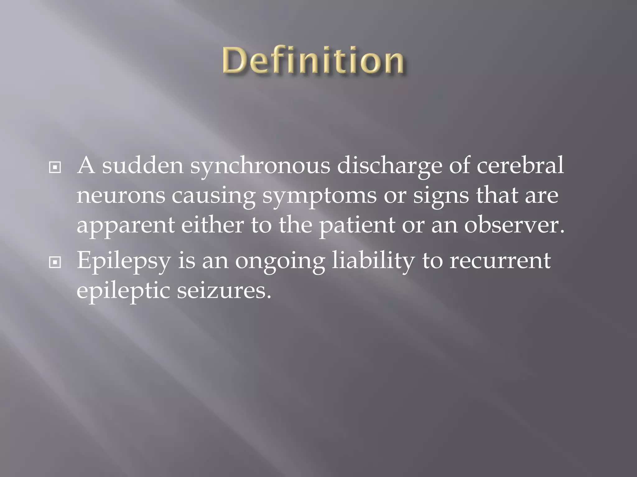
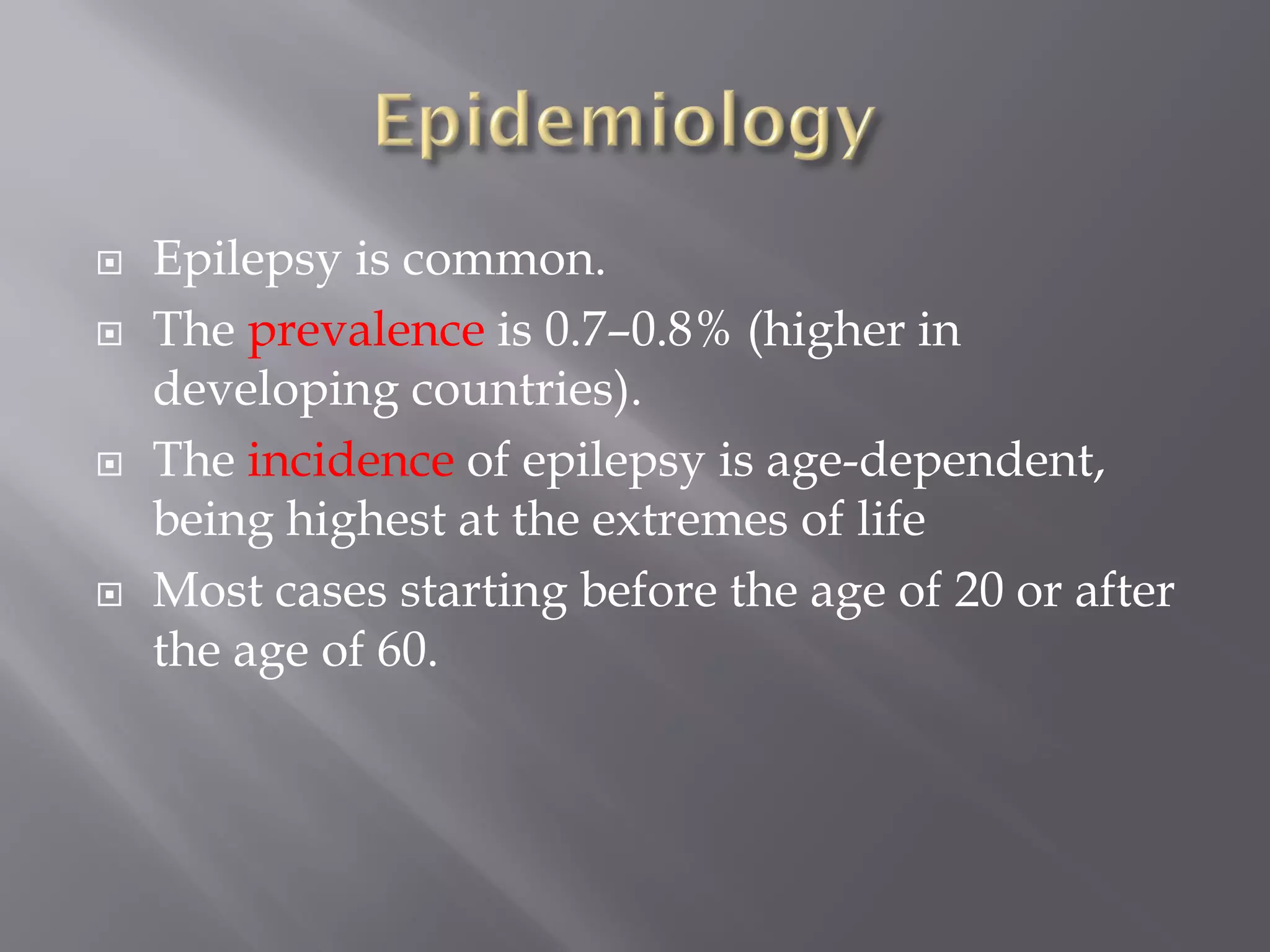
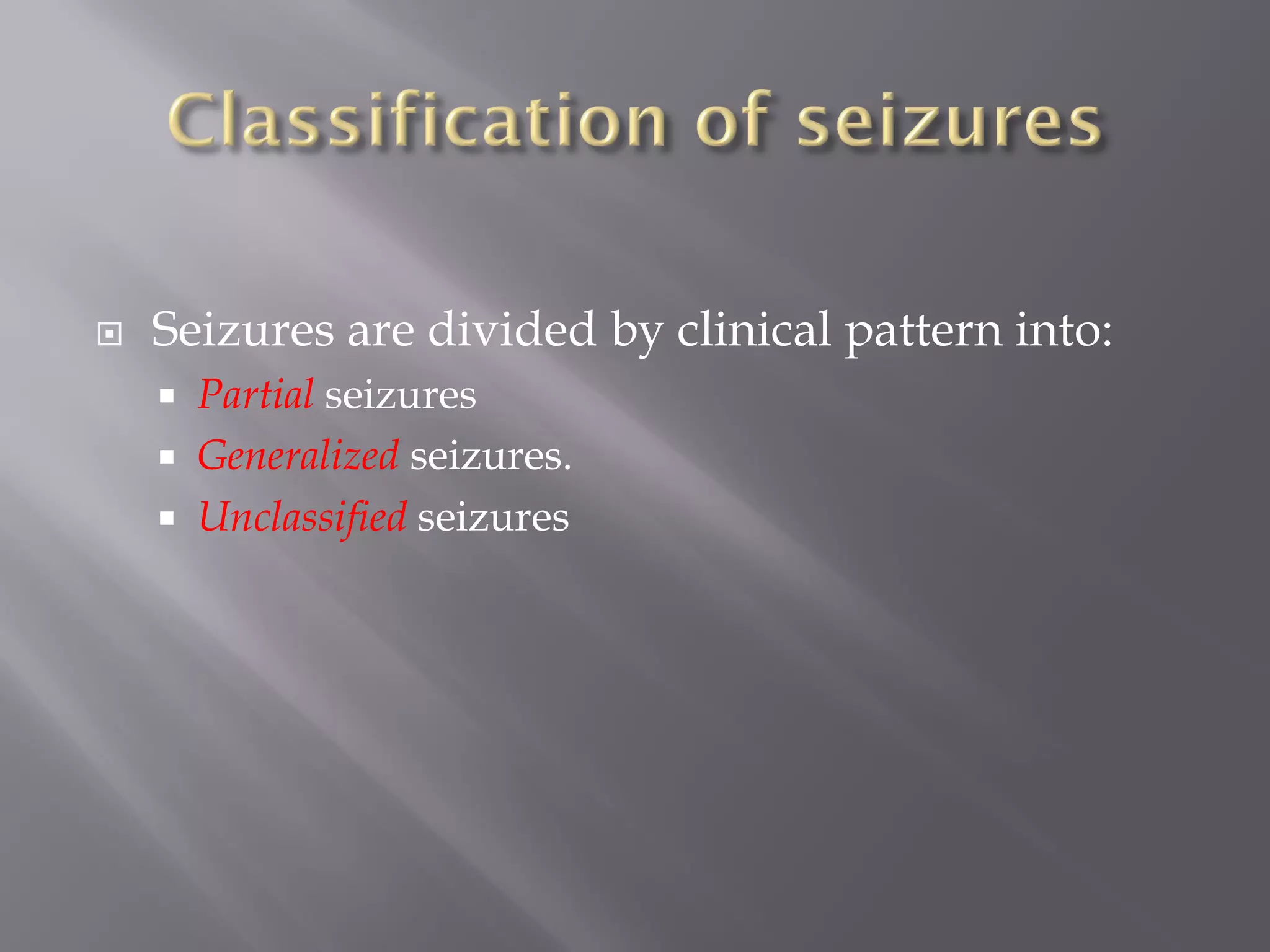
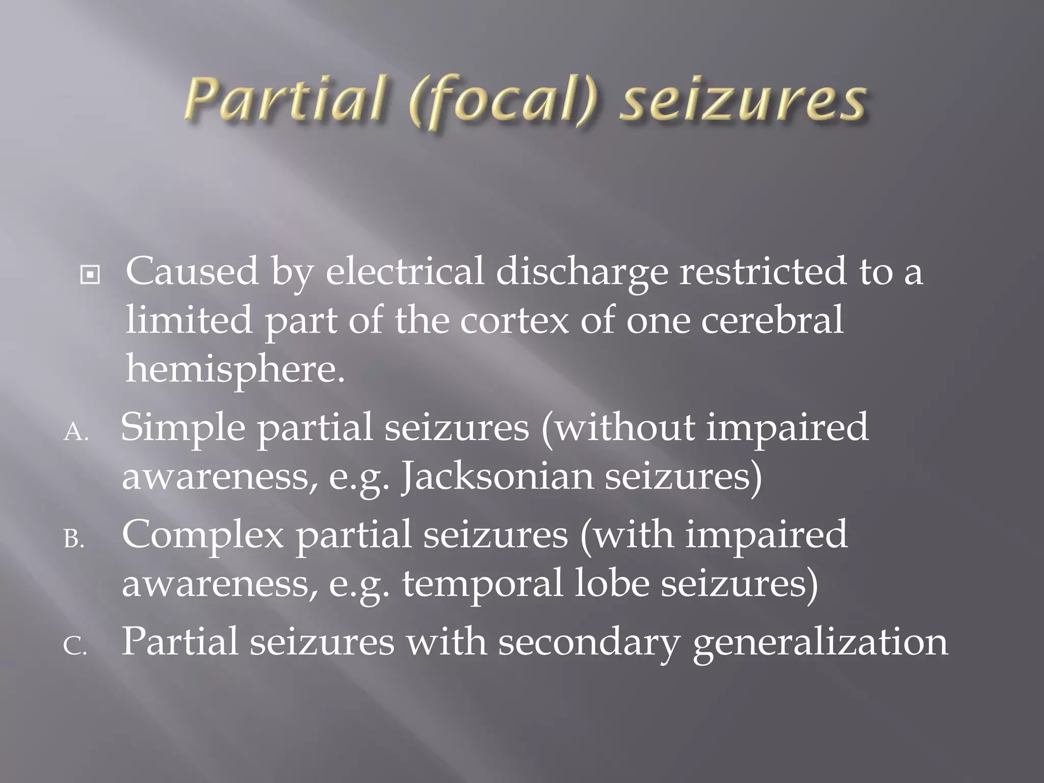
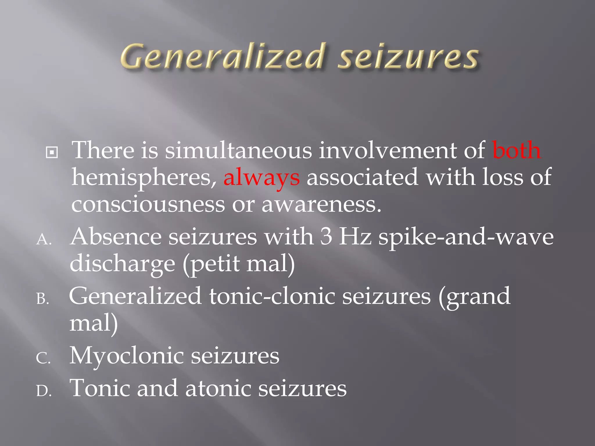
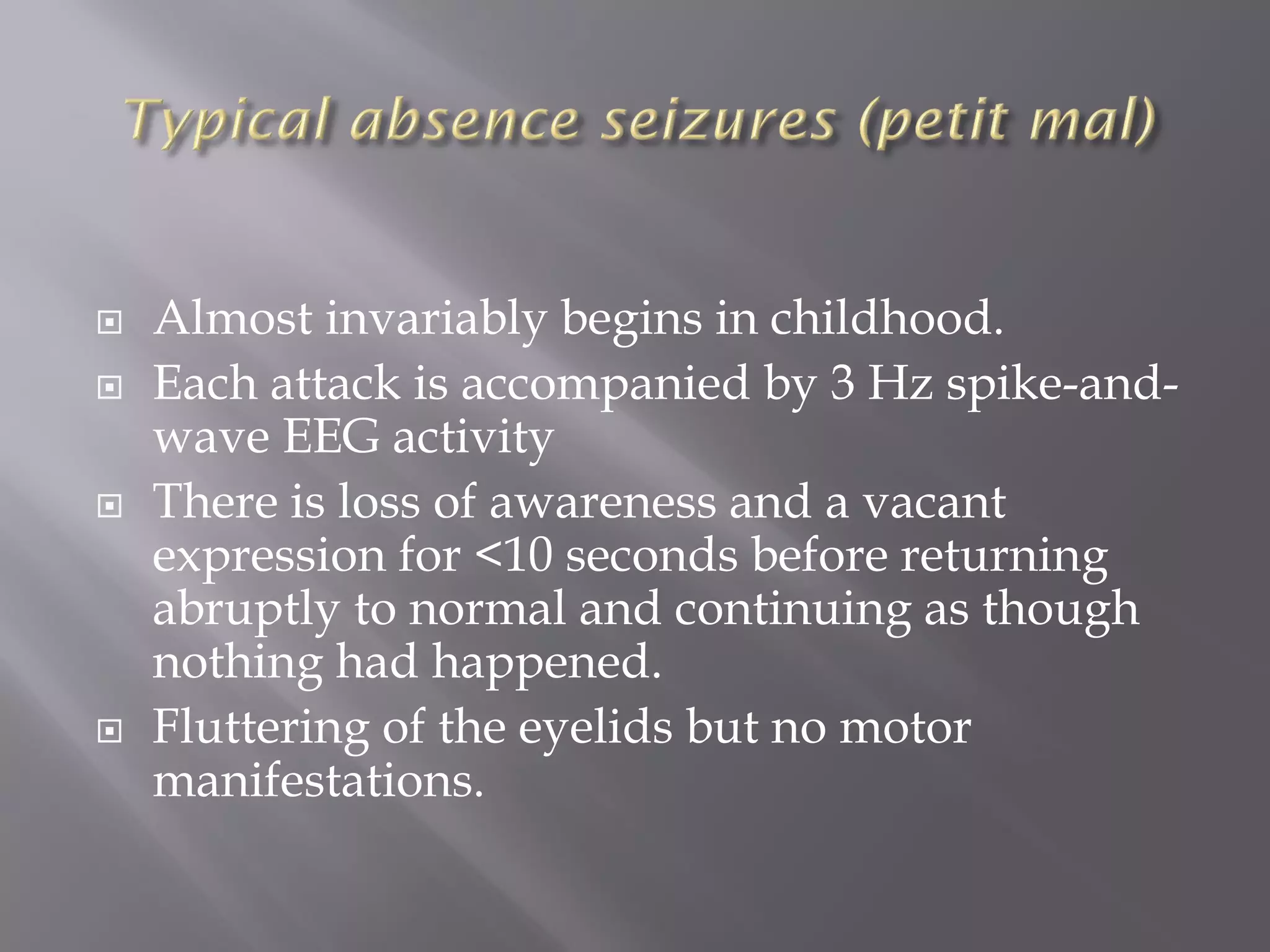
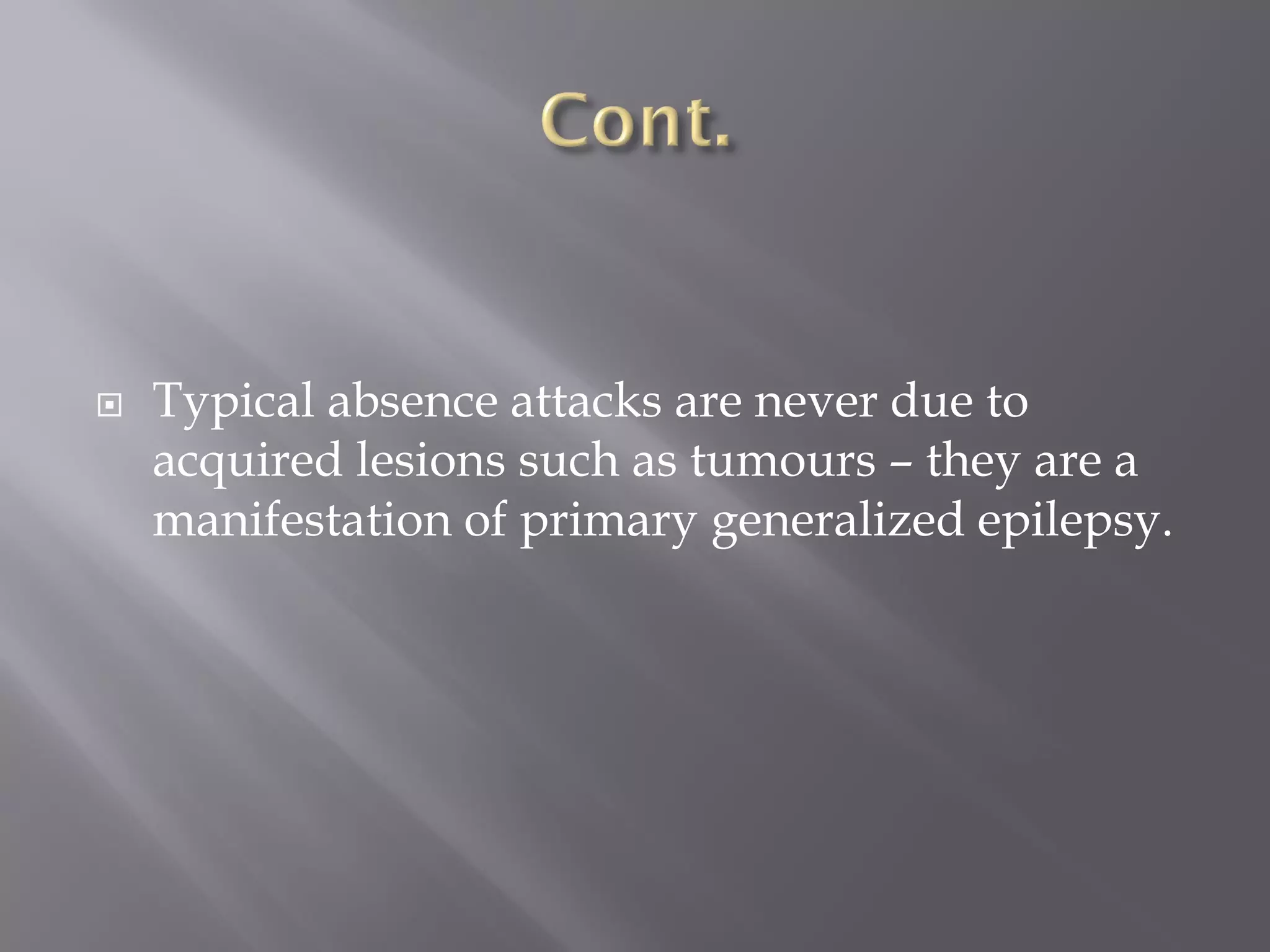
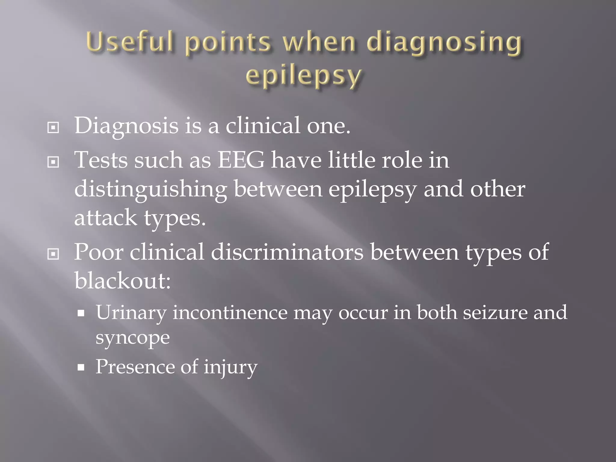
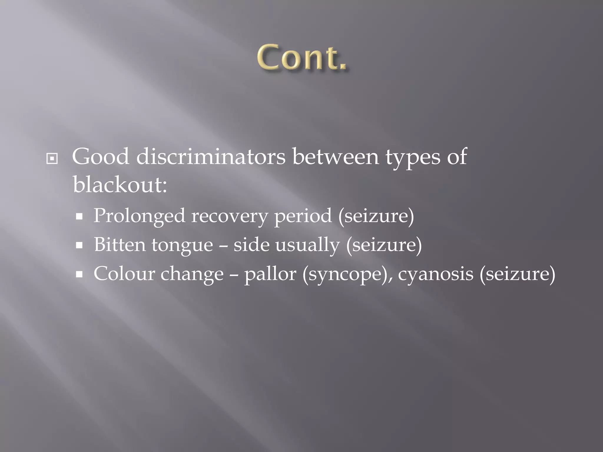
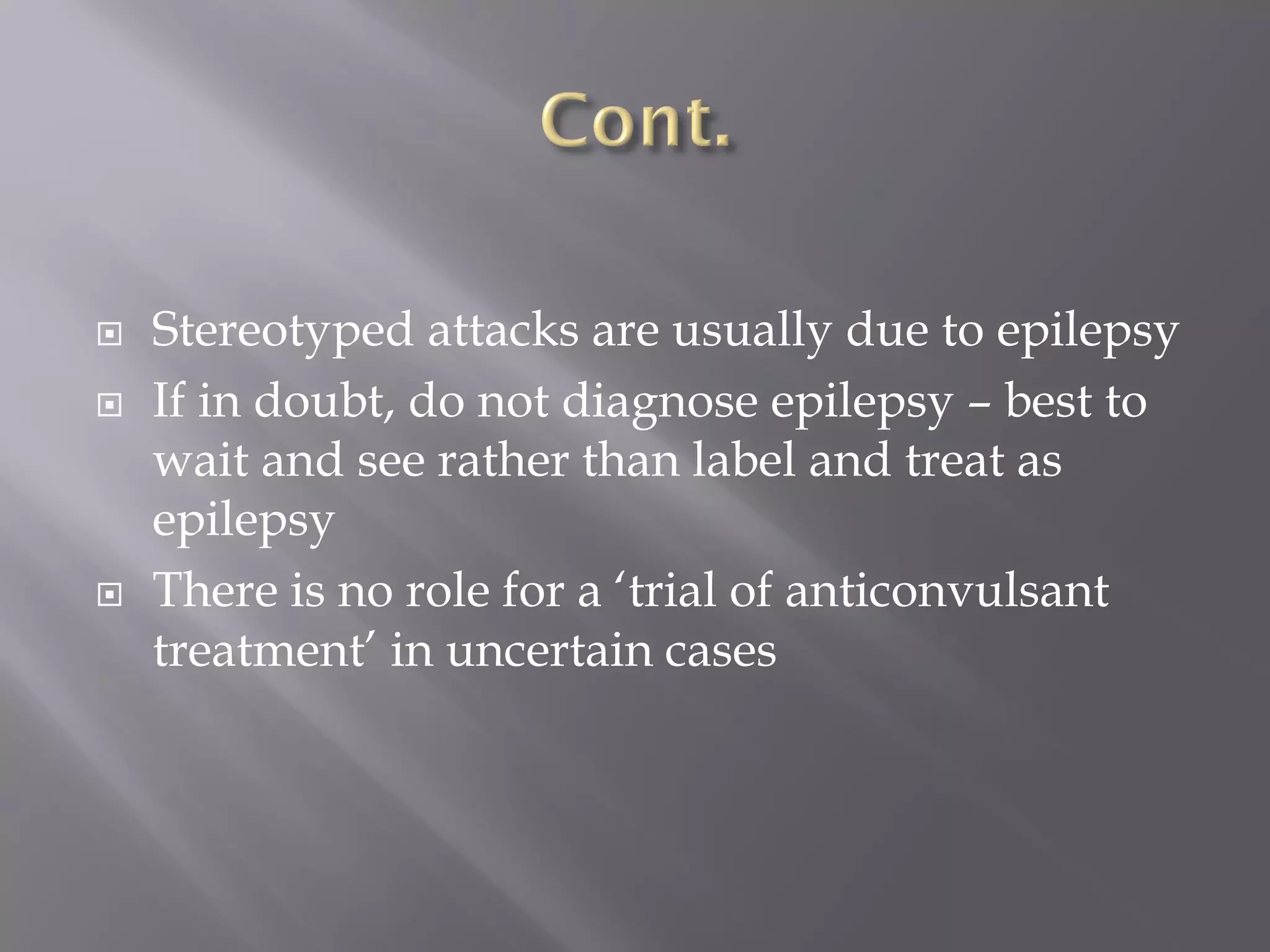
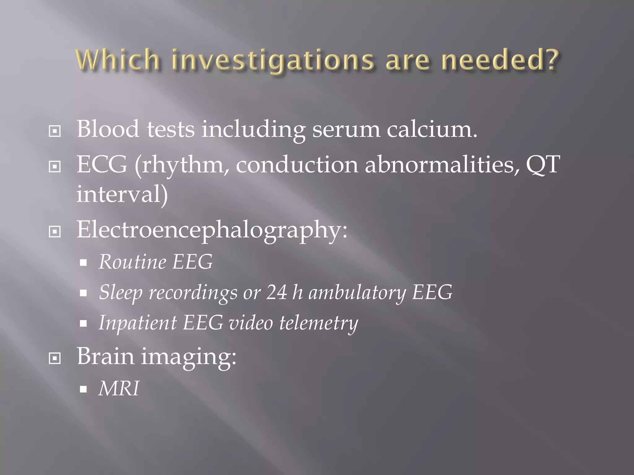
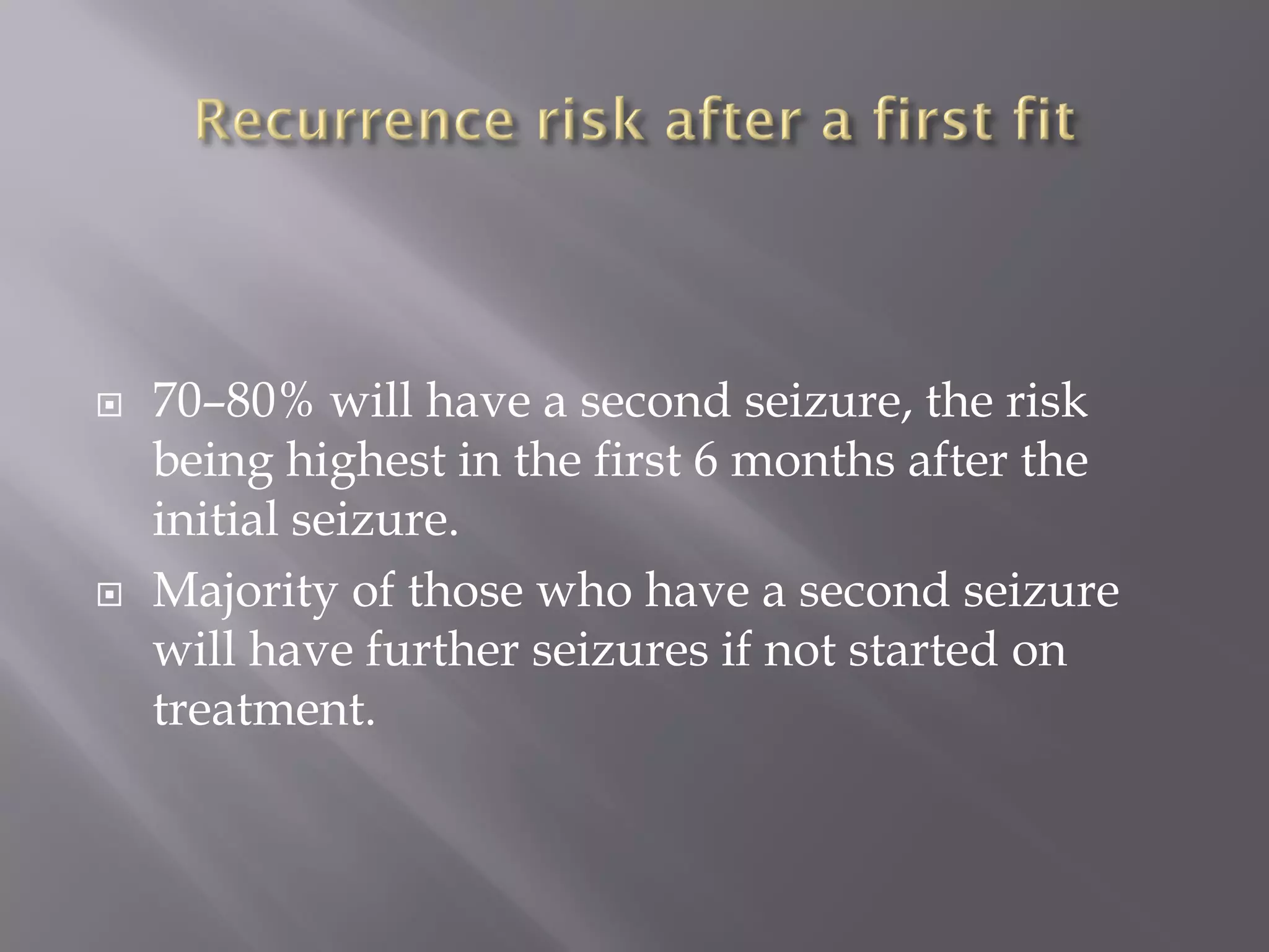
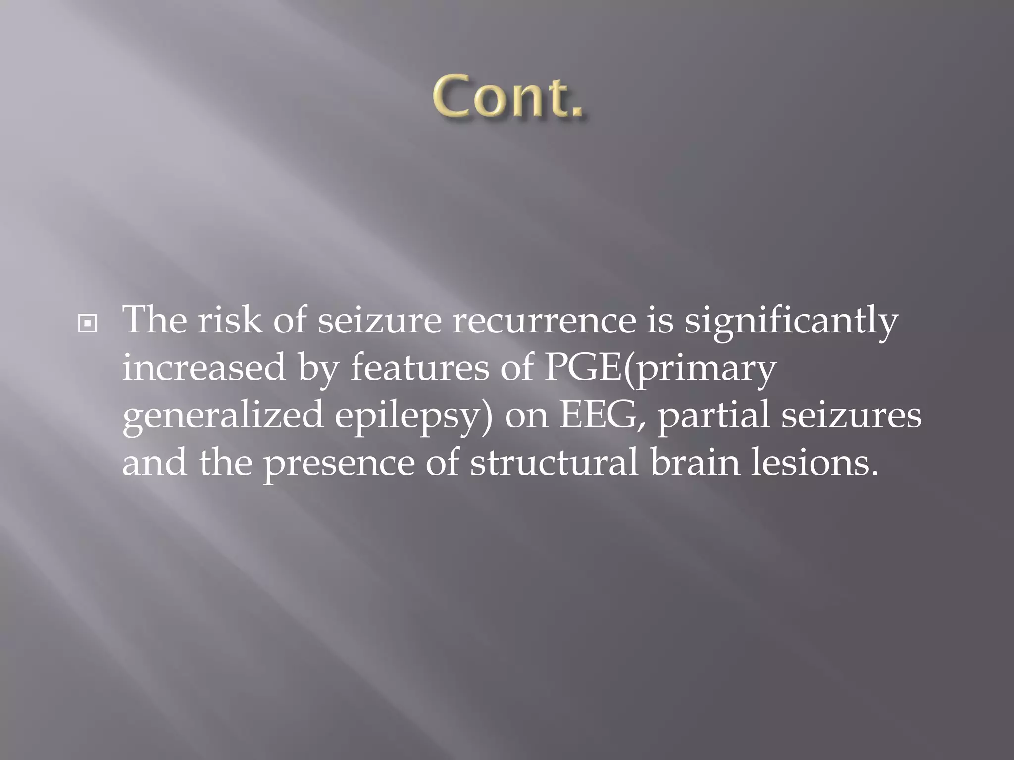
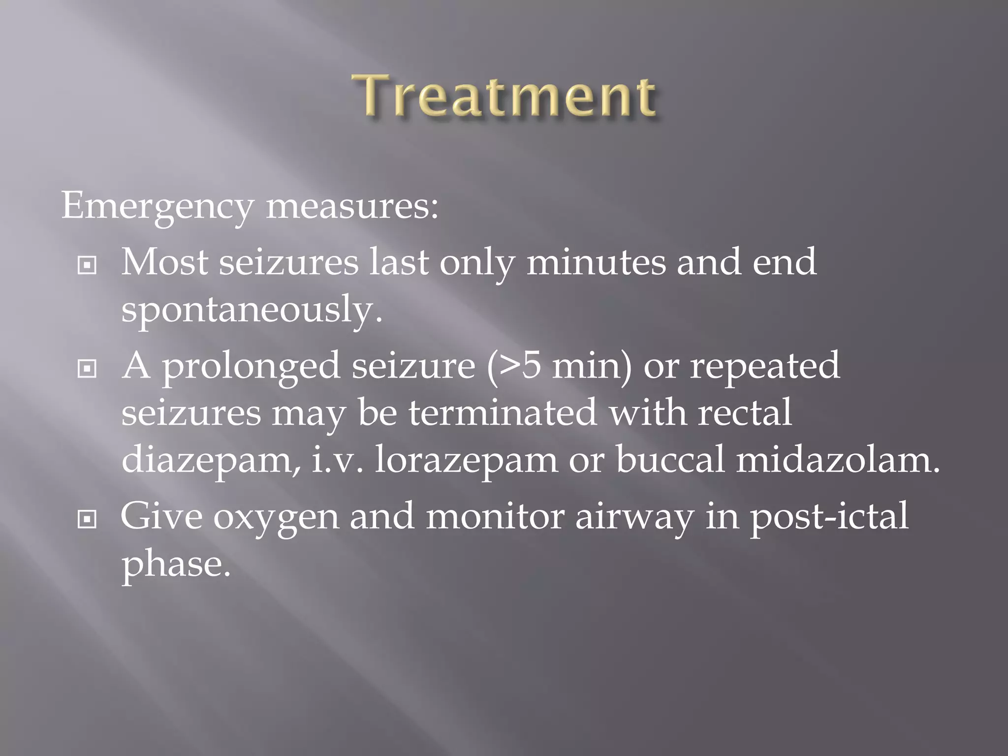
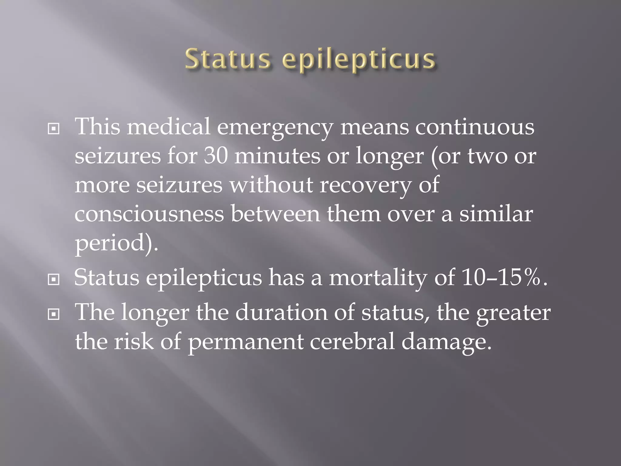
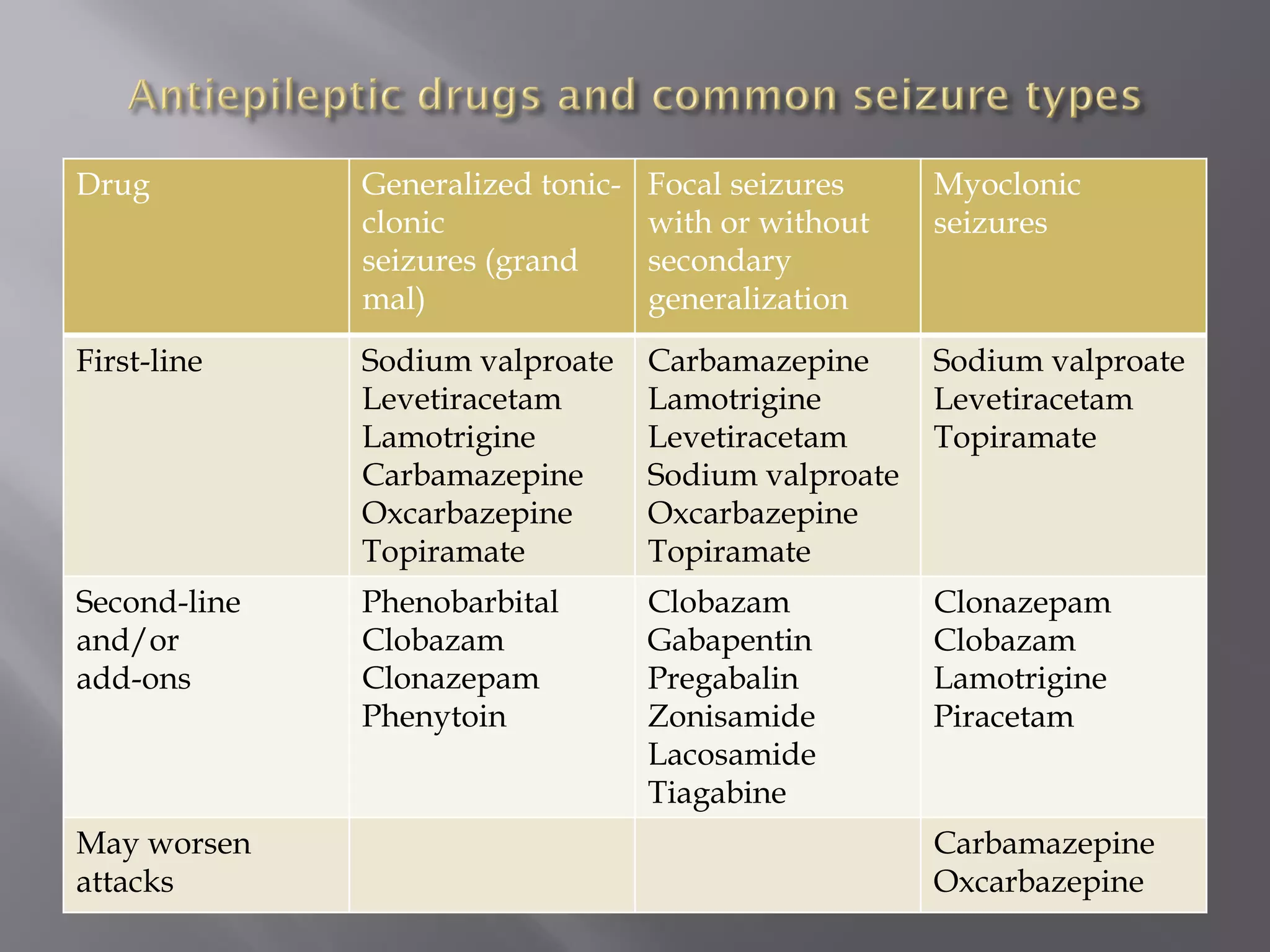
Dr. Abdelrahman Abukanna
Associate Prof. Of Int. Medicine
 A sudden synchronous discharge of cerebral
neurons causing symptoms or signs that are
apparent either to the patient or an observer.
 Epilepsy is an ongoing liability to recurrent
epileptic seizures.
 Epilepsy is common.
 The prevalence is 0.7–0.8% (higher in
developing countries).
 The incidence of epilepsy is age-dependent,
being highest at the extremes of life
 Most cases starting before the age of 20 or after
the age of 60.
 Seizures are divided by clinical pattern into:
 Partial seizures
 Generalized seizures.
 Unclassified seizures
 Caused by electrical discharge restricted to a
limited part of the cortex of one cerebral
hemisphere.
A. Simple partial seizures (without impaired
awareness, e.g. Jacksonian seizures)
B. Complex partial seizures (with impaired
awareness, e.g. temporal lobe seizures)
C. Partial seizures with secondary generalization
 There is simultaneous involvement of both
hemispheres, always associated with loss of
consciousness or awareness.
A. Absence seizures with 3 Hz spike-and-wave
discharge (petit mal)
B. Generalized tonic-clonic seizures (grand
mal)
C. Myoclonic seizures
D. Tonic and atonic seizures
 Seizures that do not fit a category
 Almost invariably begins in childhood.
 Each attack is accompanied by 3 Hz spike-and-
wave EEG activity
 There is loss of awareness and a vacant
expression for <10 seconds before returning
abruptly to normal and continuing as though
nothing had happened.
 Fluttering of the eyelids but no motor
manifestations.
 Typical absence attacks are never due to
acquired lesions such as tumours – they are a
manifestation of primary generalized epilepsy.
 Prodrome:
There is often no warning or there may be an aura
prior to a secondary generalized seizure.
 Tonic-clonic phase.
 An initial tonic stiffening is followed by the
clonic phase with synchronous jerking of the
limbs, reducing in frequency over about 2
minutes until the convulsion stops.
 The patient may utter an initial cry and falls,
sometimes suffering serious injury.
 The eyes remain open and the tongue is often
bitten.
 There may be incontinence of urine or faeces.
 Post-ictal phase.
 A period of flaccid unresponsiveness is
followed by gradual return of awareness with
confusion and drowsiness lasting 15 minutes to
an hour or longer.
 Headache is common after a GTCS.
Myoclonic seizures:
 Momentary brief contractions of a muscle or
muscle groups, e.g. causing a sudden
involuntary twitch of a finger or hand.
Tonic seizures:
 Consist of stiffening of the body, not followed
by jerking.
Atonic seizure.
A sudden collapse with loss of muscle tone and
consciousness.
Simple partial seizures:
 Originate within the motor cortex.
 Jerking typically begins on one side of the
mouth or in one hand or the entire side.
 This visible spread of activity is called the
march of a seizure.
 Local temporary paralysis of the limbs affected
sometimes follows (Todd’s paralysis).
 With some frontal seizures, conjugate gaze
deviates away from the epileptic focus and the
head turns; this is known as an (adversive
seizure).
 Arise from the temporal lobe (60%) or the
frontal lobe.
 The aura often includes a rising epigastric
sensation and nausea with a wide variety of
possible psychic phenomena (often hard for the
patient to describe) or hallucinations.
Hallucinations:
 Déjà vu or jamais vu
 Olfactory hallucinations
 Formed visual hallucinations or
misperceptions, e.g. micropsia or macropsia
(objects appear small or large, respectively)
 Fear (may be mistaken for panic attacks).
 There follows a period of complete or partial
loss of awareness of surroundings, lasting for
1–2 minutes
 This stage is accompanied by speech arrest and
often by automatisms.
 followed by a short period of post-ictal
confusion or may develop into a secondary
generalized convulsive seizure.
 Primary generalized epilepsy, e.g. JME
 Developmental, e.g. hamartomas, neuronal
migration abnormalities
 Hippocampal sclerosis
 Brain trauma and surgery
 Intracranial mass lesions, e.g. tumour,
neurocysticercosis.
 Vascular, e.g. cerebral infarction, AVM
 Encephalitis and inflammatory conditions, e.g.
herpes simplex, MS.
 Metabolic abnormalities
 Hypocalcaemia, hypoglycaemia, hyponatraemia
 Acute hypoxia
 Uraemia, hepatic encephalopathy
 Porphyria
 Neurodegenerative disorders, e.g. Alzheimer’s
 Drugs, e.g. ciclosporin, lidocaine, quinolones,
tricyclic antidepressants, antipsychotics,
lithium, stimulant drugs, e.g. cocaine.
 Alcohol withdrawal.
Diagnosis
 The diagnosis of a seizure is essentially a
clinical one based on taking a history from the
patient and any witnesses
 Investigations have a limited role in
distinguishing between a seizure and other
causes of a blackout or attack .
 The majority of patients referred to a first fit
clinic have not had a seizure.
 The commonest error is to misdiagnose a
syncopal blackout for a seizure.
 Witness account is crucial
What happened:
 Before: aura vs presyncopal prodrome
 During: convulsion, automatisms vs brief syncopal
blackout and pallor
 After: post-ictal confusion and headache vs rapid
recovery in syncope
Circumstances:
 Seizure triggers? Sleep deprivation, alcohol binge
or drugs
 Syncope triggers? Pain, heat, prolonged standing,
etc.
Epilepsy risk factors?
 Childhood febrile convulsions
 Significant head injury
 Meningitis or encephalitis
 Family history of epilepsy
Previous unrecognized seizures?
 Myoclonic jerks
 Absences
 Auras (simple partial seizures)
Alcohol excess?
Medication lowering seizure threshold?
Hold a driving license?
 Diagnosis is a clinical one.
 Tests such as EEG have little role in
distinguishing between epilepsy and other
attack types.
 Poor clinical discriminators between types of
blackout:
 Urinary incontinence may occur in both seizure and
syncope
 Presence of injury
 Good discriminators between types of
blackout:
 Prolonged recovery period (seizure)
 Bitten tongue – side usually (seizure)
 Colour change – pallor (syncope), cyanosis (seizure)
 Stereotyped attacks are usually due to epilepsy
 If in doubt, do not diagnose epilepsy – best to
wait and see rather than label and treat as
epilepsy
 There is no role for a ‘trial of anticonvulsant
treatment’ in uncertain cases
 Blood tests including serum calcium.
 ECG (rhythm, conduction abnormalities, QT
interval)
 Electroencephalography:
 Routine EEG
 Sleep recordings or 24 h ambulatory EEG
 Inpatient EEG video telemetry
 Brain imaging:
 MRI
 70–80% will have a second seizure, the risk
being highest in the first 6 months after the
initial seizure.
 Majority of those who have a second seizure
will have further seizures if not started on
treatment.
 The risk of seizure recurrence is significantly
increased by features of PGE(primary
generalized epilepsy) on EEG, partial seizures
and the presence of structural brain lesions.
Emergency measures:
 Most seizures last only minutes and end
spontaneously.
 A prolonged seizure (>5 min) or repeated
seizures may be terminated with rectal
diazepam, i.v. lorazepam or buccal midazolam.
 Give oxygen and monitor airway in post-ictal
phase.
 This medical emergency means continuous
seizures for 30 minutes or longer (or two or
more seizures without recovery of
consciousness between them over a similar
period).
 Status epilepticus has a mortality of 10–15%.
 The longer the duration of status, the greater
the risk of permanent cerebral damage.
 Not all status epilepticus is convulsive.
 Absence status is non-convulsive – the patient is
in a continuous, distant, stuporose state.
 Focal status epilepticus also occurs.
 Epilepsia partialis continua is continuous seizure
activity in one part of the body, such as a finger
or a limb, without loss of consciousness.
Antiepileptic drugs (AEDs):
 AEDs are indicated when there is a firm clinical
diagnosis of epilepsy and a substantial risk of
recurrent seizures.
 Some general principles apply:
 Introduce AEDs at low dose and slowly titrate
upwards until the seizures are controlled or side
effects become unacceptable
 Aim for monotherapy – 70% of patients will have
good seizure control with a single AED.
 If seizures not controlled with first AED, gradually
introduce second agent and then slowly withdraw
the first AED. If still not seizure free then
combination therapy is required.
 Epilepsy is one of the few disorders where non-
generic (‘brand name’) prescribing is justified to
ensure consistent drug levels.
 Routine monitoring of AED levels is not needed and
should be reserved for assessing compliance and
toxicity.
 There are interactions between AEDs (and with
other medications), e.g. between sodium valproate
and lamotrigine.
 New generation AEDs have fewer interactions.
 Phenytoin is no longer considered a first-line AED; it
is now principally used in emergency control of
seizures.
 Levetiracetam is increasingly used in most types of
epilepsy.
Myoclonic
seizures
Focal seizures
with or without
secondary
generalization
Generalized tonic-
clonic
seizures (grand
mal)
Drug
Sodium valproate
Levetiracetam
Topiramate
Carbamazepine
Lamotrigine
Levetiracetam
Sodium valproate
Oxcarbazepine
Topiramate
Sodium valproate
Levetiracetam
Lamotrigine
Carbamazepine
Oxcarbazepine
Topiramate
First-line
Clonazepam
Clobazam
Lamotrigine
Piracetam
Clobazam
Gabapentin
Pregabalin
Zonisamide
Lacosamide
Tiagabine
Phenobarbital
Clobazam
Clonazepam
Phenytoin
Second-line
and/or
add-ons
Carbamazepine
Oxcarbazepine
May worsen
attacks
 Intoxication with most AEDs causes
unsteadiness, nystagmus and drowsiness.
 Side effects are commoner with multiple AEDs.
 Skin rashes are seen particularly with
lamotrigine, carbamazepine and phenytoin.
 A wide variety of idiosyncratic drug reactions
may occur, e.g. blood dyscrasias with
carbamazepine.
Birth defects:
 The overall risk of birth defects in babies of
mothers who take one AED is around 7%, as
compared with 3% in women without epilepsy.
 The risk to the fetus of uncontrolled seizures is
greater than the risks of continuing AED
treatment.
 Folic acid (5 mg/day) supplements should be
taken before conception and throughout the first
trimester.
 Vitamin K 20 mg orally should also be taken
during the month before delivery to prevent
neonatal haemorrhage.
Contraception:
 AEDs inducing hepatic enzymes (e.g.
carbamazepine, phenytoin and phenobarbital)
reduce efficacy of oral contraceptives.
 A combined contraceptive pill containing a
higher dose of oestrogen or the progesterone
only pill provides greater contraceptive
security.
 An IUCD or barrier methods of contraception
are often used in preference to oral
contraceptives.
Breast-feeding:
 Mothers taking AEDs need not in general be
discouraged from breast-feeding, though
manufacturers are often hesitant in assuring
that there is no risk to the baby.
 Patients should be asked to stop driving after a
seizure and to inform the regulatory authorities
if they hold a driving license.
 After a seizure, a temporary driving ban until
seizure free is usual.
 People with epilepsy should be encouraged to
lead lives as unrestricted as reasonably
possible.
 Simple safety measures:
 Avoid swimming
 Avoid dangerous sports such as rock climbing.
 Advice at home:
 leaving bathroom and lavatory doors unlocked
 Taking showers rather than baths.
 Avoid sleep deprivation
 Avoid excess alcohol
 Avoid excess drugs
 Avoid strobe lighting
 Withdrawal of AEDs should be considered
after a seizure-free period of at least 2–3 years.
 There is a 50% seizure recurrence rate after
withdrawal so careful discussion and
explanation are essential.
 Seizures may persist despite treatment,
especially with temporal lobe partial epilepsy.
 Re-evaluate the diagnosis.
 Consider concordance (compliance).
 Combine AEDs and use maximum tolerated
dose.
 Refer to a specialist unit for consideration of
epilepsy surgery.
 Temporal lobectomy will result in seizure
freedom in 50–70% of selected patients with
uncontrolled seizures caused by hippocampal
sclerosis (defined by imaging and confirmed by
EEG).
Lecture 24 ( Epilepsy ).pdf

Lecture 24 ( Epilepsy ).pdf

  • 1.
    Dr. Abdelrahman Abukanna AssociateProf. Of Int. Medicine
  • 2.
     A suddensynchronous discharge of cerebral neurons causing symptoms or signs that are apparent either to the patient or an observer.  Epilepsy is an ongoing liability to recurrent epileptic seizures.
  • 3.
     Epilepsy iscommon.  The prevalence is 0.7–0.8% (higher in developing countries).  The incidence of epilepsy is age-dependent, being highest at the extremes of life  Most cases starting before the age of 20 or after the age of 60.
  • 4.
     Seizures aredivided by clinical pattern into:  Partial seizures  Generalized seizures.  Unclassified seizures
  • 5.
     Caused byelectrical discharge restricted to a limited part of the cortex of one cerebral hemisphere. A. Simple partial seizures (without impaired awareness, e.g. Jacksonian seizures) B. Complex partial seizures (with impaired awareness, e.g. temporal lobe seizures) C. Partial seizures with secondary generalization
  • 6.
     There issimultaneous involvement of both hemispheres, always associated with loss of consciousness or awareness. A. Absence seizures with 3 Hz spike-and-wave discharge (petit mal) B. Generalized tonic-clonic seizures (grand mal) C. Myoclonic seizures D. Tonic and atonic seizures
  • 7.
     Seizures thatdo not fit a category
  • 8.
     Almost invariablybegins in childhood.  Each attack is accompanied by 3 Hz spike-and- wave EEG activity  There is loss of awareness and a vacant expression for <10 seconds before returning abruptly to normal and continuing as though nothing had happened.  Fluttering of the eyelids but no motor manifestations.
  • 9.
     Typical absenceattacks are never due to acquired lesions such as tumours – they are a manifestation of primary generalized epilepsy.
  • 10.
     Prodrome: There isoften no warning or there may be an aura prior to a secondary generalized seizure.
  • 11.
     Tonic-clonic phase. An initial tonic stiffening is followed by the clonic phase with synchronous jerking of the limbs, reducing in frequency over about 2 minutes until the convulsion stops.  The patient may utter an initial cry and falls, sometimes suffering serious injury.  The eyes remain open and the tongue is often bitten.  There may be incontinence of urine or faeces.
  • 12.
     Post-ictal phase. A period of flaccid unresponsiveness is followed by gradual return of awareness with confusion and drowsiness lasting 15 minutes to an hour or longer.  Headache is common after a GTCS.
  • 13.
    Myoclonic seizures:  Momentarybrief contractions of a muscle or muscle groups, e.g. causing a sudden involuntary twitch of a finger or hand. Tonic seizures:  Consist of stiffening of the body, not followed by jerking. Atonic seizure. A sudden collapse with loss of muscle tone and consciousness.
  • 14.
    Simple partial seizures: Originate within the motor cortex.  Jerking typically begins on one side of the mouth or in one hand or the entire side.  This visible spread of activity is called the march of a seizure.
  • 15.
     Local temporaryparalysis of the limbs affected sometimes follows (Todd’s paralysis).  With some frontal seizures, conjugate gaze deviates away from the epileptic focus and the head turns; this is known as an (adversive seizure).
  • 16.
     Arise fromthe temporal lobe (60%) or the frontal lobe.  The aura often includes a rising epigastric sensation and nausea with a wide variety of possible psychic phenomena (often hard for the patient to describe) or hallucinations.
  • 17.
    Hallucinations:  Déjà vuor jamais vu  Olfactory hallucinations  Formed visual hallucinations or misperceptions, e.g. micropsia or macropsia (objects appear small or large, respectively)  Fear (may be mistaken for panic attacks).
  • 18.
     There followsa period of complete or partial loss of awareness of surroundings, lasting for 1–2 minutes  This stage is accompanied by speech arrest and often by automatisms.  followed by a short period of post-ictal confusion or may develop into a secondary generalized convulsive seizure.
  • 19.
     Primary generalizedepilepsy, e.g. JME  Developmental, e.g. hamartomas, neuronal migration abnormalities  Hippocampal sclerosis  Brain trauma and surgery  Intracranial mass lesions, e.g. tumour, neurocysticercosis.
  • 20.
     Vascular, e.g.cerebral infarction, AVM  Encephalitis and inflammatory conditions, e.g. herpes simplex, MS.  Metabolic abnormalities  Hypocalcaemia, hypoglycaemia, hyponatraemia  Acute hypoxia  Uraemia, hepatic encephalopathy  Porphyria
  • 21.
     Neurodegenerative disorders,e.g. Alzheimer’s  Drugs, e.g. ciclosporin, lidocaine, quinolones, tricyclic antidepressants, antipsychotics, lithium, stimulant drugs, e.g. cocaine.  Alcohol withdrawal.
  • 22.
    Diagnosis  The diagnosisof a seizure is essentially a clinical one based on taking a history from the patient and any witnesses  Investigations have a limited role in distinguishing between a seizure and other causes of a blackout or attack .  The majority of patients referred to a first fit clinic have not had a seizure.  The commonest error is to misdiagnose a syncopal blackout for a seizure.
  • 23.
     Witness accountis crucial What happened:  Before: aura vs presyncopal prodrome  During: convulsion, automatisms vs brief syncopal blackout and pallor  After: post-ictal confusion and headache vs rapid recovery in syncope Circumstances:  Seizure triggers? Sleep deprivation, alcohol binge or drugs  Syncope triggers? Pain, heat, prolonged standing, etc.
  • 24.
    Epilepsy risk factors? Childhood febrile convulsions  Significant head injury  Meningitis or encephalitis  Family history of epilepsy
  • 25.
    Previous unrecognized seizures? Myoclonic jerks  Absences  Auras (simple partial seizures) Alcohol excess? Medication lowering seizure threshold? Hold a driving license?
  • 26.
     Diagnosis isa clinical one.  Tests such as EEG have little role in distinguishing between epilepsy and other attack types.  Poor clinical discriminators between types of blackout:  Urinary incontinence may occur in both seizure and syncope  Presence of injury
  • 27.
     Good discriminatorsbetween types of blackout:  Prolonged recovery period (seizure)  Bitten tongue – side usually (seizure)  Colour change – pallor (syncope), cyanosis (seizure)
  • 28.
     Stereotyped attacksare usually due to epilepsy  If in doubt, do not diagnose epilepsy – best to wait and see rather than label and treat as epilepsy  There is no role for a ‘trial of anticonvulsant treatment’ in uncertain cases
  • 29.
     Blood testsincluding serum calcium.  ECG (rhythm, conduction abnormalities, QT interval)  Electroencephalography:  Routine EEG  Sleep recordings or 24 h ambulatory EEG  Inpatient EEG video telemetry  Brain imaging:  MRI
  • 30.
     70–80% willhave a second seizure, the risk being highest in the first 6 months after the initial seizure.  Majority of those who have a second seizure will have further seizures if not started on treatment.
  • 31.
     The riskof seizure recurrence is significantly increased by features of PGE(primary generalized epilepsy) on EEG, partial seizures and the presence of structural brain lesions.
  • 32.
    Emergency measures:  Mostseizures last only minutes and end spontaneously.  A prolonged seizure (>5 min) or repeated seizures may be terminated with rectal diazepam, i.v. lorazepam or buccal midazolam.  Give oxygen and monitor airway in post-ictal phase.
  • 33.
     This medicalemergency means continuous seizures for 30 minutes or longer (or two or more seizures without recovery of consciousness between them over a similar period).  Status epilepticus has a mortality of 10–15%.  The longer the duration of status, the greater the risk of permanent cerebral damage.
  • 34.
     Not allstatus epilepticus is convulsive.  Absence status is non-convulsive – the patient is in a continuous, distant, stuporose state.  Focal status epilepticus also occurs.  Epilepsia partialis continua is continuous seizure activity in one part of the body, such as a finger or a limb, without loss of consciousness.
  • 35.
    Antiepileptic drugs (AEDs): AEDs are indicated when there is a firm clinical diagnosis of epilepsy and a substantial risk of recurrent seizures.  Some general principles apply:  Introduce AEDs at low dose and slowly titrate upwards until the seizures are controlled or side effects become unacceptable  Aim for monotherapy – 70% of patients will have good seizure control with a single AED.
  • 36.
     If seizuresnot controlled with first AED, gradually introduce second agent and then slowly withdraw the first AED. If still not seizure free then combination therapy is required.  Epilepsy is one of the few disorders where non- generic (‘brand name’) prescribing is justified to ensure consistent drug levels.  Routine monitoring of AED levels is not needed and should be reserved for assessing compliance and toxicity.
  • 37.
     There areinteractions between AEDs (and with other medications), e.g. between sodium valproate and lamotrigine.  New generation AEDs have fewer interactions.  Phenytoin is no longer considered a first-line AED; it is now principally used in emergency control of seizures.  Levetiracetam is increasingly used in most types of epilepsy.
  • 38.
    Myoclonic seizures Focal seizures with orwithout secondary generalization Generalized tonic- clonic seizures (grand mal) Drug Sodium valproate Levetiracetam Topiramate Carbamazepine Lamotrigine Levetiracetam Sodium valproate Oxcarbazepine Topiramate Sodium valproate Levetiracetam Lamotrigine Carbamazepine Oxcarbazepine Topiramate First-line Clonazepam Clobazam Lamotrigine Piracetam Clobazam Gabapentin Pregabalin Zonisamide Lacosamide Tiagabine Phenobarbital Clobazam Clonazepam Phenytoin Second-line and/or add-ons Carbamazepine Oxcarbazepine May worsen attacks
  • 39.
     Intoxication withmost AEDs causes unsteadiness, nystagmus and drowsiness.  Side effects are commoner with multiple AEDs.  Skin rashes are seen particularly with lamotrigine, carbamazepine and phenytoin.  A wide variety of idiosyncratic drug reactions may occur, e.g. blood dyscrasias with carbamazepine.
  • 40.
    Birth defects:  Theoverall risk of birth defects in babies of mothers who take one AED is around 7%, as compared with 3% in women without epilepsy.  The risk to the fetus of uncontrolled seizures is greater than the risks of continuing AED treatment.  Folic acid (5 mg/day) supplements should be taken before conception and throughout the first trimester.  Vitamin K 20 mg orally should also be taken during the month before delivery to prevent neonatal haemorrhage.
  • 41.
    Contraception:  AEDs inducinghepatic enzymes (e.g. carbamazepine, phenytoin and phenobarbital) reduce efficacy of oral contraceptives.  A combined contraceptive pill containing a higher dose of oestrogen or the progesterone only pill provides greater contraceptive security.  An IUCD or barrier methods of contraception are often used in preference to oral contraceptives.
  • 42.
    Breast-feeding:  Mothers takingAEDs need not in general be discouraged from breast-feeding, though manufacturers are often hesitant in assuring that there is no risk to the baby.
  • 43.
     Patients shouldbe asked to stop driving after a seizure and to inform the regulatory authorities if they hold a driving license.  After a seizure, a temporary driving ban until seizure free is usual.
  • 44.
     People withepilepsy should be encouraged to lead lives as unrestricted as reasonably possible.  Simple safety measures:  Avoid swimming  Avoid dangerous sports such as rock climbing.
  • 45.
     Advice athome:  leaving bathroom and lavatory doors unlocked  Taking showers rather than baths.  Avoid sleep deprivation  Avoid excess alcohol  Avoid excess drugs  Avoid strobe lighting
  • 46.
     Withdrawal ofAEDs should be considered after a seizure-free period of at least 2–3 years.  There is a 50% seizure recurrence rate after withdrawal so careful discussion and explanation are essential.
  • 47.
     Seizures maypersist despite treatment, especially with temporal lobe partial epilepsy.  Re-evaluate the diagnosis.  Consider concordance (compliance).  Combine AEDs and use maximum tolerated dose.  Refer to a specialist unit for consideration of epilepsy surgery.
  • 48.
     Temporal lobectomywill result in seizure freedom in 50–70% of selected patients with uncontrolled seizures caused by hippocampal sclerosis (defined by imaging and confirmed by EEG).