Chordee correction by corporal rotation:The Split and Roll technique
•
0 likes•1,431 views

This document describes a technique called the "split and roll technique" for correcting severe chordee (abnormal curvature of the penis) associated with hypospadias. The technique involves: 1) Splitting the septum between the corpora cavernosa with a ventral midline incision to partially separate the corpora and facilitate rotation. 2) Placing nonabsorbable sutures from the dorsolateral aspect of one corpus across the midline to the other corpus. Tying the sutures rotates the corpora toward the dorsal midline, correcting the curvature. 3) This technique avoids incisions into the corporal substance and does not require grafts or cause penile shortening

Recommended
More Related Content
What's hot
What's hot (20)
Viewers also liked
Viewers also liked (20)
Similar to Chordee correction by corporal rotation:The Split and Roll technique
Similar to Chordee correction by corporal rotation:The Split and Roll technique (20)
More from Luis Fernando Gonzalez-Llinás
More from Luis Fernando Gonzalez-Llinás (20)
Recently uploaded
Recently uploaded (20)
Chordee correction by corporal rotation:The Split and Roll technique
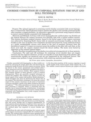
- 1. 0022-5347/99/1623-1152/0 THE JOURNAL OF UROLOGY Vol. 162, 1152–1155, September 1999 Copyright © 1999 by AMERICAN UROLOGICAL ASSOCIATION, INC. Printed in U.S.A. CHORDEE CORRECTION BY CORPORAL ROTATION: THE SPLIT AND ROLL TECHNIQUE ROSS M. DECTER From the Department of Surgery, Section of Urology, Milton S. Hershey Medical Center, Pennsylvania State Geisinger Health System, Hershey, Pennsylvania ABSTRACT Purpose: The optimal approach to correcting ventral chordee associated with severe hypospa- dias is controversial. Dorsal plication tends to shorten the phallus and ventrally positioned grafts often mandate a staged procedure. An alternative approach is presented using corporal rotation to correct ventral chordee associated with hypospadias. Materials and Methods: In 6 boys with severe hypospadias the urethral plate was divided and the septum between the corpora cavernosa was partially split with a ventral midline incision. This incision facilitates corporal rotation. Access to the dorsal aspect of the corpora cavernosa was achieved by dissecting Buck’s fascia with its encased neurovascular bundles, so that the bundles in the area of chordee were completely elevated and preserved. Using artificial erection as a guide nonabsorbable sutures were placed in the area of maximal curvature from the dorsolateral aspect of 1 corpus cavernosum across the midline to the other side such that, as the knots were tied, the corpora rotated toward the dorsal midline. The knots were buried by apposition of the rotated corporal bodies. Results: Excellent straightening of the phallus was achieved intraoperatively in these patients. Conclusions: The split and roll technique for correcting severe chordee does not require incisions into the corporal substance, involve use of grafts or cause shortening of the phallus. The neurovascular bundles are preserved and are not compressed by the rotational sutures. The surgeon may perform 1-stage reconstruction while achieving maximal penile length. KEY WORDS: penis, urethra, hypospadias, abnormalities Chordee associated with hypospadias is often readily cor- to the drop down position of the meatus, exposing a consid- rected by releasing the penile shaft skin and dissecting the erable length of the ventral aspect of the corpora (fig. 1, A). dysgenetic bands of tissue off of the corpora cavernosa, which The midline ventral septum between the corpora cavernosa lie lateral to the corpus spongiosum and urethral plate.1 These maneuvers may not adequately straighten the penis in patients with more severe chordee. Most surgeons believe that residual chordee in these patients is due to corporal disproportion. There are generally accepted techniques to correct this problem. The surgeon may choose to incise the dorsal aspect of the corpora and perform Nesbit plications,2 which straighten the penis by shortening the dorsal side of the corpora. The alternative technique is to incise the ventral aspect of the corpora cavernosa at the point of maximum curvature and place a graft in the defect created as the penis straightens.3– 6 For either procedure an incision into the cor- pora cavernosa is required, which is associated with the possible risk of corporal damage or dysfunction. A technique is described using corporal rotation to correct ventral chordee associated with hypospadias that avoids corporal incisions and penile shortening. MATERIALS AND METHODS The split and roll technique of chordee correction was ini- FIG. 1. A, ventral corpus cavernosum is cleanly dissected to ex- tially used in 6 patients with penoscrotal or scrotal hypospa- pose midline ventral septum. When urethral plate is not divided, this dias and severe chordee due to corporal disproportion. In is achieved by simply dissecting corpus spongiosum and urethral plate off of underlying corpora cavernosa. NVB, neurovascular bun- these patients dorsal plications would have been used previ- dle. B, midline ventral incision into corporal septum need only cause ously to treat this type of chordee. In these cases division of partial separation of corpora to facilitate subsequent rotation. C, the urethral plate was required to correct chordee ade- plane of section under Buck’s fascia. This dissection allows neuro- quately. vascular bundles to be completely mobilized. D, neurovascular bun- dle is elevated with vessel loops and dorsally positioned rotational To perform the split and roll technique the ventral aspect suture is placed. E, each dorsal corpus cavernosum is rotated in of the corpora cavernosa is exposed after the urethral plate is midline. Artificial erection confirms adequate straightening of phal- divided. The urethral plate and distal corpus spongiosum are lus. Vessel loops are removed and Buck’s fascia drops back to normal dissected cleanly off of the corpora cavernosa from the glans anatomical position. 1152
- 2. 1153 CHORDEE CORRECTION BY CORPORAL ROTATION is identified and incised using a microsurgical knife. The case artificial erection revealed a straight phallus intraoper- incision partially separates the 2 corpora cavernosa (fig. 1, atively. B). This incision extends along the length of the intracorporal septum from the glans to the meatus. It is deepest in the area DISCUSSION of maximum ventral curvature. Care must be taken to avoid accidental entry into either corporal body. The septum is a Various techniques are available to the reconstructive sur- thin structure and dissection must proceed carefully or the geon for correcting chordee associated with hypospadias. It is corpora will be entered and bleeding will be excessive. Pre- clear that in the majority of boys with hypospadias releasing cise placement of the incision is facilitated by rolling the the ventral skin and its associated dartos fascia straightens the phallus.1 Some patients have persistent chordee even corpora away from the ventral midline and instilling inject- able saline into the corpora using the artificial erection tech- after the skin is released, and the dysgenetic tissue on the nique to aid in identifying the appropriate plane. It is not ventral aspect of the corpora cavernosa adjacent to the cor- necessary to separate the corpora cavernosa completely, but pus spongiosum and urethral plate is dissected. Mollard and only to incise the septum partially. Splitting the septum Castagnola suggested that excising the fibrous tissue under facilitates corporal rotation, which is done subsequently to the urethral plate almost invariably results in straightening this chordee7 but this has not been my experience. Even straighten the penis. Repeat artificial erection testing at this point reveals per- when the urethral plate has been completely divided and sistent ventral curvature due to corporal disproportion and dissection is performed on the ventrum to clean the tunica points out the area of maximum deformity. Access to the albuginea of the corpora cavernosa, chordee persists in some dorsal aspect of the corpora cavernosa is achieved by dissect- patients. The persistent curvature appears to be due to cor- ing Buck’s fascia with its encased neurovascular bundles poral disproportion. starting at the ventrolateral aspect of the corpora cavernosa Perhaps the most widely used techniques to correct this problem are variations of the Nesbit plication.2, 8, 9 Plicating on each side and proceeding toward the dorsum (fig. 1, C). This dissection is performed with fine tenotomy scissors and the dorsum of the corpora obviously shortens that aspect of it mobilizes the neurovascular bundles from the glans dis- the penis to correct curvature. In most patients shortening is tally to an appropriate position proximally on the penile not significant enough to prevent using the technique. Some shaft. After Buck’s fascia and the neurovascular bundles are surgeons incise directly through Buck’s fascia to place the plicating sutures.8 This approach risks inadvertent injury to mobilized they are elevated with vessel loops to allow easy access to the dorsum (fig. 1, D). Each corpus cavernosum is the neurovascular bundles, which are located on either side then rotated toward the dorsal midline by positioning a of the dorsal midline with branches ramifying distally transverse nonabsorbable suture on the dorsal aspect of 1 around the corpora cavernosa to the ventral side of the phal- lus.10 When the corpora cavernosa are plicated, Buck’s fascia corpus across the midline to the other corpus. The suture is placed so that, as it is tied, the knot is buried between the is elevated with its encased neurovascular bundle as de- corpora as they roll toward each other (fig. 1, E). Usually 2 or scribed in the split and roll technique to avoid any direct 3 such sutures placed in the region of maximum curvature injury to these nerves. To my knowledge it is not known suffice. Repeat artificial erection guides suture placement whether there are perforating branches of the bundles into and confirms penile straightening (fig. 2). Urethroplasty then the corpora along the length of the mobilized Buck’s fascia proceeds according to surgeon preference. but none is discernible with loupe magnification. Other po- Initially the split and roll technique was performed in tential pitfalls of the technique are that the incision through patients who required division of the urethral plate to correct the tunica albuginea may enter the erectile tissue and ad- chordee. This technique now has been applied to patients in versely affect its function. Although this risk may be consid- whom the urethral plate has not been divided. In these cases ered only theoretical, to my knowledge there are no published the corpus spongiosum proximal to the meatus and the ure- studies describing the long-term followup of patients with thral plate distal to the meatus are elevated off of the under- severe chordee who underwent plication. lying corpora cavernosa using sharp dissection. This dissec- An alternative to plicating or shortening the long side of tion allows access to the ventral midline and the septum may the curved penis is to increase the length of the short or be split. Elevation of the neurovascular bundles and rota- ventral aspect of the corpora cavernosa. The surgeon incises tional suture placement then proceed as described. In each the tunica albuginea of the ventral corpus cavernosum in the region of maximum curvature and places a graft into the defect that is created as the penis straightens. Various ma- terials have been used as the grafting material, although dermal grafts have probably been used most frequently.3– 6, 11 Most suggest that this technique necessitates staged hypos- padias repair,6 although Hendren and Keating noted that a 1-stage procedure may be performed in certain cases.4 The concept of corporal rotation to correct ventral chordee associated with hypospadias has been described in the past.12–14 Koff and Eakins noted that an incision along the ventral corporal septum allows the corpora to rotate and straighten during erection.12 Snow described a technique of making an initial ventral midline incision in the corpora cavernosa and placing sutures into the dorsal lateral corpus cavernosum to rotate the corpora.13 Kass also placed dorsally positioned sutures to rotate the corpora, which straightened the phallus.14 The dorsal rotational sutures of Snow13 and Kass14 were positioned so that the neurovascular bundles lay under the sutures when the knots were tied. In this situation the neurovascular bundles are subject to the risk of compres- FIG. 2. Artificial erection. A, chordee persists after division of sion injury caused by these sutures. The split and roll tech- urethral plate, splitting of septum and clean dissection of corpus nique involves a ventral septal incision, which facilitates the spongiosum off of corpus cavernosum. B, straight phallus after place- corporal rotation provided by the dorsally positioned suture. ment of dorsal rotational sutures.
- 3. 1154 CHORDEE CORRECTION BY CORPORAL ROTATION Corporal rotation created by straightening the ventral penile 4. Hendren, W. H. and Keating, M. A.: Use of dermal graft and free urethral graft in penile reconstruction. J. Urol., 140: 1265, curvature allows the penis to achieve its full potential length. 1988. In the technique described the dorsal rotational sutures lie 5. Horton, C. E., Jr., Gearhart, J. P. and Jeffs, R. D.: Dermal grafts under the neurovascular bundles and the knots are buried for correction of severe chordee associated with hypospadias. between the corpora cavernosa when tied. These factors J. Urol., 150: 452, 1993. should obviate the risk of injury to the neurovascular bundles 6. Pope, J. C., IV, Kropp, B. P., McLaughlin, K. P., Adams, M. C., in the long term. Rink, R. C., Keating, M. A. and Brock, J. W., III.: Penile orthoplasty using dermal grafts in the outpatient setting. CONCLUSIONS Urology, 48: 124, 1996. 7. Mollard, P. and Castagnola, C.: Hypospadias: the release of The split and roll technique allows the correction of chor- chordee without dividing the urethral plate and onlay island dee due to corporal disproportion without requiring incisions flap (92 cases). J. Urol., 152: 1238, 1994. into the corporal substance. It avoids the penile shortening 8. Daskalopoulos, E. I., Baskin, L., Duckett, J. W. and Snyder, that may be caused by dorsal plication, and during erection it H. M., III.: Congenital penile curvature (chordee without hy- allows the shortened ventral aspect of the corpora to stretch pospadias). Urology, 42: 708, 1993. to the length of the dorsal corpus. The technique avoids the 9. Rehman, J., Benet, A., Minsky, L. S. and Melman, A.: Results of use of grafts and allows the surgeon to proceed with 1-stage surgical treatment for abnormal penile curvature: Peyronie’s disease and congenital deviation by modified Nesbit plication repair. Good intraoperative results have been achieved but (tunical shaving and plication). J. Urol., 157: 1288, 1997. further followup is required to confirm long-term outcomes. 10. Baskin, L. S., Erol, A., Ying, W. L. and Cunha, G. R.: Anatomical studies of hypospadias. J. Urol., 160: 1108, 1998. REFERENCES 11. Perlmutter, A. D., Montgomery, B. T. and Steinhardt, G. F.: Tunica vaginalis free graft for the correction of chordee. 1. King, L. R.: Hypospadias: a one-stage repair without skin graft J. Urol., 134: 311, 1985. based on a new principle: chordee is sometimes produced by 12. Koff, S. A. and Eakins, M.: The treatment of penile chordee using skin alone. J. Urol., 103: 660, 1970. corporal rotation. J. Urol., 131: 931, 1984. 2. Nesbit, R. M.: Congenital curvature of the phallus: report of 13. Snow, B. W.: Transverse corporal plication for persistent chor- three cases with description of corrective operation. J. Urol., dee. Urology, 34: 360, 1989. 93: 230, 1965. 14. Kass, E. J.: Dorsal corporal rotation: an alternative technique for 3. Devine, C. J., Jr. and Horton, C. E.: Use of dermal graft to correct chordee. J. Urol., 113: 56, 1975. the management of severe chordee. J. Urol., 150: 635, 1993. DISCUSSION Dr. Antoine E. Khoury. Are you concerned about lifting the neurovascular bundle along the lateral edges? Are there no nerve perforators that enter the corpora at that point, which may impact on sensation or erectile function on a long-term basis? Dr. Ross M. Decter. The technique that we use, wherein we start our dissection ventrolaterally, basically allows us to lift up the neurovascular bundles even as they spread around the lateral aspects of the phallus. You do not see perforating nerves when you are doing the dissection. There may well be some tiny ones but you do not see them. You can do the dissection atraumatically and get nice access to the dorsum of the penis. Doctor Khoury. Those lateral nerve endings coming around the sides are entering the tunica albuginea and corpora? Doctor Decter. They may be but you do not perceive it when you are doing it. Dr. Sava V. Perovic. In my hands the split and roll technique in the septal region is a good method and decreases the severity of penile chordee. Rotation of the corpora cavernosa in my hands is not successful. What do you do when chordee is in the distal part of the corpora cavernosa near the glans? Doctor Decter. We have limited experience but we have straightened the phallus in each situation that I mentioned using dorsal rotational sutures. In all cases that I described the main part of the curvature was in the shaft of the phallus. There was not as much in the way of distal curvature under the glans. Two weeks ago I had a case in which there was some glans tilt that I was not happy with after I put in some dorsal rotational sutures. In fact, I applied your technique. I mobilized the glans completely off of the corpora cavernosa with the blood supply coming from the neurovascular bundle and urethral plate which exposed the end of the corpora cavernosa. Then I put a rotational suture in the distal end of the glans, which resolved the situation. Dr. Laurence S. Baskin. I want to comment about this concept of lifting up the neurovascular bundle. The neurovascular bundle does something. It does not just innervate the glans. There are all these piercing nerves that specifically go into the tunica. Dr. John Duckett showed me how to do the tunica albuginea plication procedure, which I did for many years. When we lifted up the neurovascular bundle, I am convinced that we were cutting these little perforating nerves. Does it make a difference? We do not know but I think that we should probably try to minimize it. I would not advocate lifting the neurovascular bundle. Doctor Decter. When you did that procedure, you incised Buck’s fascia right over the major part of the neurovascular bundles. I think that this procedure has a much greater chance of not injuring the bundles because we are elevating them and not incising through them. I advocate a technique starting with dissection ventro- laterally to try to preserve all of Buck’s fascia with its encased neurovascular bundle. Dr. Mark Zaontz. I agree with Doctor Baskin. The nerves span out all over the dorsum of the penis and laterally. When we make a lateral incision, we tend to cut 1 or 2 nerves but so what? My colleagues who treat Peyronie’s disease in adults have told me that these patients have no functional or sensory deficits. Several adults have been referred to me for hypospadias repair with chordee release. I have probably nicked a few nerves
- 4. 1155 CHORDEE CORRECTION BY CORPORAL ROTATION myself and have not seen any deficit. I think that a few nerves cut here and there is not going to make a difference. Doctor Baskin. It makes a difference in San Francisco. The technique that we are using in children is taken from observations made by Dr. Tom Lue in adults. He started his technique because of complaints of patients who underwent a plication or Nesbit type procedure that was done laterally. These patients had decreased sensation in the glans and skin. Doctor Lue, a penile anatomist, started to put sutures in the dorsal midline or near the urethra. Based on fetal studies that made a lot of sense. When I showed him my fetal studies, he indicated that he had had similar findings in adult cadaver penises. We do not know what the long-term outcome will be of placing midline sutures but I think that it is going to be good.