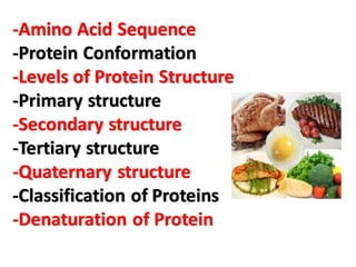
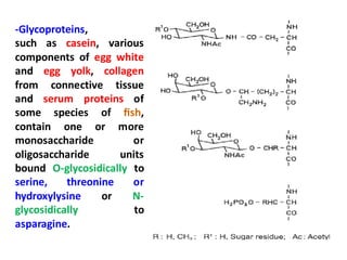
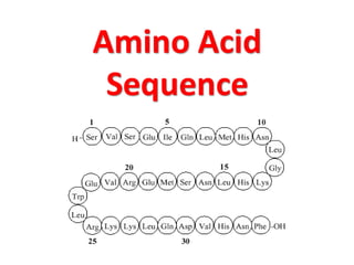
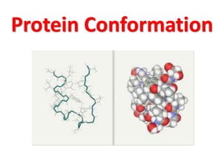
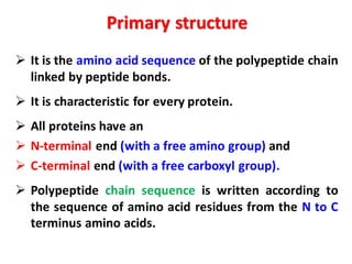
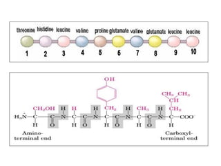
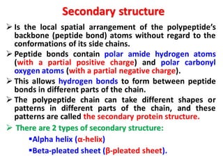
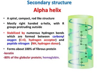
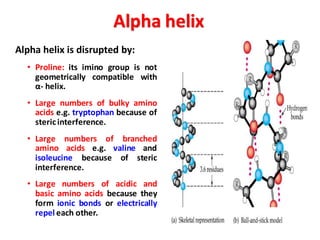
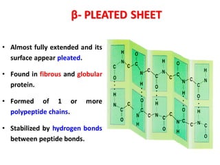
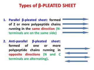
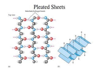
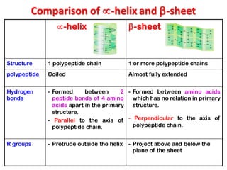
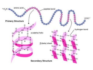
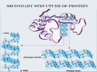
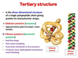
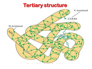
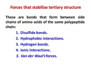
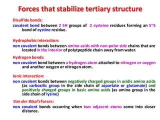
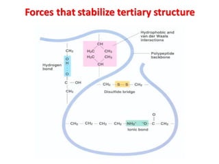
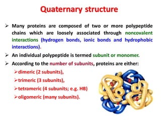
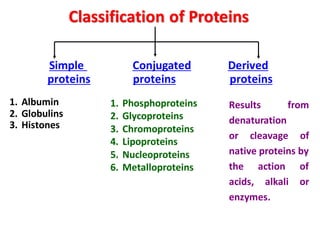
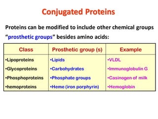
‫الغذاء‬ ‫بروتينات‬
Food Proteins
Prof. Dr. Mohamed Fawzy Ramadan Hassanien
Zagazig University, Egypt
-Amino Acid Sequence
-Protein Conformation
-Levels of Protein Structure
-Primary structure
-Secondary structure
-Tertiary structure
-Quaternary structure
-Classification of Proteins
-Denaturation of Protein
Peptide Linkage Formation
Peptides and Proteins
➢ Peptides and proteins are polymers of twenty amino
acids connected to each other by peptide bonds.
➢ Oligopeptide is formed of (2 –10) amino acids:
2 amino acids dipeptide,
3 amino acids tripeptide,
4 amino acids tetrapeptide ….etc.
➢ Polypeptide is formed of more than 10 amino acids.
In proteins,
almost all carboxyl and amino groups
are
combined in peptide linkage
and
not available for chemical reaction
(except for hydrogen bond formation).
-Like peptides, proteins are formed from amino acids through
amide linkages.
-Covalently bound hetero constituents can also be incorporated
into proteins. For example, phosphoproteins such as milk casein or
phosvitin of egg yolk contain phosphoric acid esters of serine and
threonine residues.
-The structure of a protein is dependent on the amino acid
sequence (the primary structure) which determines the molecular
conformation (secondary and tertiary structures).
-Proteins sometimes occur as molecular aggregates which are
arranged in an orderly geometric fashion (quaternary structure).
-The sequences and conformations of a large number of proteins
have been elucidated and recorded in several data bases.
Food Proteins
-Glycoproteins,
such as casein, various
components of egg white
and egg yolk, collagen
from connective tissue
and serum proteins of
some species of fish,
contain one or more
monosaccharide or
oligosaccharide units
bound O-glycosidically to
serine, threonine or
hydroxylysine or N-
glycosidically to
asparagine.
Amino Acid
Sequence
-Sequence analysis can only be conducted on a pure protein.
-First, the amino acid compositionis determinedafter acidic hydrolysis.
-The procedure (separation on a single cation-exchange resin column and color
development with ninhydrin reagent) has been standardized and automated
(amino acid analyzers).
-As an alternative to these established methods, the derivatization of amino
acids with the subsequent separation and detection of derivatives is possible
(pre-column derivatization).
Various derivatizationreagents can be selected, such as:
• 9-Fluorenylmethylchloroformate(FMOC)
• Phenylisothiocyanate(PITC)
• Dimethylaminoazobenzenesulfonylchloride (DABS-Cl)
• Dimethylaminonaphthalenesulfonylchloride (DANS-Cl)
• 7-Fluoro-4-nitrobenzo-2-oxa-1,3-diazole(NBDF)
• 7-Chloro-4-nitrobenzo-2-oxa-1,3-diazole(NBDCl)
• o-Phthaldialdehyde (OPA)
1-Amino Acid Composition, Subunits
Amino acid chromatogram.
Separation of a mixture of amino acids (10
nmol/amino acid) by an amino acid analyzer.
-It is also necessary to know the molecular weight(MW) of the protein.
-MW could be determined by gel column chromatography, ultracentrifugation
or electrophoresis.
-It is necessary to determine whether the protein is a single molecule or
consists of a number of different polypeptide chains (subunits) associated
through disulfide bonds or non-covalentforces.
-Dissociation into subunits can be accomplished by a change in pH, by chemical
modification of the protein, such as succinylation, or with denaturing agents
(urea, guanidine hydrochloride, sodium dodecyl sulfate SDS).
-Disulfide bonds, which are also found in proteins which consist of only one
peptide chain, can be cleaved by oxidation of cystine to cysteic acid or by
reduction to cysteine with subsequent alkylation of thiol group to prevent re-
oxidation.
-Separation of subunits is achieved by chromatographic or electrophoretic
methods.
Amino Acid Composition, Subunits
-N-terminal amino acids can be determined by treating a protein
with l-fluoro-2,4-dinitrobenzene (Sanger’s reagent) or 5-
dimethylaminonaphthalene-1-sulfonyl chloride (dansyl chloride).
-Another possibility is the reaction with cyanate, followed by
elimination of the N-terminal amino acid in the form of hydantoin,
and separation and recovery of the amino acid by cleavage of the
hydantoin .
-The N-terminal amino acid (and the amino acid sequence close to
the N-terminal) is accessible by hydrolysis with aminopeptidase, in
which case it should be remembered that the hydrolysis rate is
dependent on amino acid side chains and that proline residues are
not cleaved.
-A special procedure is required when the N-terminal residue is
acylated (N-formyl- or N-acetyl amino acids).
2-Terminal Groups
-Determination of C-terminal amino acids is possible via the
hydrazinolysis procedure recommended by Akabori:
-The C-terminal amino acid could be then separated from the
amino acid hydrazides by a cation exchange resin.
-The C-terminal amino acids can be removed
enzymatically by
- Carboxypeptidase A which cleaves amino acids with
aromatic and large aliphatic side chains,
- Carboxypeptidase B which cleaves lysine, arginine and
amino acids with neutral side chains or
- Carboxypeptidase C which cleaves with less specificity
but cleaves proline.
Terminal Groups
-Long peptide chains are usually fragmented. The fragments are then analyzed
for amino acid sequences.
-Selective enzymaticcleavage of peptide bonds is accomplished primarily with
Trypsin, which cleaves exclusivelyLys-X- and Arg-X-bonds,and
Chymotrypsin, which cleaves peptide bonds with less specificity (Tyr-X, Phe-X,
Trp-X and Leu-X).
-The enzymatic attack can be influenced by modificationof protein.For example,
-Acylation of the amino group of lysine limits tryptic hydrolysis to Arg-X,
-Substitution of the SH-group of cysteine residue with an aminoethyl group
introduces a new cleavage position for trypsin into the molecule “pseudolysine
residue”
3- Partial Hydrolysis
-Also suited for the specific
enzymatic hydrolysis of
peptide chains is the
endoproteinase Glu-C from
Staphylococcus aureus. It
cleaves Glu-X bonds as well
as Glu-X plus Asp-X bonds.
-The most important
chemical method for
selective cleavage uses
cyanogen bromide (BrCN)
to attack Met-X-linkages.
Partial Hydrolysis
-Hydrolysis of proteins with strong
acids reveals a difference in the rates
of hydrolysis of peptide bonds
depending on the next amino acid
side chain.
-Bonds involving amino groups of
serine and threonine are particularly
sensitive to hydrolysis.
-This effect is due to N→O-acyl
migration via oxazoline and
subsequent hydrolysis of the ester
bond.
-Hydrolysis of proteins with dilute
acids cleaves aspartyl-X-bonds.
Partial Hydrolysis
-Separation of peptide fragments is achieved by gel and ion-
exchange column chromatography using a volatile buffer as eluent
(pyridine) which can be removed by freeze-drying of the fractions.
-The separation of peptides and proteins by reversed-phase HPLC
has gained great importance, using volatile buffers mixed with
organic, water-soluble solvents as the mobile phase.
-The fragmentation of the protein is performed by different
enzymic and/or chemical techniques, at least by two enzymes of
different specifity.
-The arrangement of the obtained peptides in the same order as
they found in the protein is accomplished with the aid of
overlapping sequences.
Partial Hydrolysis
-The classical method is the Edman degradation reaction.
-It involves stepwise degradation of peptides with
phenylisothiocyanate.
-The resultant phenylthiohydantoin is identified directly.
-The stepwise reactions are performed in solution or on peptide
bound to a carrier, i. e. to a solid phase.
-Both approaches have been automated (“sequencer”). Carriers
used include resins containing amino groups (e.g., amino
polystyrene) or glass beads treated with amino alkylsiloxane:
4- Sequence Analysis
-The peptides are then attached to the carrier by
carboxyl groups (activation with carbodiimide as
in peptide synthesis) or by amino groups.
-For example, a peptide segment from the
hydrolysis of protein by trypsin has lysine as its
C-terminal amino acid. It is attached to the
carrier with phenylene-diisothiocyanate through
amino groups.
-Mild acidic treatment of the carrier under
conditions of the Edman degradation splits the
first peptide bond.
-The Edman procedure is then performed on
the shortened peptide through second, third and
subsequent repetitivereactions:
Sequence Analysis
Protein Conformation
Protein molecule can be formed of
one or more
polypeptide chains
which may vary in the number
and sequence of amino acid residues.
-Information about conformation is available
through X-ray crystallographic analysis of
protein crystals and by measuring the distance
between selected protons of the peptide chain
by means of H-NMR spectroscopy in solution.
-X-ray structural analysis of a fully extended
peptide chain reveal the lengths and angles of
bonds
-The peptide bond has partial (40%) double
bond character with electrons shared between
the C-O and C-N bonds.
-The resonance energy is about 83.6 kJ/mole
Structure of an elongated
peptide chain.
Extended Peptide Chains
Levels of Protein Structure
➢ Primary structure
➢ Secondary structure
➢ Tertiary structure
➢ Quaternary structure
➢ It is the amino acid sequence of the polypeptide chain
linked by peptide bonds.
➢ It is characteristic for every protein.
➢ All proteins have an
➢ N-terminal end (with a free amino group) and
➢ C-terminal end (with a free carboxyl group).
➢ Polypeptide chain sequence is written according to
the sequence of amino acid residues from the N to C
terminus amino acids.
Primary structure
➢ Is the local spatial arrangement of the polypeptide’s
backbone (peptide bond) atoms without regard to the
conformations of its side chains.
➢ Peptide bonds contain polar amide hydrogen atoms
(with a partial positive charge) and polar carbonyl
oxygen atoms (with a partial negative charge).
➢ This allows hydrogen bonds to form between peptide
bonds in different parts of the chain.
➢ The polypeptide chain can take different shapes or
patterns in different parts of the chain, and these
patterns are called the secondary protein structure.
➢ There are 2 types of secondary structure:
▪Alpha helix (α-helix)
▪Beta-pleated sheet (β-pleated sheet).
Secondary structure
Alpha helix
• A spiral, compact, rod like structure
• Mostly right handed α-helix, with R
groups protruding outside
• Stabilized by numerous hydrogen bonds
which are formed between carbonyl
oxygen (C=O, hydrogen acceptor) and
peptide nitrogen (NH, hydrogen donor).
• Forms about 100% of fibrous protein
-keratin
-80% of the globular protein; hemoglobin.
Secondary structure
Alpha helix
Alpha helix is disrupted by:
• Proline: its imino group is not
geometrically compatible with
α- helix.
• Large numbers of bulky amino
acids e.g. tryptophan because of
stericinterference.
• Large numbers of branched
amino acids e.g. valine and
isoleucine because of steric
interference.
• Large numbers of acidic and
basic amino acids because they
form ionic bonds or electrically
repel each other.
β- PLEATED SHEET
• Almost fully extended and its
surface appear pleated.
• Found in fibrous and globular
protein.
• Formed of 1 or more
polypeptide chains.
• Stabilized by hydrogen bonds
between peptide bonds.
Types of β-PLEATED SHEET
1. Parallel β-pleated sheet: formed
of 2 or more polypeptide chains
running in the same direction (N-
terminals are on the same side)
2. Anti-parallel β-pleated sheet:
formed of one or more
polypeptide chains running in
opposite directions (N and C
terminals are alternating).
Comparison of -helix and -sheet
-helix -sheet
Structure 1 polypeptide chain 1 or more polypeptide chains
polypeptide Coiled Almost fully extended
Hydrogen
bonds
- Formed between 2
peptide bonds of 4 amino
acids apart in the primary
structure.
- Parallel to the axis of
polypeptide chain.
- Formed between amino acids
which has no relation in primary
structure.
- Perpendicular to the axis of
polypeptide chain.
R groups - Protrude outside the helix - Project above and below the
plane of the sheet
SECONDARY STRUCTURE OF PROTEIN
α- helix
Tertiary structure
• Is the three dimensional structure
of a single polypeptide chain giving
protein its characteristic shape.
I- Globular proteins (enzymes)
• Approximately spherical shape- water
Soluble.
II- Fibrous proteins (structural
proteins)
• Rod-like shape
• Poor water solubility.
• Cross links and bonds in 3ry structure:
• S-S bond, Ionic, Hydrophobicinteractions
and H-bonding.
Fibrous
protein
Globular
protein
Tertiary structure
Forces that stabilize tertiary structure
These are bonds that form between side
chains of amino acids of the same polypeptide
chain:
1. Disulfide bonds.
2. Hydrophobic interactions.
3. Hydrogen bonds.
4. Ionic interactions.
5. Van der Waal’s forces.
Disulfide bonds:
covalent bond between 2 SH groups of 2 cysteine residues forming an S~S
bond of cystine residue.
Hydrophobicinteraction:
non covalent bonds between amino acids with non-polar side chains that are
locatedin the interior of polytpeptide chain away from water.
Hydrogenbonds:
non covalent bond between a hydrogen atom attached to nitrogen or oxygen
and another oxygen or nitrogenatom.
Ionic interaction:
non covalent bonds between negatively charged groups in acidic amino acids
(as carboxilic group in the side chain of aspartate or glutamate) and
positively charged groups in basic amino acids (as amino group in the
side chain of lysine)
Van der Waal’s forces:
non covalent bonds occurring when two adjacent atoms come into closer
distance.
Forces that stabilize tertiary structure
➢ Many proteins are composed of two or more polypeptide
chains which are loosely associated through noncovalent
interactions (hydrogen bonds, ionic bonds and hydrophobic
interactions).
➢ An individual polypeptide is termed subunit or monomer.
➢ According to the number of subunits, proteins are either:
➢dimeric (2 subunits),
➢trimeric (3 subunits),
➢tetrameric (4 subunits; e.g. HB)
➢oligomeric (many subunits).
Quaternary structure
Examples of globular proteins
Classification of Proteins
Simple Conjugated Derived
proteins proteins proteins
1. Albumin
2. Globulins
3. Histones
1. Phosphoproteins
2. Glycoproteins
3. Chromoproteins
4. Lipoproteins
5. Nucleoproteins
6. Metalloproteins
Results from
denaturation
or cleavage of
native proteins by
the action of
acids, alkali or
enzymes.
Proteins can be modified to include other chemical groups
“prosthetic groups” besides amino acids:
Class Prosthetic group (s) Example
•Lipoproteins
•Glycoproteins
•Phosphoproteins
•hemoproteins
•Lipids
•Carbohydrates
•Phosphate groups
•Heme (iron porphyrin)
•VLDL
•Immunoglobulin G
•Casinogen of milk
•Hemoglobin
Conjugated Proteins
Denaturation of Protein
-The term denaturation denotes a reversible or irreversible change
of native conformation (tertiary structure) without cleavage of
covalent bonds (except for disulfide bridges).
➢ The primary structure of the protein is not changed because the
peptide bonds are not affected
➢ Denaturing agents include:
1. Heat
2. Changes in pH (concentrated acids or alkali)
3. Ultraviolet rays
4. X ray
5. High salt concentration
6. Heavy metals.
Denaturation
-Denaturation is possible with any treatment that cleaves
hydrogen bridges, ionic or hydrophobic bonds. This can be
accomplished by: changing the temperature, adjusting the pH,
increasing the interface area, or adding organic solvents, salts,
urea, or detergents such as sodium dodecyl sulfate.
-Denaturation is generally reversible when the peptide chain is
stabilized in its unfolded state by the denaturing agent and native
conformation can be re-established after removal of the agent.
-Irreversible denaturation occurs when the unfolded peptide chain
is stabilized by interaction with other chains (as occurs for instance
with egg proteins during boiling). During unfolding reactive groups,
such as thiol groups, that were blocked, may be exposed. Their
participation in the formation of disulfide bonds may also cause an
irreversible denaturation.
Denaturation
Effects of Denaturation
-Denaturation destroys the native conformation of protein.
-Denaturation destroys the biologic activity of protein, there is
loss of hormonal, enzymatic and antibody activity.
Applications of protein denaturing
1- Boiling eggs: Change in albumin shape and solubility.
2- Cooking meat: Easily chewable, digestible.
3- Swabbing skin with alcohol (disinfectant):
Denatures/kills bacteria and viruses.
4- HCl in our stomach: denatures proteins and making it easily
digestible by enzymes
- So, eating cooked eggs, meat and liver is more useful to
humans than eating them raw
-An aggregation of the peptide
chains caused by the folding of
globular proteins is connected with
reduced solubility or swellability.
-Thus the part of wheat gluten that
is soluble in acetic acid diminishes
as heat stress increases.
-As a result of the reduced rising
capacity of gluten caused by the
pre-treatment, the volume of
bread made of recombined flours
is smaller.
Solubility of gluten
(wheat) in diluted acetic
acid after various forms of
thermal stress
Denaturation of Protein:
Examples in Food
-In the case of fibrous proteins, denaturation, through
destruction of the highly ordered structure, generally
leads to increased solubility or rising capacity. One
example is the thermally caused collagen-to-gelatin
conversion, which occurs when meat is cooked.
-The thermal denaturation of the whey proteins β-
lactoglobulin and α-lactalbumin has been well-studied.
-Denaturation of biologically active proteins is usually
associated with loss of activity. The fact that denatured
proteins are more readily digested by proteolytic
enzymes is also of interest.
Denaturation of Protein:
Examples in Food

بروتينات الغذاء

  • 1.
    ‫الغذاء‬ ‫بروتينات‬ Food Proteins Prof.Dr. Mohamed Fawzy Ramadan Hassanien Zagazig University, Egypt
  • 2.
    -Amino Acid Sequence -ProteinConformation -Levels of Protein Structure -Primary structure -Secondary structure -Tertiary structure -Quaternary structure -Classification of Proteins -Denaturation of Protein
  • 3.
  • 4.
    Peptides and Proteins ➢Peptides and proteins are polymers of twenty amino acids connected to each other by peptide bonds. ➢ Oligopeptide is formed of (2 –10) amino acids: 2 amino acids dipeptide, 3 amino acids tripeptide, 4 amino acids tetrapeptide ….etc. ➢ Polypeptide is formed of more than 10 amino acids.
  • 5.
    In proteins, almost allcarboxyl and amino groups are combined in peptide linkage and not available for chemical reaction (except for hydrogen bond formation).
  • 6.
    -Like peptides, proteinsare formed from amino acids through amide linkages. -Covalently bound hetero constituents can also be incorporated into proteins. For example, phosphoproteins such as milk casein or phosvitin of egg yolk contain phosphoric acid esters of serine and threonine residues. -The structure of a protein is dependent on the amino acid sequence (the primary structure) which determines the molecular conformation (secondary and tertiary structures). -Proteins sometimes occur as molecular aggregates which are arranged in an orderly geometric fashion (quaternary structure). -The sequences and conformations of a large number of proteins have been elucidated and recorded in several data bases. Food Proteins
  • 7.
    -Glycoproteins, such as casein,various components of egg white and egg yolk, collagen from connective tissue and serum proteins of some species of fish, contain one or more monosaccharide or oligosaccharide units bound O-glycosidically to serine, threonine or hydroxylysine or N- glycosidically to asparagine.
  • 8.
  • 9.
    -Sequence analysis canonly be conducted on a pure protein. -First, the amino acid compositionis determinedafter acidic hydrolysis. -The procedure (separation on a single cation-exchange resin column and color development with ninhydrin reagent) has been standardized and automated (amino acid analyzers). -As an alternative to these established methods, the derivatization of amino acids with the subsequent separation and detection of derivatives is possible (pre-column derivatization). Various derivatizationreagents can be selected, such as: • 9-Fluorenylmethylchloroformate(FMOC) • Phenylisothiocyanate(PITC) • Dimethylaminoazobenzenesulfonylchloride (DABS-Cl) • Dimethylaminonaphthalenesulfonylchloride (DANS-Cl) • 7-Fluoro-4-nitrobenzo-2-oxa-1,3-diazole(NBDF) • 7-Chloro-4-nitrobenzo-2-oxa-1,3-diazole(NBDCl) • o-Phthaldialdehyde (OPA) 1-Amino Acid Composition, Subunits
  • 10.
    Amino acid chromatogram. Separationof a mixture of amino acids (10 nmol/amino acid) by an amino acid analyzer.
  • 11.
    -It is alsonecessary to know the molecular weight(MW) of the protein. -MW could be determined by gel column chromatography, ultracentrifugation or electrophoresis. -It is necessary to determine whether the protein is a single molecule or consists of a number of different polypeptide chains (subunits) associated through disulfide bonds or non-covalentforces. -Dissociation into subunits can be accomplished by a change in pH, by chemical modification of the protein, such as succinylation, or with denaturing agents (urea, guanidine hydrochloride, sodium dodecyl sulfate SDS). -Disulfide bonds, which are also found in proteins which consist of only one peptide chain, can be cleaved by oxidation of cystine to cysteic acid or by reduction to cysteine with subsequent alkylation of thiol group to prevent re- oxidation. -Separation of subunits is achieved by chromatographic or electrophoretic methods. Amino Acid Composition, Subunits
  • 12.
    -N-terminal amino acidscan be determined by treating a protein with l-fluoro-2,4-dinitrobenzene (Sanger’s reagent) or 5- dimethylaminonaphthalene-1-sulfonyl chloride (dansyl chloride). -Another possibility is the reaction with cyanate, followed by elimination of the N-terminal amino acid in the form of hydantoin, and separation and recovery of the amino acid by cleavage of the hydantoin . -The N-terminal amino acid (and the amino acid sequence close to the N-terminal) is accessible by hydrolysis with aminopeptidase, in which case it should be remembered that the hydrolysis rate is dependent on amino acid side chains and that proline residues are not cleaved. -A special procedure is required when the N-terminal residue is acylated (N-formyl- or N-acetyl amino acids). 2-Terminal Groups
  • 13.
    -Determination of C-terminalamino acids is possible via the hydrazinolysis procedure recommended by Akabori: -The C-terminal amino acid could be then separated from the amino acid hydrazides by a cation exchange resin.
  • 14.
    -The C-terminal aminoacids can be removed enzymatically by - Carboxypeptidase A which cleaves amino acids with aromatic and large aliphatic side chains, - Carboxypeptidase B which cleaves lysine, arginine and amino acids with neutral side chains or - Carboxypeptidase C which cleaves with less specificity but cleaves proline. Terminal Groups
  • 15.
    -Long peptide chainsare usually fragmented. The fragments are then analyzed for amino acid sequences. -Selective enzymaticcleavage of peptide bonds is accomplished primarily with Trypsin, which cleaves exclusivelyLys-X- and Arg-X-bonds,and Chymotrypsin, which cleaves peptide bonds with less specificity (Tyr-X, Phe-X, Trp-X and Leu-X). -The enzymatic attack can be influenced by modificationof protein.For example, -Acylation of the amino group of lysine limits tryptic hydrolysis to Arg-X, -Substitution of the SH-group of cysteine residue with an aminoethyl group introduces a new cleavage position for trypsin into the molecule “pseudolysine residue” 3- Partial Hydrolysis
  • 16.
    -Also suited forthe specific enzymatic hydrolysis of peptide chains is the endoproteinase Glu-C from Staphylococcus aureus. It cleaves Glu-X bonds as well as Glu-X plus Asp-X bonds. -The most important chemical method for selective cleavage uses cyanogen bromide (BrCN) to attack Met-X-linkages. Partial Hydrolysis
  • 17.
    -Hydrolysis of proteinswith strong acids reveals a difference in the rates of hydrolysis of peptide bonds depending on the next amino acid side chain. -Bonds involving amino groups of serine and threonine are particularly sensitive to hydrolysis. -This effect is due to N→O-acyl migration via oxazoline and subsequent hydrolysis of the ester bond. -Hydrolysis of proteins with dilute acids cleaves aspartyl-X-bonds. Partial Hydrolysis
  • 18.
    -Separation of peptidefragments is achieved by gel and ion- exchange column chromatography using a volatile buffer as eluent (pyridine) which can be removed by freeze-drying of the fractions. -The separation of peptides and proteins by reversed-phase HPLC has gained great importance, using volatile buffers mixed with organic, water-soluble solvents as the mobile phase. -The fragmentation of the protein is performed by different enzymic and/or chemical techniques, at least by two enzymes of different specifity. -The arrangement of the obtained peptides in the same order as they found in the protein is accomplished with the aid of overlapping sequences. Partial Hydrolysis
  • 19.
    -The classical methodis the Edman degradation reaction. -It involves stepwise degradation of peptides with phenylisothiocyanate. -The resultant phenylthiohydantoin is identified directly. -The stepwise reactions are performed in solution or on peptide bound to a carrier, i. e. to a solid phase. -Both approaches have been automated (“sequencer”). Carriers used include resins containing amino groups (e.g., amino polystyrene) or glass beads treated with amino alkylsiloxane: 4- Sequence Analysis
  • 20.
    -The peptides arethen attached to the carrier by carboxyl groups (activation with carbodiimide as in peptide synthesis) or by amino groups. -For example, a peptide segment from the hydrolysis of protein by trypsin has lysine as its C-terminal amino acid. It is attached to the carrier with phenylene-diisothiocyanate through amino groups. -Mild acidic treatment of the carrier under conditions of the Edman degradation splits the first peptide bond. -The Edman procedure is then performed on the shortened peptide through second, third and subsequent repetitivereactions: Sequence Analysis
  • 21.
  • 22.
    Protein molecule canbe formed of one or more polypeptide chains which may vary in the number and sequence of amino acid residues.
  • 23.
    -Information about conformationis available through X-ray crystallographic analysis of protein crystals and by measuring the distance between selected protons of the peptide chain by means of H-NMR spectroscopy in solution. -X-ray structural analysis of a fully extended peptide chain reveal the lengths and angles of bonds -The peptide bond has partial (40%) double bond character with electrons shared between the C-O and C-N bonds. -The resonance energy is about 83.6 kJ/mole Structure of an elongated peptide chain. Extended Peptide Chains
  • 24.
    Levels of ProteinStructure ➢ Primary structure ➢ Secondary structure ➢ Tertiary structure ➢ Quaternary structure
  • 25.
    ➢ It isthe amino acid sequence of the polypeptide chain linked by peptide bonds. ➢ It is characteristic for every protein. ➢ All proteins have an ➢ N-terminal end (with a free amino group) and ➢ C-terminal end (with a free carboxyl group). ➢ Polypeptide chain sequence is written according to the sequence of amino acid residues from the N to C terminus amino acids. Primary structure
  • 27.
    ➢ Is thelocal spatial arrangement of the polypeptide’s backbone (peptide bond) atoms without regard to the conformations of its side chains. ➢ Peptide bonds contain polar amide hydrogen atoms (with a partial positive charge) and polar carbonyl oxygen atoms (with a partial negative charge). ➢ This allows hydrogen bonds to form between peptide bonds in different parts of the chain. ➢ The polypeptide chain can take different shapes or patterns in different parts of the chain, and these patterns are called the secondary protein structure. ➢ There are 2 types of secondary structure: ▪Alpha helix (α-helix) ▪Beta-pleated sheet (β-pleated sheet). Secondary structure
  • 28.
    Alpha helix • Aspiral, compact, rod like structure • Mostly right handed α-helix, with R groups protruding outside • Stabilized by numerous hydrogen bonds which are formed between carbonyl oxygen (C=O, hydrogen acceptor) and peptide nitrogen (NH, hydrogen donor). • Forms about 100% of fibrous protein -keratin -80% of the globular protein; hemoglobin. Secondary structure
  • 29.
    Alpha helix Alpha helixis disrupted by: • Proline: its imino group is not geometrically compatible with α- helix. • Large numbers of bulky amino acids e.g. tryptophan because of stericinterference. • Large numbers of branched amino acids e.g. valine and isoleucine because of steric interference. • Large numbers of acidic and basic amino acids because they form ionic bonds or electrically repel each other.
  • 30.
    β- PLEATED SHEET •Almost fully extended and its surface appear pleated. • Found in fibrous and globular protein. • Formed of 1 or more polypeptide chains. • Stabilized by hydrogen bonds between peptide bonds.
  • 31.
    Types of β-PLEATEDSHEET 1. Parallel β-pleated sheet: formed of 2 or more polypeptide chains running in the same direction (N- terminals are on the same side) 2. Anti-parallel β-pleated sheet: formed of one or more polypeptide chains running in opposite directions (N and C terminals are alternating).
  • 33.
    Comparison of -helixand -sheet -helix -sheet Structure 1 polypeptide chain 1 or more polypeptide chains polypeptide Coiled Almost fully extended Hydrogen bonds - Formed between 2 peptide bonds of 4 amino acids apart in the primary structure. - Parallel to the axis of polypeptide chain. - Formed between amino acids which has no relation in primary structure. - Perpendicular to the axis of polypeptide chain. R groups - Protrude outside the helix - Project above and below the plane of the sheet
  • 35.
    SECONDARY STRUCTURE OFPROTEIN α- helix
  • 36.
    Tertiary structure • Isthe three dimensional structure of a single polypeptide chain giving protein its characteristic shape. I- Globular proteins (enzymes) • Approximately spherical shape- water Soluble. II- Fibrous proteins (structural proteins) • Rod-like shape • Poor water solubility. • Cross links and bonds in 3ry structure: • S-S bond, Ionic, Hydrophobicinteractions and H-bonding. Fibrous protein Globular protein
  • 37.
  • 38.
    Forces that stabilizetertiary structure These are bonds that form between side chains of amino acids of the same polypeptide chain: 1. Disulfide bonds. 2. Hydrophobic interactions. 3. Hydrogen bonds. 4. Ionic interactions. 5. Van der Waal’s forces.
  • 39.
    Disulfide bonds: covalent bondbetween 2 SH groups of 2 cysteine residues forming an S~S bond of cystine residue. Hydrophobicinteraction: non covalent bonds between amino acids with non-polar side chains that are locatedin the interior of polytpeptide chain away from water. Hydrogenbonds: non covalent bond between a hydrogen atom attached to nitrogen or oxygen and another oxygen or nitrogenatom. Ionic interaction: non covalent bonds between negatively charged groups in acidic amino acids (as carboxilic group in the side chain of aspartate or glutamate) and positively charged groups in basic amino acids (as amino group in the side chain of lysine) Van der Waal’s forces: non covalent bonds occurring when two adjacent atoms come into closer distance. Forces that stabilize tertiary structure
  • 40.
    Forces that stabilizetertiary structure
  • 41.
    ➢ Many proteinsare composed of two or more polypeptide chains which are loosely associated through noncovalent interactions (hydrogen bonds, ionic bonds and hydrophobic interactions). ➢ An individual polypeptide is termed subunit or monomer. ➢ According to the number of subunits, proteins are either: ➢dimeric (2 subunits), ➢trimeric (3 subunits), ➢tetrameric (4 subunits; e.g. HB) ➢oligomeric (many subunits). Quaternary structure
  • 42.
  • 43.
    Classification of Proteins SimpleConjugated Derived proteins proteins proteins 1. Albumin 2. Globulins 3. Histones 1. Phosphoproteins 2. Glycoproteins 3. Chromoproteins 4. Lipoproteins 5. Nucleoproteins 6. Metalloproteins Results from denaturation or cleavage of native proteins by the action of acids, alkali or enzymes.
  • 44.
    Proteins can bemodified to include other chemical groups “prosthetic groups” besides amino acids: Class Prosthetic group (s) Example •Lipoproteins •Glycoproteins •Phosphoproteins •hemoproteins •Lipids •Carbohydrates •Phosphate groups •Heme (iron porphyrin) •VLDL •Immunoglobulin G •Casinogen of milk •Hemoglobin Conjugated Proteins
  • 45.
  • 46.
    Denaturation of Protein -Theterm denaturation denotes a reversible or irreversible change of native conformation (tertiary structure) without cleavage of covalent bonds (except for disulfide bridges). ➢ The primary structure of the protein is not changed because the peptide bonds are not affected ➢ Denaturing agents include: 1. Heat 2. Changes in pH (concentrated acids or alkali) 3. Ultraviolet rays 4. X ray 5. High salt concentration 6. Heavy metals.
  • 47.
    Denaturation -Denaturation is possiblewith any treatment that cleaves hydrogen bridges, ionic or hydrophobic bonds. This can be accomplished by: changing the temperature, adjusting the pH, increasing the interface area, or adding organic solvents, salts, urea, or detergents such as sodium dodecyl sulfate. -Denaturation is generally reversible when the peptide chain is stabilized in its unfolded state by the denaturing agent and native conformation can be re-established after removal of the agent. -Irreversible denaturation occurs when the unfolded peptide chain is stabilized by interaction with other chains (as occurs for instance with egg proteins during boiling). During unfolding reactive groups, such as thiol groups, that were blocked, may be exposed. Their participation in the formation of disulfide bonds may also cause an irreversible denaturation.
  • 48.
  • 49.
    Effects of Denaturation -Denaturationdestroys the native conformation of protein. -Denaturation destroys the biologic activity of protein, there is loss of hormonal, enzymatic and antibody activity. Applications of protein denaturing 1- Boiling eggs: Change in albumin shape and solubility. 2- Cooking meat: Easily chewable, digestible. 3- Swabbing skin with alcohol (disinfectant): Denatures/kills bacteria and viruses. 4- HCl in our stomach: denatures proteins and making it easily digestible by enzymes - So, eating cooked eggs, meat and liver is more useful to humans than eating them raw
  • 50.
    -An aggregation ofthe peptide chains caused by the folding of globular proteins is connected with reduced solubility or swellability. -Thus the part of wheat gluten that is soluble in acetic acid diminishes as heat stress increases. -As a result of the reduced rising capacity of gluten caused by the pre-treatment, the volume of bread made of recombined flours is smaller. Solubility of gluten (wheat) in diluted acetic acid after various forms of thermal stress Denaturation of Protein: Examples in Food
  • 51.
    -In the caseof fibrous proteins, denaturation, through destruction of the highly ordered structure, generally leads to increased solubility or rising capacity. One example is the thermally caused collagen-to-gelatin conversion, which occurs when meat is cooked. -The thermal denaturation of the whey proteins β- lactoglobulin and α-lactalbumin has been well-studied. -Denaturation of biologically active proteins is usually associated with loss of activity. The fact that denatured proteins are more readily digested by proteolytic enzymes is also of interest. Denaturation of Protein: Examples in Food