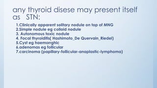
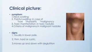
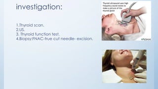
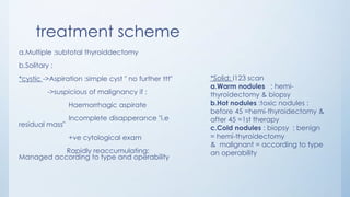
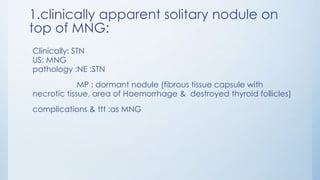
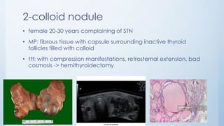
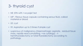
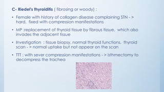
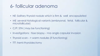
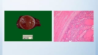
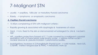
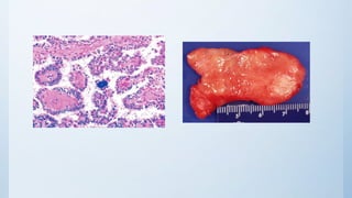
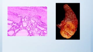
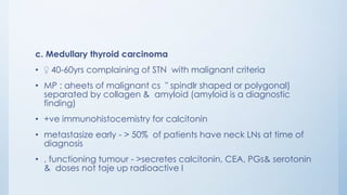
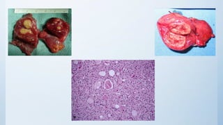
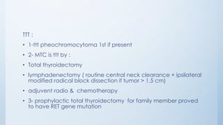
Solitary thyroid nodule
By: Yehia Matter EL-ashcker
any thyroid disese may present itself
as STN:
1.Clinically apparent solitary nodule on top of MNG
2.Simple nodule eg colloid nodule
3. Autonomous toxic nodule
4. Focal thyroiditis( Hashimoto_De Quervain_Riedel)
5.Cyst eg haemorrghic
6.adenomas eg follicular
7.carcinoma (papillary-follicular-anaplastic-lymphoma)
Clinical picture:
• symptom:
1.Neck swelling
2. Painful swelling: in case of
*hae *thyroiditis *malignancy
3. Toxic manifestation :in toxic nodules
4.Criteria of malignany:in malignant nodules
• signs:
1. Usually in lower pole.
2. Firm ,hard or cystic.
3.moves up and down with deglutition
investigation:
1.Thyroid scan.
2.US.
3. Thyroid function test.
4.Biopsy:FNAC-true cut needle- excision.
treatment scheme
a.Multiple :subtotal thyroiddectomy
b.Solitary :
*cystic ->Aspiration :simple cyst " no further ttt"
->suspicious of malignancy if :
Haemorrhagic aspirate
Incomplete disapperance "i.e
residual mass"
+ve cytological exam
Rapidly reaccumulating:
Managed according to type and operability
*Solid: I123 scan
a.Warm nodules : hemi-
thyroidectomy & biopsy
b.Hot nodules :toxic nodules :
before 45 =hemi-thyroidectomy &
after 45 =1st therapy
c.Cold nodules : biopsy : benign
= hemi-thyroidectomy
& malignant = according to type
an operability
1.clinically apparent solitary nodule on
top of MNG:
Clinically: STN
US: MNG
pathology :NE :STN
MP : dormant nodule (fibrous tissue capsule with
necrotic tissue, area of Haemorrhage & destroyed thyroid follicles)
complications & ttt :as MNG
2-colloid nodule
• female 20-30 years complaining of STN
• MP: fibrous tissue with capsule surrounding inactive thyroid
follicles filled with colloid
• ttt: with compression manifestations, retrosternal extension, bad
cosmosis -> hemithyroidectomy
3- thyroid cyst
• NE :STN with +ve pajet test
• MP : Fibrous tissue capsule containing serous fluid, colloid
material or blood
• US :cyst
• ttt: Aspiration up to 3 times if simple cyst
• suspicious of malignancy (Haemorrhagic aspirate, residual tissue
mass, rapidly reaccumulating, +ve cytology) ->
hemithyroidectomy or total thyroidectomy according to
pathology
4-autonomous toxic nodule "ATN"
• Etiology: autonomous activity of solitary nodule "with no thyroid Ab.)
• pathology :
• NE : STN + toxic manifestations
• ME : over activity nodule surrounded by suppressed thyroid tissue
• this over active nodule consists of autonomously active thyroid follicles
surrounded by fibrous tissue
• Investigation :increase in T3,T4 & decrease in TSH,
• U. S :Solitary solid nodule
• thyroid scan -> hot nodule
• ttt : <45yrs - >hemi-thyroidectomy, >45yrs - >radioactive I131
5.Thyroiditis
A- autoimmune (Hashimoto) thyroiditis :
• 30 - 50 yrs, may give a family history of thyroiditis
• NE :STN +/- Pain & tenderness , rubbery or firm in consistency. Initially - > toxic manifestations, later
on - > manifestations if hypothyroidism
• MP : infiltration of the thyroid gland in the affected area by lymphocytes & plasma cells.
• Complications : Hypothyroidism , Malignant change (malignant lymphoma) - > 70 fold increase in
the risk O (papillary carcinoma)
• Investigation :
• Thytoid function tests : initial hyperthyroidism then hypothyroidism ( increas in TSH & decrease in T3
& T4)
• Thyroid Antibodies : anti microsomal Ab. , anti thyroglobulin Ab.
• Thyroid biopsy
• TTT : hemi-thyroidectomy with retrosternal extension, compression manifestations or suspicious of
malignancy
B-subacute thyroiditis (Granulomatous - de Quervain's thyroiditis) :
• Female 30-40 years, history of URTI 2 weeks ago, followed by neck pain
& toxic manifestations
• NE:tender STN
• MP : degeneration of thyroid follicles that become surrounded by
giant cs forming granuloma
• Investigation : increase in Esr, T3,T4 & decrease in TSH, radio active
iodine uptake
• TTT: the disease is self-limited so ttt is primarily symptomatic
(Prednisolone & NSAID)
C- Riedel's thyroiditis ( fibrosing or woody) :
• Female with history of collagen disease complaining STN - >
hard, fixed with compression manifestations
• MP :replacement of thyroid tissue by fibrous tissue, which also
invades the adjacent tissue
• Investigation : tissue biopsy, normal thyroid functions, thyroid
scan - > normal uptake but not appear on the scan
• TTT : with sever compression manifestations - > isthmectomy to
decompress the trachea
6- follicular adenoma
• NE :Solitary thyroid nodule which is firm & well encapsulated
• ME :several histological variants (embryonal, fetal, follicular &
microfollicular)
• C/P: STN ( may be functioning)
• Investigations : tisse biopsy - >no anglo capsular invasion
• Thyroid scan - > warm nodules (if functioning)
• TTT :hemi-thyroidectomy
7-Malignant STN
• usually - > papillary, follicular or medullary thyroid carcinoma
• Rarely - > lymphoma or anaplaatic carcinoma
A -Papillary thyroid carcinoma
• 15-40yrs complaining of STN with malignant criteria :
• Rapidly growing & associated with dysphagia & hoarseness of voice
• Hard, >1cm, fixed to the skin or sternomastoid wit enlarged Cx. LNs & +ve berr's
sign
• MP : papillary projections formed of C. T core covered by a malignant cuboidal
epithelium with a characteristic pale empty nuclei - > orphan Annie nuclei +
nuclear drooves & pseudo-inclusion + psammoma bodies.
• Investigations :increasw in S.thyroglobulin thyroid scan - > cold nodule, neck U/S,
CT&MRI, indirect larygoscope & FNAC + metastatic work up
B. follicular carcinoma
• female 40-60yrs complaining of STN with malignant criteria
• MP : thyroid follicles with variable degree of differentiation (solid sheets of malignant
cells may be present).
• angiocapsular invasion is a characteristic feature & according to its degree, follicular
carcinoma may be :
• non-invasive : if angiocapsular invasion is minimal
• invasive :if angio capsular is moderate or marked
• spread mainly by blood, differentiated tumour & may be functioning
• investigations :
• increase in S.thyroglobulin , Thyroid scan - >cold nodule , Neck U/S, CT&MRI
• Indirect larygoscope & trucut or open surgical biopsy + metastatic work up
•
ttt of differentiated thyroid carcinoma (papillary & follicular)
• 1- total (in high risk group) or near total thyroidectomy (in low risk
group) to avoid injury of parathyroids & RLN at least on one side
• 2- Lymphadectomy : for involved Cx. LNs -> cherry 🍒 picking or
modified block dissection "spare sternomastoid, accessory nerve &
Internal jugular vein"
• 3-thyroid tissue suppression :post operative thyroxin in a full suppressive
dose (making TSH < 1 micro micro /L) as tge tumour is TSH dependant
• 4-radiactive iodin scanning and ablation : post operative whole body
scan using radioactive I131 to detect metastasis (done after stoppage
of T4 for 6 weeks in order to permit a high stimulating level of TSH on
the metastasis) - > if metastasis is found - >theraputic dosses of radio
active iodine (I131)
c. Medullary thyroid carcinoma
• ♀ 40-60yrs complaining of STN with malignant criteria
• MP : aheets of malignant cs " spindlr shaped or polygonal)
separated by collagen & amyloid (amyloid is a diagnostic
finding)
• +ve immunohistocemistry for calcitonin
• metastasize early - > 50% of patients have neck LNs at time of
diagnosis
• , functioning tumour - >secretes calcitonin, CEA, PGs& serotonin
& doses not taje up radioactive I
TTT :
• 1-ttt pheochromocytoma 1st if present
• 2- MTC is ttt by :
• Total thyroidectomy
• lymphadenectomy ( routine central neck clearance + ipsilateral
modified radical block dissection if tumor > 1.5 cm)
• adjuvent radio & chemotherapy
• 3- prophylactic total thyroidectomy for family member proved
to have RET gene mutation
Thank you

Solitary thyroid nodule

  • 1.
    Solitary thyroid nodule By:Yehia Matter EL-ashcker
  • 2.
    any thyroid disesemay present itself as STN: 1.Clinically apparent solitary nodule on top of MNG 2.Simple nodule eg colloid nodule 3. Autonomous toxic nodule 4. Focal thyroiditis( Hashimoto_De Quervain_Riedel) 5.Cyst eg haemorrghic 6.adenomas eg follicular 7.carcinoma (papillary-follicular-anaplastic-lymphoma)
  • 3.
    Clinical picture: • symptom: 1.Neckswelling 2. Painful swelling: in case of *hae *thyroiditis *malignancy 3. Toxic manifestation :in toxic nodules 4.Criteria of malignany:in malignant nodules • signs: 1. Usually in lower pole. 2. Firm ,hard or cystic. 3.moves up and down with deglutition
  • 4.
    investigation: 1.Thyroid scan. 2.US. 3. Thyroidfunction test. 4.Biopsy:FNAC-true cut needle- excision.
  • 5.
    treatment scheme a.Multiple :subtotalthyroiddectomy b.Solitary : *cystic ->Aspiration :simple cyst " no further ttt" ->suspicious of malignancy if : Haemorrhagic aspirate Incomplete disapperance "i.e residual mass" +ve cytological exam Rapidly reaccumulating: Managed according to type and operability *Solid: I123 scan a.Warm nodules : hemi- thyroidectomy & biopsy b.Hot nodules :toxic nodules : before 45 =hemi-thyroidectomy & after 45 =1st therapy c.Cold nodules : biopsy : benign = hemi-thyroidectomy & malignant = according to type an operability
  • 6.
    1.clinically apparent solitarynodule on top of MNG: Clinically: STN US: MNG pathology :NE :STN MP : dormant nodule (fibrous tissue capsule with necrotic tissue, area of Haemorrhage & destroyed thyroid follicles) complications & ttt :as MNG
  • 7.
    2-colloid nodule • female20-30 years complaining of STN • MP: fibrous tissue with capsule surrounding inactive thyroid follicles filled with colloid • ttt: with compression manifestations, retrosternal extension, bad cosmosis -> hemithyroidectomy
  • 8.
    3- thyroid cyst •NE :STN with +ve pajet test • MP : Fibrous tissue capsule containing serous fluid, colloid material or blood • US :cyst • ttt: Aspiration up to 3 times if simple cyst • suspicious of malignancy (Haemorrhagic aspirate, residual tissue mass, rapidly reaccumulating, +ve cytology) -> hemithyroidectomy or total thyroidectomy according to pathology
  • 9.
    4-autonomous toxic nodule"ATN" • Etiology: autonomous activity of solitary nodule "with no thyroid Ab.) • pathology : • NE : STN + toxic manifestations • ME : over activity nodule surrounded by suppressed thyroid tissue • this over active nodule consists of autonomously active thyroid follicles surrounded by fibrous tissue • Investigation :increase in T3,T4 & decrease in TSH, • U. S :Solitary solid nodule • thyroid scan -> hot nodule • ttt : <45yrs - >hemi-thyroidectomy, >45yrs - >radioactive I131
  • 10.
    5.Thyroiditis A- autoimmune (Hashimoto)thyroiditis : • 30 - 50 yrs, may give a family history of thyroiditis • NE :STN +/- Pain & tenderness , rubbery or firm in consistency. Initially - > toxic manifestations, later on - > manifestations if hypothyroidism • MP : infiltration of the thyroid gland in the affected area by lymphocytes & plasma cells. • Complications : Hypothyroidism , Malignant change (malignant lymphoma) - > 70 fold increase in the risk O (papillary carcinoma) • Investigation : • Thytoid function tests : initial hyperthyroidism then hypothyroidism ( increas in TSH & decrease in T3 & T4) • Thyroid Antibodies : anti microsomal Ab. , anti thyroglobulin Ab. • Thyroid biopsy • TTT : hemi-thyroidectomy with retrosternal extension, compression manifestations or suspicious of malignancy
  • 12.
    B-subacute thyroiditis (Granulomatous- de Quervain's thyroiditis) : • Female 30-40 years, history of URTI 2 weeks ago, followed by neck pain & toxic manifestations • NE:tender STN • MP : degeneration of thyroid follicles that become surrounded by giant cs forming granuloma • Investigation : increase in Esr, T3,T4 & decrease in TSH, radio active iodine uptake • TTT: the disease is self-limited so ttt is primarily symptomatic (Prednisolone & NSAID)
  • 13.
    C- Riedel's thyroiditis( fibrosing or woody) : • Female with history of collagen disease complaining STN - > hard, fixed with compression manifestations • MP :replacement of thyroid tissue by fibrous tissue, which also invades the adjacent tissue • Investigation : tissue biopsy, normal thyroid functions, thyroid scan - > normal uptake but not appear on the scan • TTT : with sever compression manifestations - > isthmectomy to decompress the trachea
  • 14.
    6- follicular adenoma •NE :Solitary thyroid nodule which is firm & well encapsulated • ME :several histological variants (embryonal, fetal, follicular & microfollicular) • C/P: STN ( may be functioning) • Investigations : tisse biopsy - >no anglo capsular invasion • Thyroid scan - > warm nodules (if functioning) • TTT :hemi-thyroidectomy
  • 16.
    7-Malignant STN • usually- > papillary, follicular or medullary thyroid carcinoma • Rarely - > lymphoma or anaplaatic carcinoma A -Papillary thyroid carcinoma • 15-40yrs complaining of STN with malignant criteria : • Rapidly growing & associated with dysphagia & hoarseness of voice • Hard, >1cm, fixed to the skin or sternomastoid wit enlarged Cx. LNs & +ve berr's sign • MP : papillary projections formed of C. T core covered by a malignant cuboidal epithelium with a characteristic pale empty nuclei - > orphan Annie nuclei + nuclear drooves & pseudo-inclusion + psammoma bodies. • Investigations :increasw in S.thyroglobulin thyroid scan - > cold nodule, neck U/S, CT&MRI, indirect larygoscope & FNAC + metastatic work up
  • 18.
    B. follicular carcinoma •female 40-60yrs complaining of STN with malignant criteria • MP : thyroid follicles with variable degree of differentiation (solid sheets of malignant cells may be present). • angiocapsular invasion is a characteristic feature & according to its degree, follicular carcinoma may be : • non-invasive : if angiocapsular invasion is minimal • invasive :if angio capsular is moderate or marked • spread mainly by blood, differentiated tumour & may be functioning • investigations : • increase in S.thyroglobulin , Thyroid scan - >cold nodule , Neck U/S, CT&MRI • Indirect larygoscope & trucut or open surgical biopsy + metastatic work up •
  • 20.
    ttt of differentiatedthyroid carcinoma (papillary & follicular) • 1- total (in high risk group) or near total thyroidectomy (in low risk group) to avoid injury of parathyroids & RLN at least on one side • 2- Lymphadectomy : for involved Cx. LNs -> cherry 🍒 picking or modified block dissection "spare sternomastoid, accessory nerve & Internal jugular vein" • 3-thyroid tissue suppression :post operative thyroxin in a full suppressive dose (making TSH < 1 micro micro /L) as tge tumour is TSH dependant • 4-radiactive iodin scanning and ablation : post operative whole body scan using radioactive I131 to detect metastasis (done after stoppage of T4 for 6 weeks in order to permit a high stimulating level of TSH on the metastasis) - > if metastasis is found - >theraputic dosses of radio active iodine (I131)
  • 21.
    c. Medullary thyroidcarcinoma • ♀ 40-60yrs complaining of STN with malignant criteria • MP : aheets of malignant cs " spindlr shaped or polygonal) separated by collagen & amyloid (amyloid is a diagnostic finding) • +ve immunohistocemistry for calcitonin • metastasize early - > 50% of patients have neck LNs at time of diagnosis • , functioning tumour - >secretes calcitonin, CEA, PGs& serotonin & doses not taje up radioactive I
  • 23.
    TTT : • 1-tttpheochromocytoma 1st if present • 2- MTC is ttt by : • Total thyroidectomy • lymphadenectomy ( routine central neck clearance + ipsilateral modified radical block dissection if tumor > 1.5 cm) • adjuvent radio & chemotherapy • 3- prophylactic total thyroidectomy for family member proved to have RET gene mutation
  • 24.