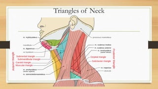
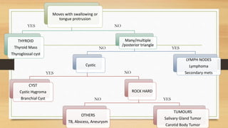
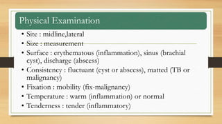
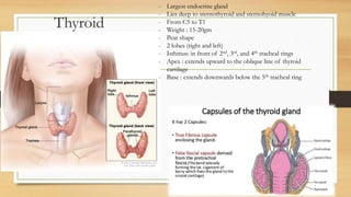
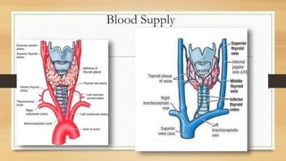
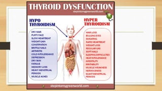
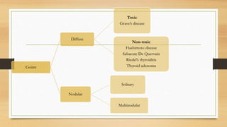
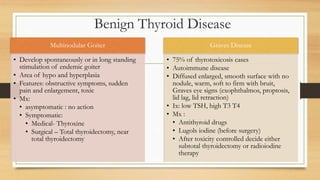
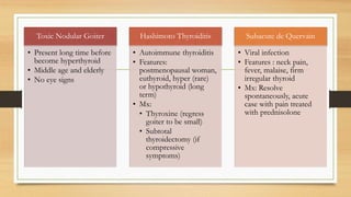
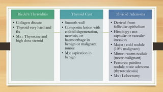
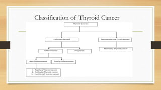
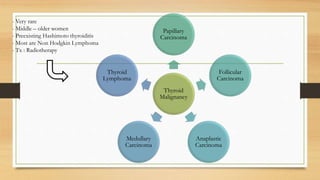
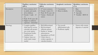
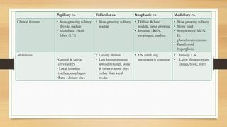
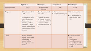
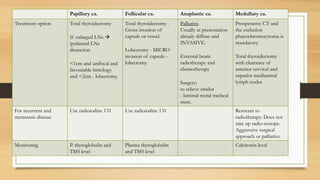
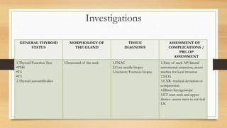
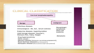
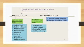
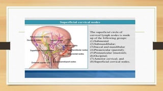
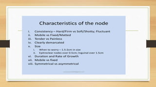
NECK SWELLING
(THYROID,CERVICAL LYMPH NODE)
By :SHAVINA PANIKKAR PARAMESWARAN
Outline
• Definition
• Differential Diagnoses
• Approach to Neck Swelling
• Thyroid Swelling
• Benign
• Malignant
• Thyroglossal Duct Cyst
• Cervical Lymph Node
Definition of Neck Lump
• Any congenital or acquired mass arising in the anterior or posterior triangles
of the neck between the clavicles inferiorly and mandible and base of skull
superiorly.
Triangles of Neck
Moves with swallowing or
tongue protrusion
THYROID
Thyroid Mass
Thyroglossal cyst
Many/multiple
/posterior triangle
Cystic
CYST
Cystic Hygroma
Branchial Cyst
ROCK HARD
OTHERS
TB, Abscess, Aneurysm
TUMOURS
Salivary Gland Tumor
Carotid Body Tumor
LYMPH NODES
Lymphoma
Secondary mets
YES NO
YES
YES
YES
NO
NO
NO
Approach
History
• Detailed of mass : onset(birth), progression (slow, rapid
growing).
• Associated symptoms :pain, fever, LOA/LOW, thyroid status
• Complication : dyspnea, dysphagia, choking, hoarseness of
voices
• Family history of thyroid disease
• Previous head and neck radiation
Physical Examination
• Site : midline,lateral
• Size : measurement
• Surface : erythematous (inflammation), sinus (brachial
cyst), discharge (abscess)
• Consistency : fluctuant (cyst or abscess), matted (TB or
malignancy)
• Fixation : mobility (fix-malignancy)
• Temperature : warm (inflammation) or normal
• Tenderness : tender (inflammatory)
Thyroid
- Largest endocrine gland
- Lies deep to sternothyroid and sternohyoid muscle
- From C5 to T1
- Weight : 15-20gm
- Pear shape
- 2 lobes (right and left)
- Isthmus: in front of 2nd, 3rd, and 4th tracheal rings
- Apex : extends upward to the oblique line of thyroid
cartilage
- Base : extends downwards below the 5th tracheal ring
Blood Supply
Goitre
Diffuse
Toxic
Grave’s disease
Non-toxic
Hashimoto disease
Sabacute De Quervain
Riedel’s thyroiditis
Thyroid adenoma
Nodular
Solitary
Multinodular
Benign Thyroid Disease
Multinodular Goiter
• Develop spontaneously or in long standing
stimulation of endemic goiter
• Area of hypo and hyperplasia
• Features: obstructive symptoms, sudden
pain and enlargement, toxic
• Mx:
• asymptomatic : no action
• Symptomatic:
• Medical- Thyroxine
• Surgical – Total thyroidectomy, near
total thyroidectomy
Graves Disease
• 75% of thyrotoxicosis cases
• Autoimmune disease
• Diffused enlarged, smooth surface with no
nodule, warm, soft to firm with bruit,
Graves eye signs (exophthalmos, proptosis,
lid lag, lid retraction)
• Ix: low TSH, high T3 T4
• Mx :
• Antithyroid drugs
• Lugols iodine (before surgery)
• After toxicity controlled decide either
subtotal thyroidectomy or radioiodine
therapy
Toxic Nodular Goiter
• Present long time before
become hyperthyroid
• Middle age and elderly
• No eye signs
Hashimoto Thyroiditis
• Autoimmune thyroiditis
• Features:
postmenopausal woman,
euthyroid, hyper (rare)
or hypothyroid (long
term)
• Mx:
• Thyroxine (regress
goiter to be small)
• Subtotal
thyroidectomy (if
compressive
symptoms)
Subacute de Quervain
• Viral infection
• Features : neck pain,
fever, malaise, firm
irregular thyroid
• Mx: Resolve
spontaneously, acute
case with pain treated
with prednisolone
Riedel’s Thyroiditis
• Collagen disease
• Thyroid very hard and
fix
• Mx : Thyroxine and
high dose steroid
Thyroid Cyst
• Smooth wall
• Composite lesion with
colloid degeneration,
necrosis, or
haemorrhage in
benign or malignant
tumor
• Mx: aspiration in
benign
Thyroid Adenoma
• Derived from
follicular epithelium
• Histology : not
capsular or vascular
invasion
• Major : cold nodule
(10% malignant)
• Minor : warm nodule
(never malignant)
• Features: painless
nodule, toxic adenoma
(thyrotoxicosis)
• Mx : Lobectomy
Classification of Thyroid Cancer
Thyroid
Malignancy
Papillary
Carcinoma
Follicular
Carcinoma
Anaplastic
Carcinoma
Medullary
Carcinoma
Thyroid
Lymphoma
- Very rare
- Middle – older women
- Preexisting Hashimoto thyroiditis
- Most are Non Hodgkin Lymphoma
- Tx : Radiotherapy
Papillary carcinoma
(50%)
Follicular carcinoma
(30%)
Anaplastic carcinoma Medullary carcinoma
(7%)
Description • Most common (2/3
in adults and nearly
all in children)
• Female > male (3
:1)
• Peak: 30-45 years old
• Higher in radiation
exposure - chernobyl
• 2nd most common
• Female >male (3:1)
• Peak: 40-50 years old
• Variant - Hurtle cell
tumor
• Uncommon
• More in elderly.
• Rare
• Etiology:
 Sporadic
 Familial - MEN II
Histology • Complex papillary
folds lined by severa;
cuboidal cells project
into cystic spaces.
• Psammoma bodies
(calcified area)
• Orphan Anne-eye
nuclei (pale/empty
nuclei)
• Well differentiated
• Well developed
follicular pattern
• Similar to -benign
adenomatous
hyperplasia on FNAC
• Unless there is
evident capsular or
vascular invasion
• Very poorly
differentiated cells.
• Proliferate rapidly
• Tumor cells contain
deposits of amyloid
Papillary ca. Follicular ca. Anaplastic ca. Medullary ca.
Clinical features • Slow-growing solitary
thyroid nodule
• Multifocal - both
lobes (1/3)
• Slow-growing solitary
nodule
• Diffuse & hard
nodule, rapid growing
• Invasive - RLN,
esophagus, trachea.
• Slow growing solitary.
• Stony hard
• Symptom of MEN
II:
pheochromocytoma
• Parathyroid
hyperplasia
Metastasis
•Central & lateral
cervical LN
• Local invasion
trachea, esophagus
•Rare - distant sites
• Usually distant
• Late hematogenous
spread to lungs, bone
& other remote sites
rather than local
nodes
• LN and Lung
metastasis is common
• Intially: LN
• Later: distant organs
(lungs, bone, liver)
Papillary ca. Follicular ca. Anaplastic ca. Medullary ca.
Tissue Diagnosis FNAC Lobectomy and
histology
FNAC FNAC
Prognosis • 10 years survival rate
- 90% :
• 10% pts dying in 10
years : distant mets
• Much better for
‘minimal papillary
carcinoma’ - single
tumour less than 1 cm
young patient with no
invasion.
• The 10-year survival
rate - 75%
• Depends on degree
of vascular invasion
• No invasion: 100% 10
years survival rate
• Extensive invasion: 30
%
• Very poor
• Most die within a year
• No mets: curative
• LN involvement: 10
years survival rate -
50%
Others • ‘Lateral aberrant
thyroid’ – only node
enlargement is
present and the
histology is close to
normal
• Raise in calcitonin
level .
• Prophylactic
thyroidectomy done
for familial depends
on types of mutation
n risk.
Papillary ca. Follicular ca. Anaplastic ca. Medullary ca.
Treatment option Total thyroidectomy
If enlarged LNs 
ipsilateral LNs
dissection
<1cm and unifocal and
favourable histology
and <2cm - lobectomy.
Total thyroidectomy -
Gross invasion of
capsule or vessel.
Lobectomy - MICRO
invasion of capsule -
lobectomy.
Palliative.
Usually at presentation
already diffuse and
INVASIVE.
External beam
radiotherapy and
chemotherapy
Surgery:
to relieve stridor
- luminal metal tracheal
stent.
Preoperative CT and
the exclusion
phaeochromocytoma is
mandatory.
Total thyroidectomy
with clearance of
anterior cervical and
superior mediastinal
lymph nodes
For recurrent and
metastasis disease
Use radioiodine 131 Use radioiodine 131 Resistant to
radiotherapy. Does not
take up radio-isotope.
Aggressive surgical
approach or palliative.
Monitoring P. thyroglobulin and
TSH level
Plasma thyroglobulin
and TSH level -
Calcitonin level
Investigations
GENERAL THYROID
STATUS
MORPHOLOGY OF
THE GLAND
TISSUE
DIAGNOSIS
ASSESSMENT OF
COMPLICATIONS /
PRE-OP
ASSESSMENT
1.Thyroid Function Test
•TSH
•T4
•T3
2.Thyroid autoantibodies
Ultrasound of the neck 1.FNAC
2.Core needle biopsy
3.Incision/Excision biopsy
1.Xray of neck AP/lateral-
retrosternal extension, assess
trachea for local invasion
2.ECG
3.CXR- tracheal deviation or
compression
4.Direct laryngoscope
5.CT scan neck and upper
thorax- assess mets to cervical
LN
ULTRASONOGRAPHY
THYROID NODULE WITH FEW,
EASILY COUNTABLE
MICROCALCIFICATIONS
SOLID, HYPOECHOIC, AND COARSE
CENTRAL
CALCIFICATIONS
• LATER PROVED TO BE MEDULLARY
CARCINOMA
RADIOIODINE STUDIES
• Recommended in patients having Follicular CA on FNAB and suppressed TSH.
• Determine functional status of a nodule
• • Based on radioisotope studies nodule can be →
• Hot
• Autonomous toxic nodule
• Warm
• Normally functioning
• Cold
• Non functioning nodule (likely to be malignant but not always)
• Limitations of Thyroid scan
• • Two dimensional scanning technique
• • Inability to measure the size of a nodule accurately
• • Missed malignant thyroid nodules
Thyroid Scan showing
cold nodule
Thyroid scan showing hot nodule
• X-RAYS
CXR to rule out metastatic deposits
• Skull metastasis more likely in
Follicular carcinoma
CT SCANNING
& MRI
• Used to evaluate soft-tissue
extension of large or suspicious thyroid masses
into the neck, trachea, or oesophagus
• To assess metastases to the
cervical lymph nodes
Images of a large, asymmetric multinodular
goiter. (A) Chest radiography shows marked
tracheal deviation to the right (arrow). (B) Chest
CT confirmed the presence of a large substernal
goiter on the left to the level of tracheal
bifurcation.
Thyroid Surgery
• Indications:
a) Large : compressive symptoms
b) Locally aggressive tumour
c) Hyperthyroidism
d) Cosmetic
f) MNG
g) Grave’s disease (diffuse toxic goitre)
• Relapse on antithyroid drugs
• Large goitre
• Difficult control
• High T4 concentrations (>70pmol/l)
• Eye signs
Preoperative Preparation
• Consent
• Indirect laryngoscope (IDL)
• Normalize thyroid function by anti thyroid
• Carbimazole 30-40 mg per day or 5mg TDS for 8-12
weeks
• Propranolol 30/80 mg TDS
• Lugol’s iodine- reduce vascularity
Types of Thyroid Surgery
Clinical Importance
Enlarged thyroid tends to grow
backwards and medially because of the
direction of pretracheal fascia
Cancer of thyroid may invade:
- Trachea : difficulty in breathing
- Esophagus : dysphagia
- Carotid sheath : bleeding
- RLN : hoarseness of voice
- Symphatethic trunk : loss of voice
During Thyroidectomy:
- Sup. Thyroid artery is ligated close to the apex (to avoid injury to ELN)
- Inf. Thyroid artery is ligated away from the gland (to avoid injury to RLN) –nerve
bruised causes temporary aphonia, complete cut of nerve cause whisper
- In subtotal thyroidectomy, post part of the gland is preserved to avoid removal of
PT gland.
Complications
INTRAOPERATIVELY • Hemorrhage
• RLN damage - temporary paralysis of a VC in 1% but
recovery within 3 months.
- Unilateral - voice is altered in time, hoarse, weak and
breathy.
- Bilateral - stridor.
• Damage surrounding structures - trachea, esophagus,
laryngeal muscle
EARLY POST-OP (12H) •Major hemorrhage - emergency surgical exploration.
•Mediastinal hemorrhage
•Laryngeal edema - need ETT
•Thyrotoxic crisis - emergency beta-adrenergic blockade, IV
hydrocortisone and potassium iodide therapy.
•Tracheomalacia - removal of a longstanding lesion
compressing the trachea may lead to tracheal collapse and
stridor
Complications
LATER POST-OP •Hypoparathyroidism - hypocalcemia symptoms and serum
calcium 24 H post op.
- Require calcium supplements, if severe vitamin D therapy.
•External laryngeal nerve damage - damages the quality of
voice.
LONG TERM •Hypothyroidism- monitor TFT if subtotal.
•Recurrent thyrotoxicosis - due to insufficiency removal.
• Scar - hypertrophic or keloid
Follow Up
• Asses response and complication to therapy :
• Change in serum thyroglobulin over time
• Results of follow up Neck US
• Results of RAI scanning
• Other cross sectional imaging
• Results of FDG PET imaging
• Physical examination
• Follow up - 6 monthly for 2 years.
Thyroglossal Duct Cyst
• The most common congenital neck cyst.
• They are typically located in the midline and are the most common midline neck mass in
young patients.
• Thyroglossal duct cysts typically present during childhood (90% before the age of 10), or
remain asymptomatic until they become infected, in which case they can present at any time.
• Thyroglossal duct cysts are epithelial lined cysts. They result from failure of normal
developmental obliteration of the thyroglossal duct during development (8th-10thgestational
week), and can thus occur anywhere along the course of the duct.
• Location
• suprahyoid: 20-25% (less common in adults ~5%)
• at the level of hyoid bone: ~30% (range 15-50%)
• infrahyoid: ~45% (range 25-65%)
• They can be diagnosed with FNAC and multiple imaging modalities, including
ultrasound, CT, and MRI.
• Presentation is typically either as a painless rounded midline anterior neck swelling or, if
infected, as a red warm painful lump. It classically elevates on tongue protrusion.
• Complete resection of the cyst and duct up to the foramen caecum is curative.
The Sistrunk procedure includes resection of the middle third of the hyoid bone.
There is small risk of recurrence (<5%).
• Complications
• Infection
• Malignancies do occur but are rare seen in <1% of cysts when they do occur they are most
frequently papillary thyroid carcinoma
Definition
• Enlarged lymph nodes is one of the common presentation in neck of
patients.
• Cervical lymphadenopathy refers to enlarged lymph nodes of neck
Investigations
Investigations Details
FBC -normochromic normocytic anemia
-leukoerythroblastic anemia
-neutrophilia
-eosoniphilia
-lymphopenia (late stage disease)
ESR, CRP -raised
-monitor disease
Serum lactate dehydrogenase -raised (30%-40%)
-adverse prognostic factor
HIV, Hep B, Hep C -screening, treatment
Lymph node biopsy -FNAC
-Excisional biopsy
Bone marrow aspirate and trephine
biopsy
-Involvement in advanced disease
-Seldom done
MANAGEMENT
ROLE OF SURGERY
• Radiotherapy and chemotherapy for the treatment of both Hodgkin's disease and non-
Hodgkin's lymphomas have resulted in a greater number of cures with fewer treatment-
related complications.
• The role of surgery in the management of these malignancies has become more peripheral,
and is most often called upon for diagnosis rather than therapy.
• Traditionally, a staging laparotomy was a standard part of the evaluation of patients with
early stage Hodgkin's disease, as it was the best method to distinguish those patients who
had limited disease (disease confined to above the diaphragm) from those with more
extensive involvement (disease both above and below the diaphragm, involvement of
extranodal sites).
• However it may not be crucial to accurately stage most patients with a laparotomy, as
radiation therapy, chemotherapy, or a combination of both will effectively treat the majority
of patients with early stage Hodgkin's disease, and recurrent disease can be successfully
treated with salvage chemotherapy without a decrease in overall survival.
• In Non-Hodgkin's lymphoma, splenectomy is used more for palliation and has little
role in therapy.
• One of the most common indications for splenectomy in non-Hodgkin's
lymphoma is the palliation of symptoms, (left upper quadrant pain, early satiety,
weakness and fatigue) that accompany the marked splenomegaly often seen in this
disease.
• Another common indication for splenectomy is for the treatment of cytopenias
resulting from hypersplenism.
• Splenectomy can be viewed as a primary therapy only in rare cases of primary
splenic lymphoma.
THANK YOU

attachment(1).pptx

  • 1.
    NECK SWELLING (THYROID,CERVICAL LYMPHNODE) By :SHAVINA PANIKKAR PARAMESWARAN
  • 2.
    Outline • Definition • DifferentialDiagnoses • Approach to Neck Swelling • Thyroid Swelling • Benign • Malignant • Thyroglossal Duct Cyst • Cervical Lymph Node
  • 3.
    Definition of NeckLump • Any congenital or acquired mass arising in the anterior or posterior triangles of the neck between the clavicles inferiorly and mandible and base of skull superiorly.
  • 4.
  • 5.
    Moves with swallowingor tongue protrusion THYROID Thyroid Mass Thyroglossal cyst Many/multiple /posterior triangle Cystic CYST Cystic Hygroma Branchial Cyst ROCK HARD OTHERS TB, Abscess, Aneurysm TUMOURS Salivary Gland Tumor Carotid Body Tumor LYMPH NODES Lymphoma Secondary mets YES NO YES YES YES NO NO NO
  • 6.
    Approach History • Detailed ofmass : onset(birth), progression (slow, rapid growing). • Associated symptoms :pain, fever, LOA/LOW, thyroid status • Complication : dyspnea, dysphagia, choking, hoarseness of voices • Family history of thyroid disease • Previous head and neck radiation
  • 7.
    Physical Examination • Site: midline,lateral • Size : measurement • Surface : erythematous (inflammation), sinus (brachial cyst), discharge (abscess) • Consistency : fluctuant (cyst or abscess), matted (TB or malignancy) • Fixation : mobility (fix-malignancy) • Temperature : warm (inflammation) or normal • Tenderness : tender (inflammatory)
  • 8.
    Thyroid - Largest endocrinegland - Lies deep to sternothyroid and sternohyoid muscle - From C5 to T1 - Weight : 15-20gm - Pear shape - 2 lobes (right and left) - Isthmus: in front of 2nd, 3rd, and 4th tracheal rings - Apex : extends upward to the oblique line of thyroid cartilage - Base : extends downwards below the 5th tracheal ring
  • 9.
  • 11.
    Goitre Diffuse Toxic Grave’s disease Non-toxic Hashimoto disease SabacuteDe Quervain Riedel’s thyroiditis Thyroid adenoma Nodular Solitary Multinodular
  • 12.
    Benign Thyroid Disease MultinodularGoiter • Develop spontaneously or in long standing stimulation of endemic goiter • Area of hypo and hyperplasia • Features: obstructive symptoms, sudden pain and enlargement, toxic • Mx: • asymptomatic : no action • Symptomatic: • Medical- Thyroxine • Surgical – Total thyroidectomy, near total thyroidectomy Graves Disease • 75% of thyrotoxicosis cases • Autoimmune disease • Diffused enlarged, smooth surface with no nodule, warm, soft to firm with bruit, Graves eye signs (exophthalmos, proptosis, lid lag, lid retraction) • Ix: low TSH, high T3 T4 • Mx : • Antithyroid drugs • Lugols iodine (before surgery) • After toxicity controlled decide either subtotal thyroidectomy or radioiodine therapy
  • 13.
    Toxic Nodular Goiter •Present long time before become hyperthyroid • Middle age and elderly • No eye signs Hashimoto Thyroiditis • Autoimmune thyroiditis • Features: postmenopausal woman, euthyroid, hyper (rare) or hypothyroid (long term) • Mx: • Thyroxine (regress goiter to be small) • Subtotal thyroidectomy (if compressive symptoms) Subacute de Quervain • Viral infection • Features : neck pain, fever, malaise, firm irregular thyroid • Mx: Resolve spontaneously, acute case with pain treated with prednisolone
  • 14.
    Riedel’s Thyroiditis • Collagendisease • Thyroid very hard and fix • Mx : Thyroxine and high dose steroid Thyroid Cyst • Smooth wall • Composite lesion with colloid degeneration, necrosis, or haemorrhage in benign or malignant tumor • Mx: aspiration in benign Thyroid Adenoma • Derived from follicular epithelium • Histology : not capsular or vascular invasion • Major : cold nodule (10% malignant) • Minor : warm nodule (never malignant) • Features: painless nodule, toxic adenoma (thyrotoxicosis) • Mx : Lobectomy
  • 15.
  • 16.
    Thyroid Malignancy Papillary Carcinoma Follicular Carcinoma Anaplastic Carcinoma Medullary Carcinoma Thyroid Lymphoma - Very rare -Middle – older women - Preexisting Hashimoto thyroiditis - Most are Non Hodgkin Lymphoma - Tx : Radiotherapy
  • 17.
    Papillary carcinoma (50%) Follicular carcinoma (30%) Anaplasticcarcinoma Medullary carcinoma (7%) Description • Most common (2/3 in adults and nearly all in children) • Female > male (3 :1) • Peak: 30-45 years old • Higher in radiation exposure - chernobyl • 2nd most common • Female >male (3:1) • Peak: 40-50 years old • Variant - Hurtle cell tumor • Uncommon • More in elderly. • Rare • Etiology:  Sporadic  Familial - MEN II Histology • Complex papillary folds lined by severa; cuboidal cells project into cystic spaces. • Psammoma bodies (calcified area) • Orphan Anne-eye nuclei (pale/empty nuclei) • Well differentiated • Well developed follicular pattern • Similar to -benign adenomatous hyperplasia on FNAC • Unless there is evident capsular or vascular invasion • Very poorly differentiated cells. • Proliferate rapidly • Tumor cells contain deposits of amyloid
  • 18.
    Papillary ca. Follicularca. Anaplastic ca. Medullary ca. Clinical features • Slow-growing solitary thyroid nodule • Multifocal - both lobes (1/3) • Slow-growing solitary nodule • Diffuse & hard nodule, rapid growing • Invasive - RLN, esophagus, trachea. • Slow growing solitary. • Stony hard • Symptom of MEN II: pheochromocytoma • Parathyroid hyperplasia Metastasis •Central & lateral cervical LN • Local invasion trachea, esophagus •Rare - distant sites • Usually distant • Late hematogenous spread to lungs, bone & other remote sites rather than local nodes • LN and Lung metastasis is common • Intially: LN • Later: distant organs (lungs, bone, liver)
  • 19.
    Papillary ca. Follicularca. Anaplastic ca. Medullary ca. Tissue Diagnosis FNAC Lobectomy and histology FNAC FNAC Prognosis • 10 years survival rate - 90% : • 10% pts dying in 10 years : distant mets • Much better for ‘minimal papillary carcinoma’ - single tumour less than 1 cm young patient with no invasion. • The 10-year survival rate - 75% • Depends on degree of vascular invasion • No invasion: 100% 10 years survival rate • Extensive invasion: 30 % • Very poor • Most die within a year • No mets: curative • LN involvement: 10 years survival rate - 50% Others • ‘Lateral aberrant thyroid’ – only node enlargement is present and the histology is close to normal • Raise in calcitonin level . • Prophylactic thyroidectomy done for familial depends on types of mutation n risk.
  • 20.
    Papillary ca. Follicularca. Anaplastic ca. Medullary ca. Treatment option Total thyroidectomy If enlarged LNs  ipsilateral LNs dissection <1cm and unifocal and favourable histology and <2cm - lobectomy. Total thyroidectomy - Gross invasion of capsule or vessel. Lobectomy - MICRO invasion of capsule - lobectomy. Palliative. Usually at presentation already diffuse and INVASIVE. External beam radiotherapy and chemotherapy Surgery: to relieve stridor - luminal metal tracheal stent. Preoperative CT and the exclusion phaeochromocytoma is mandatory. Total thyroidectomy with clearance of anterior cervical and superior mediastinal lymph nodes For recurrent and metastasis disease Use radioiodine 131 Use radioiodine 131 Resistant to radiotherapy. Does not take up radio-isotope. Aggressive surgical approach or palliative. Monitoring P. thyroglobulin and TSH level Plasma thyroglobulin and TSH level - Calcitonin level
  • 21.
    Investigations GENERAL THYROID STATUS MORPHOLOGY OF THEGLAND TISSUE DIAGNOSIS ASSESSMENT OF COMPLICATIONS / PRE-OP ASSESSMENT 1.Thyroid Function Test •TSH •T4 •T3 2.Thyroid autoantibodies Ultrasound of the neck 1.FNAC 2.Core needle biopsy 3.Incision/Excision biopsy 1.Xray of neck AP/lateral- retrosternal extension, assess trachea for local invasion 2.ECG 3.CXR- tracheal deviation or compression 4.Direct laryngoscope 5.CT scan neck and upper thorax- assess mets to cervical LN
  • 22.
    ULTRASONOGRAPHY THYROID NODULE WITHFEW, EASILY COUNTABLE MICROCALCIFICATIONS SOLID, HYPOECHOIC, AND COARSE CENTRAL CALCIFICATIONS • LATER PROVED TO BE MEDULLARY CARCINOMA
  • 23.
    RADIOIODINE STUDIES • Recommendedin patients having Follicular CA on FNAB and suppressed TSH. • Determine functional status of a nodule • • Based on radioisotope studies nodule can be → • Hot • Autonomous toxic nodule • Warm • Normally functioning • Cold • Non functioning nodule (likely to be malignant but not always) • Limitations of Thyroid scan • • Two dimensional scanning technique • • Inability to measure the size of a nodule accurately • • Missed malignant thyroid nodules
  • 24.
    Thyroid Scan showing coldnodule Thyroid scan showing hot nodule
  • 25.
    • X-RAYS CXR torule out metastatic deposits • Skull metastasis more likely in Follicular carcinoma CT SCANNING & MRI • Used to evaluate soft-tissue extension of large or suspicious thyroid masses into the neck, trachea, or oesophagus • To assess metastases to the cervical lymph nodes Images of a large, asymmetric multinodular goiter. (A) Chest radiography shows marked tracheal deviation to the right (arrow). (B) Chest CT confirmed the presence of a large substernal goiter on the left to the level of tracheal bifurcation.
  • 26.
    Thyroid Surgery • Indications: a)Large : compressive symptoms b) Locally aggressive tumour c) Hyperthyroidism d) Cosmetic f) MNG g) Grave’s disease (diffuse toxic goitre) • Relapse on antithyroid drugs • Large goitre • Difficult control • High T4 concentrations (>70pmol/l) • Eye signs
  • 27.
    Preoperative Preparation • Consent •Indirect laryngoscope (IDL) • Normalize thyroid function by anti thyroid • Carbimazole 30-40 mg per day or 5mg TDS for 8-12 weeks • Propranolol 30/80 mg TDS • Lugol’s iodine- reduce vascularity
  • 28.
  • 31.
    Clinical Importance Enlarged thyroidtends to grow backwards and medially because of the direction of pretracheal fascia Cancer of thyroid may invade: - Trachea : difficulty in breathing - Esophagus : dysphagia - Carotid sheath : bleeding - RLN : hoarseness of voice - Symphatethic trunk : loss of voice During Thyroidectomy: - Sup. Thyroid artery is ligated close to the apex (to avoid injury to ELN) - Inf. Thyroid artery is ligated away from the gland (to avoid injury to RLN) –nerve bruised causes temporary aphonia, complete cut of nerve cause whisper - In subtotal thyroidectomy, post part of the gland is preserved to avoid removal of PT gland.
  • 32.
    Complications INTRAOPERATIVELY • Hemorrhage •RLN damage - temporary paralysis of a VC in 1% but recovery within 3 months. - Unilateral - voice is altered in time, hoarse, weak and breathy. - Bilateral - stridor. • Damage surrounding structures - trachea, esophagus, laryngeal muscle EARLY POST-OP (12H) •Major hemorrhage - emergency surgical exploration. •Mediastinal hemorrhage •Laryngeal edema - need ETT •Thyrotoxic crisis - emergency beta-adrenergic blockade, IV hydrocortisone and potassium iodide therapy. •Tracheomalacia - removal of a longstanding lesion compressing the trachea may lead to tracheal collapse and stridor
  • 33.
    Complications LATER POST-OP •Hypoparathyroidism- hypocalcemia symptoms and serum calcium 24 H post op. - Require calcium supplements, if severe vitamin D therapy. •External laryngeal nerve damage - damages the quality of voice. LONG TERM •Hypothyroidism- monitor TFT if subtotal. •Recurrent thyrotoxicosis - due to insufficiency removal. • Scar - hypertrophic or keloid
  • 34.
    Follow Up • Assesresponse and complication to therapy : • Change in serum thyroglobulin over time • Results of follow up Neck US • Results of RAI scanning • Other cross sectional imaging • Results of FDG PET imaging • Physical examination • Follow up - 6 monthly for 2 years.
  • 35.
    Thyroglossal Duct Cyst •The most common congenital neck cyst. • They are typically located in the midline and are the most common midline neck mass in young patients. • Thyroglossal duct cysts typically present during childhood (90% before the age of 10), or remain asymptomatic until they become infected, in which case they can present at any time. • Thyroglossal duct cysts are epithelial lined cysts. They result from failure of normal developmental obliteration of the thyroglossal duct during development (8th-10thgestational week), and can thus occur anywhere along the course of the duct. • Location • suprahyoid: 20-25% (less common in adults ~5%) • at the level of hyoid bone: ~30% (range 15-50%) • infrahyoid: ~45% (range 25-65%)
  • 36.
    • They canbe diagnosed with FNAC and multiple imaging modalities, including ultrasound, CT, and MRI. • Presentation is typically either as a painless rounded midline anterior neck swelling or, if infected, as a red warm painful lump. It classically elevates on tongue protrusion. • Complete resection of the cyst and duct up to the foramen caecum is curative. The Sistrunk procedure includes resection of the middle third of the hyoid bone. There is small risk of recurrence (<5%). • Complications • Infection • Malignancies do occur but are rare seen in <1% of cysts when they do occur they are most frequently papillary thyroid carcinoma
  • 38.
    Definition • Enlarged lymphnodes is one of the common presentation in neck of patients. • Cervical lymphadenopathy refers to enlarged lymph nodes of neck
  • 46.
    Investigations Investigations Details FBC -normochromicnormocytic anemia -leukoerythroblastic anemia -neutrophilia -eosoniphilia -lymphopenia (late stage disease) ESR, CRP -raised -monitor disease Serum lactate dehydrogenase -raised (30%-40%) -adverse prognostic factor HIV, Hep B, Hep C -screening, treatment Lymph node biopsy -FNAC -Excisional biopsy Bone marrow aspirate and trephine biopsy -Involvement in advanced disease -Seldom done
  • 47.
  • 48.
    ROLE OF SURGERY •Radiotherapy and chemotherapy for the treatment of both Hodgkin's disease and non- Hodgkin's lymphomas have resulted in a greater number of cures with fewer treatment- related complications. • The role of surgery in the management of these malignancies has become more peripheral, and is most often called upon for diagnosis rather than therapy. • Traditionally, a staging laparotomy was a standard part of the evaluation of patients with early stage Hodgkin's disease, as it was the best method to distinguish those patients who had limited disease (disease confined to above the diaphragm) from those with more extensive involvement (disease both above and below the diaphragm, involvement of extranodal sites). • However it may not be crucial to accurately stage most patients with a laparotomy, as radiation therapy, chemotherapy, or a combination of both will effectively treat the majority of patients with early stage Hodgkin's disease, and recurrent disease can be successfully treated with salvage chemotherapy without a decrease in overall survival.
  • 49.
    • In Non-Hodgkin'slymphoma, splenectomy is used more for palliation and has little role in therapy. • One of the most common indications for splenectomy in non-Hodgkin's lymphoma is the palliation of symptoms, (left upper quadrant pain, early satiety, weakness and fatigue) that accompany the marked splenomegaly often seen in this disease. • Another common indication for splenectomy is for the treatment of cytopenias resulting from hypersplenism. • Splenectomy can be viewed as a primary therapy only in rare cases of primary splenic lymphoma.
  • 50.

Editor's Notes

  • #6 Branchial cleft cysts are congenital epithelial cysts, which arise on the lateral part of the neck from a failure of obliteration of the second branchial cleft in embryonic development.  branchial cyst commonly presents as a solitary, painless mass in the neck of a child or a young adult. A history of intermittent swelling and tenderness of the lesion during upper respiratory tract infection may exist. Discharge may be reported if the lesion is associated with a sinus tract. A cystic hygroma, also known as cystic lymphangioma and macrocystic lymphatic malformation, is an often congenital multiloculated lymphatic lesion that can arise anywhere, but is classically found in the left posterior triangle of the neck and armpits.