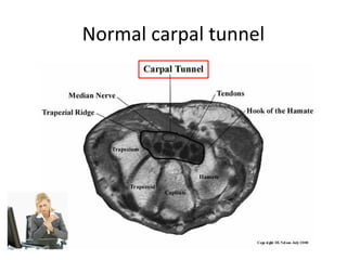
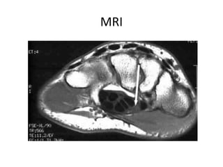
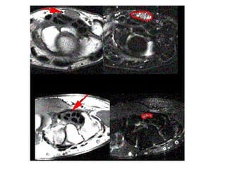
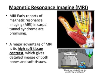
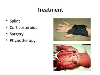
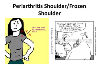
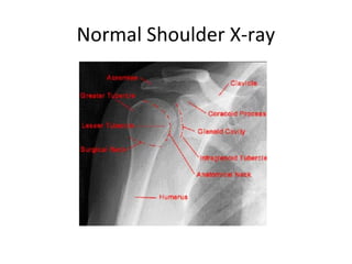
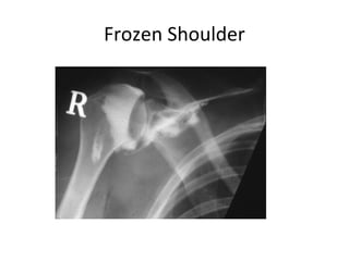
Soft Tissue Swelling
Rosshini Jegatheswaran
CARPAL
TUNNEL
SYNDROME
Normal carpal tunnel
MRI
Magnetic Resonance Imaging (MRI)
• MRI Early reports of
magnetic resonance
imaging (MRI) in carpal
tunnel syndrome are
promising.
• A major advantage of MRI
is its high soft tissue
contrast, which gives
detailed images of both
bones and soft tissues.
Treatment
•
•
•
•

Splint
Corticosteroids
Surgery
Physiotherapy
Periarthritis Shoulder/Frozen
Shoulder
Normal Shoulder X-ray
Frozen Shoulder
Treatment
• Keep the shoulder moving to have a better
chance to recover.
• Anti-inflammatory drugs and pain-killers. Your
family doctor will be able to advise on
appropriate medication.
• Physiotherapy and massage.
• Exercises are recommended to maintain
movement and strength - to keep the
shoulder mobile and avoid further stiffness.
• Injection - Hydrodilatation (hydrodistension)
where fluid is injected in the shoulder joint to
provide relief and a sensation of stretching.
This fluid is a combination of antiinflammatory, local anaesthetic and sterile
saline.
• Surgery

Soft tissue swelling

Editor's Notes

  • #5 Carpal tunnel syndrome. Normal findings on an axial spin-echo T1 MRI of the carpal tunnel showing the intermediate signal intensity of the median nerve (arrow).
  • #10 Adhesive capsulitis
  • #13 This X-Ray shows what a frozen shoulder looks like. The dye (white) outlines the tightness in the shoulder joint.
  • #14 Dense structures, such as bone, show up clearly on x-rays. X-rays may show other problems in your shoulder, such as arthritis. This X-ray showing arthritis of the shoulder.