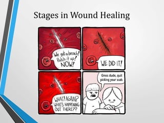
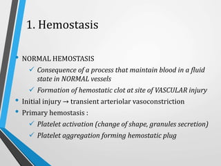
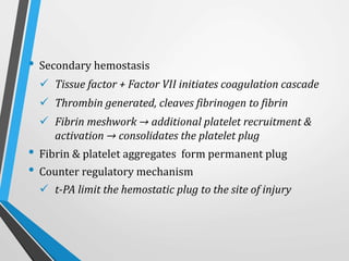
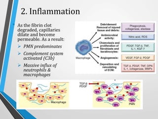
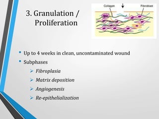
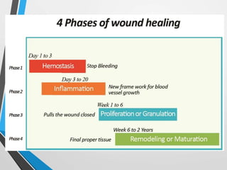
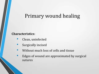
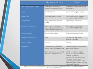
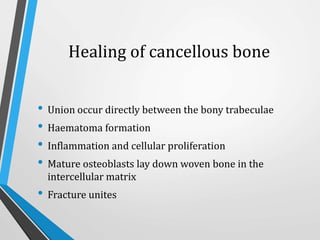
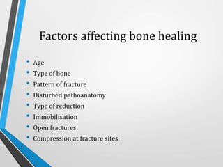
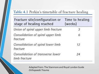
Wound and bone healing involve regeneration and repair processes. Wound healing occurs in four stages - hemostasis, inflammation, proliferation, and remodeling. There are three types of wound healing - primary, secondary, and delayed primary. Primary healing involves clean surgical wounds closed with sutures. Secondary healing involves open wounds without sutures that heal from the bottom up. Bone fracture healing similarly occurs through stages of hematoma formation, inflammation and callus formation, consolidation, and remodeling either directly or indirectly through callus formation. Factors like age, bone type, fracture pattern, and immobilization can affect bone healing.
2. HEALING
• Body’s response to injury in an attempt to restore normal
structure and function.
• 2 distinct processes:
Regeneration when healing takes place by
proliferation of parenchymal cells and usually results
in complete restoration of the original tissues.
Repair when healing takes place by proliferation of
connective tissue elements resulting in fibrosis and
scarring.
• At times, both the processes take place simultaneously.
4. Contents
• Definition
• Stages of wound healing
• Categories of wound healing
• Factors affecting wound healing
• Complications of wound healing
5. Wound Healing
Definition
• mechanism whereby the body attempts to restore the
integrity of the injured part
• combination of regeneration and repair
7. 1. Hemostasis
• NORMAL HEMOSTASIS
Consequence of a process that maintain blood in a fluid
state in NORMAL vessels
Formation of hemostatic clot at site of VASCULAR injury
• Initial injury → transient arteriolar vasoconstriction
• Primary hemostasis :
Platelet activation (change of shape, granules secretion)
Platelet aggregation forming hemostatic plug
8. • Secondary hemostasis
Tissue factor + Factor VII initiates coagulation cascade
Thrombin generated, cleaves fibrinogen to fibrin
Fibrin meshwork → additional platelet recruitment &
activation → consolidates the platelet plug
• Fibrin & platelet aggregates form permanent plug
• Counter regulatory mechanism
t-PA limit the hemostatic plug to the site of injury
9.
10.
11. 2. Inflammation
• As the fibrin clot
degraded, capillaries
dilate and become
permeable. As a result:
PMN predominates
Complement system
activated (C3b)
Massive influx of
neutrophils &
macrophages
12. 3. Granulation /
Proliferation
• Up to 4 weeks in clean, uncontaminated wound
• Subphases
Fibroplasia
Matrix deposition
Angiogenesis
Re-epithelialization
13. 4. Remodeling /
Maturation
• Collagen is degraded and deposited (in equilibrium)
• Wound contraction
• Maximum tensile strength at 12th week
• Resultant scar 80% of the original skin that has
replaced
14.
15.
16. Categories of Wound Healing
• Primary wound healing / healing by first intention
• Secondary wound healing / healing by secondary
intention
• Delayed primary wound healing / Tertiary closure
17. Primary wound healing
Characteristics:
• Clean, uninfected
• Surgically incised
• Without much loss of cells and tissue
• Edges of wound are approximated by surgical
sutures
20. Secondary wound healing
Characteristics:
• open with a large tissue defect, at times infected;
• having extensive loss of cells and tissues
• wound not approximated by surgical sutures but
left open.
23. Differences between primary and
secondary wound healing
FEATURES PRIMARY SECONDARY
CLEANLINESS CLEAN NOT CLEAN
INFECTION NOT INFECTED INFECTED
MARGINS SURGICALLY NEAT IRREGULAR
SUTURES USED NOT USED
HEALING SMALL GRANULATION
TISSUE
LARGE GRANULATION
TISSUE
OUT COME LINEAR SCAR IRREGULAR WOUND
COMPLICATIONS NOT FREQUENT FREQUENT
24. Delayed primary wound healing
• Wound edges are not reapproximated immediately
• First cleaned and observed for a few days to ensure
no infection is apparent before it is surgically closed
• Late presentation (>24 hours)
• Contaminated crush wounds
• Mammalian bites
25. Factors affecting wound healing
• Site
• Structures involved
• Mechanism
• Contamination
• Loss of tissue
• Local factors (Examples?)
• Systemic factors (Examples?)
30. Contents
• Types of healing of fractures
• Stages of healing of fractures
• Factors affecting fracture healing
31. Types of bone healing
Primary fracture healing Secondary fracture healing
Healing by direct union Healing by callus
Haematoma has been disturbed
Haematoma has NOT been
disturbed
Operative treatment Non-operative treatment
Union difficult to evaluate union on
X-rays
Union can be evaluated union on X-
rays
32. Stages of healing of fractures
- cortical bone
1. Tissue destruction and haematoma formation
2. Inflammation & cellular proliferation
3. Callus formation
4. Consolidation of callus
5. Remodeling
34. Stages of healing of fractures
Stage 1
Tissue destruction & haematoma
formation
• Vessels are torn
• Haematoma forms around and
within the fracture.
• Bone at the fracture surfaces,
deprived of a blood supply,
dies back for a millimetre or
two.
35. Stage 2
Inflammation & cellular proliferation
Within 8 hours of the fracture
• Acute inflammatory reaction with
migration of inflammatory cells and
the initiation of proliferation and
differentiation of mesenchymal stem
cells from the periosteum, the
breached medullary canal and the
surrounding muscle.
• The fragment ends are surrounded by
cellular tissue, which creates a
scaffold across the fracture site.
• The clotted haematoma is slowly
absorbed and fine new capillaries
grow into the area.
36. Stage 3
Formation of soft callus
• The differentiating stem cells
provide chrondrogenic and
osteogenic cell populations,
including osteoclasts.
• The thick cellular mass, with its
islands of immature bone and
cartilage, forms the callus.
• As the immature fibre bone (or
‘woven’ bone) becomes more
densely mineralized, movement
at the fracture site decreases
progressively and at about 4
weeks after injury the fracture
‘unites’.
37. Stage 4
Consolidation
• Woven bone is transformed
into lamellar bone.
• Osteoclasts burrow through
the debris at the fracture
line, and close behind them.
• Osteoblasts fill in the
remaining gaps between the
fragments with new bone.
• Takes several months
38. Stage 5
Remodeling
• The fracture has been bridged by
a cuff of solid bone.
• Continuous process of alternating
bone resorption and formation.
• Medullary cavity formed
• 1-2 years
39. Healing of cancellous bone
• Union occur directly between the bony trabeculae
• Haematoma formation
• Inflammation and cellular proliferation
• Mature osteoblasts lay down woven bone in the
intercellular matrix
• Fracture unites
40. Factors affecting bone healing
• Age
• Type of bone
• Pattern of fracture
• Disturbed pathoanatomy
• Type of reduction
• Immobilisation
• Open fractures
• Compression at fracture sites
42. References
• Apley’s System of Orthopaedics and Fractures 9th Edition
• Bailey & Love’s Short Practice of Surgery 26th Edition
• Harsh Mohan Textbook of Pathology 7th edition
• Maheshwari & Mhaskar Essential Orthopaedics 4th Edition
• Medscape
• Robbins and Cotran Pathology Basis of Disease 8th Edition
• Sharon Baranoski and Elizabeth A. Ayello Wound Care Essentials 3rd
edition
• The Stanmore and Royal London Guide Orthopaedic Trauma
• Wheeless Textbook of Orthopaedics
• https://meds.queensu.ca/central/assets/modules/basic_suturing/categ
ories_of_wound_closure.html
It was a Greek physician, Galen (129-199) who identified two forms of wound healing, primary and secondary intention
MEDSCAPE
The initial injury results in an outflow of blood and lymphatic fluid. This is also the process during which the initial reparative coagulum is created. Both the intrinsic and extrinsic clotting mechanisms are activated. The intrinsic mechanism is enjoined from the thrombocytes and the extrinsic mechanism from the injured tissues. Following vasoconstriction, platelets adhere to damaged endothelium and discharge adenosine diphosphate (ADP), promoting thrombocyte clumping, which dams the wound. With the short-lived vasoconstriction complete, the vessels dilate allowing the influx of more thrombocytes and other blood cells.
At this stage, one can think of the commencement of the inflammatory phase. Although some speak of a separate inflammatory phase, it commences during the hemostasis phase, again providing evidence of the overlapping nature of the healing compendium. These thrombocytes, as well as the recruited white blood cells, release numerous factors to ramp up the healing process. Alpha-granules liberate platelet-derived growth factor (PDGF), platelet factor IV, and transforming growth factor (TGF)–β). The processes of inflammation, collagen degradation and collagenogenesis, myoblastic creation from transformed fibroblasts, growth of new blood vessels, and reepithelialization have all commenced.
These processes are mediated by a host of cytokines and growth factors. The interleukins strongly influence the inflammatory process. Vascular endothelial growth factor (VEGF) and other factors enhance blood vessel formation, and some have multiple roles such as fibroblast growth factor (FGF)–2, which affects not only the process of angiogenesis but also that of reepithelialization. Vasoactive amines such as histamine and serotonin are released from dense bodies found in thrombocytes. PDGF is chemotactic for fibroblasts and, along with TGF-β, is a potent modulator of fibroblastic mitosis, leading to prolific collagen fibril construction in later phases. Fibrinogen is cleaved into fibrin, and the framework for completion of the coagulation process is formed. Fibrin provides the structural support for cellular constituents of inflammation. This process starts immediately after the insult and may continue for a few days
A, After vascular injury local neurohumoral factors induce a transient vasoconstriction.
B, Platelets bind via glycoprotein Ib (GpIb) receptors to von Willebrand factor (vWF) on exposed extracellular matrix (ECM) and are activated, undergoing a shape change and granule release. Released adenosine diphosphate (ADP) and thromboxane A2 (TxA2) induce additional platelet aggregation through platelet GpIIb-IIIa receptor binding to fibrinogen, and form the primary hemostatic plug.
C, Local activation of the coagulation cascade (involving tissue factor and platelet phospholipids) results in fibrin polymerization, “cementing” the platelets into a definitive secondary hemostatic plug.
D, Counter-regulatory mechanisms, mediated by tissue plasminogen activator (t-PA, a fibrinolytic product) and thrombomodulin, confine the hemostatic process to the site of injury.
As the hemostasis phase can be construed to consist of an early and a late phase, the early phase being bleeding and hemostasis and the late phase being coagulation, so is it also with inflammation. While the inflammatory phase commences during the hemostasis phase, the early component of the inflammatory phase is predominated by the influx of the polymorphonuclear leukocytes (PMNs) and the later component predominated by monocytes/macrophages.
Within the first 6-8 hours, the next phase of the healing process is underway, PMNs engorging the wound. TGF-β facilitates PMN migration from surrounding blood vessels, where they extrude themselves from these vessels. These cells cleanse the wound, clearing it of debris. The PMNs attain their maximal numbers in 24-48 hours and commence their departure by hour 72. Other chemotactic agents are released, including FGF, TGF-β and TGF-α, PDGF, and plasma-activated complements C3a and C5a (anaphylactic toxins). They are sequestered by macrophages or interred within the scab or eschar. [11]
As the process continues, monocytes also exude from the vessels. These are termed macrophages once they leave the vessel. The macrophages continue the cleansing process and manufacture various growth factors during days 3-4. The macrophages orchestrate the multiplication of endothelial cells with the sprouting of new blood vessels, the duplication of smooth muscle cells, and the creation of the milieu created by the fibroblast. Many factors influencing the wound healing process are secreted by macrophages. These include TGFs, cytokines and interleukin (IL)–1, tumor necrosis factor (TNF), and PDGF.
This phase consists of different subphases. These subphases do not happen in discrete time frames but constitute an overall and ongoing process. The subphases are "fibroplasia, matrix deposition, angiogenesis and re-epithelialization".
In days 5-7, fibroblasts have migrated into the wound, laying down new collagen of the subtypes I and III. Early in normal wound healing, type III collagen predominates but is later replaced by type I collagen.
Tropocollagen is the precursor of all collagen types and is transformed within the cell's rough endoplasmic reticulum, where proline and lysine are hydroxylated. Disulfide bonds are established, allowing 3 tropocollagen strands to form a triple left-handed triple helix, termed procollagen. As the procollagen is secreted into the extracellular space, peptidases in the cell wall cleave terminal peptide chains, creating true collagen fibrils.
The wound is suffused with GAGs and fibronectin produced by fibroblasts. These GAGs include heparan sulfate, hyaluronic acid, chondroitin sulfate, and keratan sulfate. Proteoglycans are GAGs that are bonded covalently to a protein core and contribute to matrix deposition.
Angiogenesis is the product of parent vessel offshoots. The formation of new vasculature requires extracellular matrix and basement membrane degradation followed by migration, mitosis, and maturation of endothelial cells. Basic FGF and vascular endothelial growth factor are believed to modulate angiogenesis.
Re-epithelization occurs with the migration of cells from the periphery of the wound and adnexal structures. This process commences with the spreading of cells within 24 hours. Division of peripheral cells occurs in hours 48-72, resulting in a thin epithelial cell layer, which bridges the wound. Epidermal growth factors are believed to play a key role in this aspect of wound healing.
This succession of subphases can last up to 4 weeks in the clean and uncontaminated wound.
After the third week, the wound undergoes constant alterations, known as remodeling, which can last for years after the initial injury occurred. Collagen is degraded and deposited in an equilibrium-producing fashion, resulting in no change in the amount of collagen present in the wound. The collagen deposition in normal wound healing reaches a peak by the third week after the wound is created. Contraction of the wound is an ongoing process resulting in part from the proliferation of the specialized fibroblasts termed myofibroblasts, which resemble contractile smooth muscle cells. Wound contraction occurs to a greater extent with secondary healing than with primary healing. Maximal tensile strength of the wound is achieved by the 12th week, and the ultimate resultant scar has only 80% of the tensile strength of the original skin that it has replaced.
Each suture track is a separate wound and incites the same phenomena as in healing of the primary wound i.e. filling the space with haemorrhage, some inflammatory cell reaction, epithelial cell proliferation along the suture track from both margins, fibroblastic proliferation and formation of young collagen.
When sutures are removed around 7th day, much of epithelialised suture track is avulsed and the remaining epithelial tissue in the track is absorbed.
However, sometimes the suture track gets infected (stitch abscess), or the epithelial cells may persist in the track (implantation or epidermal cysts).
Thus, the scar formed in a sutured wound is neat due to close apposition of the margins of wound; the use of adhesive tapes avoids removal of stitches and its complications.
The basic events in secondary union are similar to primary union but differ in having a larger tissue defect which has to be bridged. Hence healing takes place from the base upwards as well as from the margins inwards. The healing by second intention is slow and results in a large, at times ugly, scar as compared to rapid healing and neat scar of primary union.
Thus, the scar formed in a sutured wound is neat due to close apposition of the margins of wound; the use of adhesive tapes avoids removal of stitches and its complications.
By the fourth day, phagocytosis of contaminated tissues is well underway, and the processes of epithelization, collagen deposition, and maturation are occurring.
Foreign materials are walled off by macrophages that may metamorphose into epithelioid cells, which are encircled by mononuclear leukocytes, forming granulomas.
Usually the wound is closed surgically at this juncture, and if the "cleansing" of the wound is incomplete, chronic inflammation can ensue, resulting in prominent scarring.
Other local factors
Vascular insufficiency (arterial or venous)
Previous radiation
Pressure
■ Systemic factors
Malnutrition or vitamin and mineral deficiencies
Disease (e.g. diabetes mellitus)
Medications (e.g. steroids)
Immune deficiencies [e.g. chemotherapy, acquired
immunodeficiency syndrome (AIDS)]
Smoking
Thicker lamellae are laid down where the stresses are high, unwanted buttresses are carved away and the medullary cavity is reformed.
Spiral fracture in the upper limb unites in 3 weeks;
for consolidation multiply by 2; for the lower limb multiply by 2 again;
for transverse fractures multiply again by 2.
A more sophisticated formula is as follows.
A spiral fracture in the upper limb takes 6–8 weeks to consolidate;
the lower limb needs twice as long.
Add 25% if the fracture is not spiral or if it involves the femur. Children’s fractures, of course, join more quickly.
These figures are only a rough guide; there must be clinical and radiological evidence of consolidation before full stress is permitted without splintage.
Causes related to patient : Old age, associated systemic illness
Causes related to fracture : Distraction at fracture site, soft tissue interposition, Bone loss at time of fracture, infection from open fracture, damage to blood supply of fracture fragments, pathological fracture
Causes related to treatment : inadequate reduction, inadequate immobilization, distraction during treatment
Fractures in children unite faster
Fractures closer to the joints unite faster
Displacements at the fracture will remodel
Angulations in the plane of movement of the joint < 20 degrees are acceptable
Rotations will never remodel