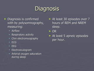
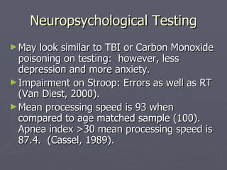
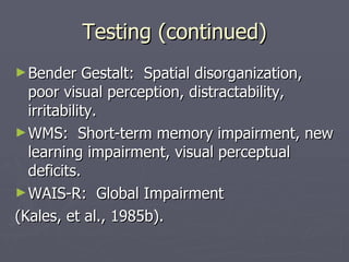
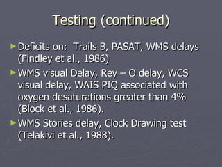
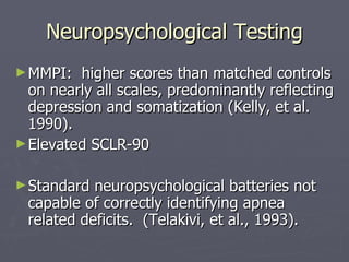
Sleep Apnea:  Neuropsychological Effects, Research, and Treatment. By:  Brooke Schauder, PhD Erie Psychological Consortium Pacific Graduate School of Psychology
What is Apnea? Sleep Apnea:  Absence of breathing for  10 seconds during sleep, either due to obstruction, central, or mixed.  Obstructive is by far the most common form. Obstructive Sleep Apnea:  1 st  described in 1965, characterized by recurrent interruptions of breathing during sleep due to temporary obstruction of the airway by tissues (soft palate, uvula, or tonsils) with resultant hypoxemia and chronic lethargy.
Hypoxia:  Decrease below normal levels of oxygen in inspired gases, arterial blood, or tissue.  Normal oxygen hemoglobin saturation is approximately 95%. Decreases up to 50% during apneic episodes Anoxia:  Absence or almost complete absence of oxygen in inspired gases, arterial blood, or tissue.
Sleep Stages and Apnea During REM motor tone is at lowest level. Collapse of upper airway. Accessory muscles of respiration are effectively paralyzed in REM.  Diaphragm is solely responsible for breathing. Typically worse at the end of the night, during longer stages of REM sleep & stages 3-4.
Sleep Apnea Symptoms Loud snoring Gasping for air with complete or partial waking Profuse sweating Nightmares or night-terrors Enuresis (in children) Nocturnal restlessness Daytime sleepiness Morning Headache Apneic episodes typically last 10-120 seconds. Episodes may be recurrent  – 1 to 100 times throughout the night.
 
Demographics Affects 4% of men and 2% of women between ages 30 and 60.  Mainly affects middle-aged adults. “ Typical” patient is overweight adult male. Up to 88% of adults patients estimated to be overweight. Incidence increases with advancing age.  1% to 3% of children are affected
Risk Factors Obesity Hypothyroidism Cigarette Smoking Alcohol use Hypnotics use (benzodiazepines)
Recognizing Sleep Apnea Consider diagnosis if patient snores AND any of the following are present: Depression Hypertension Gastro esophageal Reflux Disease Diabetes Congestive Heart Failure Obesity
Diagnosis Diagnosis is confirmed with by polysomnography, measuring: Airflow Respiratory activity Chin electromyography ECG EEG Electrooculogram Arterial oxygen saturation during sleep At least 30 episodes over 7 hours of REM and NREM sleep. OR At least 5 apneic episodes per hour.
Neuropsychological Deficits Sustained Attention and Concentration Immediate and Delayed Recall of Visual and Verbal Information Executive Functioning Processing Speed
Deficits (continued) Selective attention, mental flexibility, impulse control, behavioral and emotional regulation, and metacognition in children (Beebe, 2004). Deficits on visual memory, concentration, and auditory reaction time (Laakso, Herrala, & Rikka (1999).
Neuropsychological Deficits (continued) Apperceptive Agnosia Motor disturbances: posture, gait, involuntary movements, Parkinsonian symptoms, limb appraxia. Psychological:  Depression, personality changes, and emotional lability.
Neuropsychological Testing May look similar to TBI or Carbon Monoxide poisoning on testing:  however, less depression and more anxiety. Impairment on Stroop: Errors as well as RT (Van Diest, 2000). Mean processing speed is 93 when compared to age matched sample (100).  Apnea index >30 mean processing speed is 87.4.  (Cassel, 1989).
Testing (continued) Bender Gestalt:  Spatial disorganization, poor visual perception, distractability, irritability. WMS:  Short-term memory impairment, new learning impairment, visual perceptual deficits. WAIS-R:  Global Impairment (Kales, et al., 1985b).
Testing (continued) Deficits on:  Trails B, PASAT, WMS delays (Findley et al., 1986) WMS visual Delay, Rey – O delay, WCS visual delay, WAIS PIQ associated with oxygen desaturations greater than 4% (Block et al., 1986). WMS Stories delay, Clock Drawing test (Telakivi et al., 1988).
Neuropsychological Testing MMPI:  higher scores than matched controls on nearly all scales, predominantly reflecting depression and somatization (Kelly, et al. 1990). Elevated SCLR-90 Standard neuropsychological batteries not capable of correctly identifying apnea related deficits.  (Telakivi, et al., 1993).
Animal Testing Aptopsis in cortex and CA1 region of hippocampus. Spatial learning deficits in rats (Row, 2002). Rats 10-25 days post-natal had significantly higher susceptibility to hypoxia.
Mechanisms of Impairment Blood-Oxygen desaturations result in neural tissue loss due to ischemia, acidosis, calcium influx, formation of oxygen radicals, and excitotoxicity. Ischemia Induced neuronal loss:  Hypoxia alone may not cause neuronal apoptosis.  Hypoxia may increase neuronal p53, which increases apoptosis processes. Banasiak, 1998
Mechanisms (continued) Sleep fragmentation interrupts REM sleep; studies demonstrate REM deprivation associated decrements of learning and memory. Comparison of Sleep Apnea Patients and patients with insomnia showed no differences in neuropsychological deficit. (Verstraeten, 1996) Desaturations greater than 4% cause significantly more impairment (Telakivi et al., 1988).
Mechanisms (continued) Decreased hippocampal volume, resulting in memory impairment. Areas more vulnerable:  neocortex, basal ganglia, cerebellar Perkinje cells, primary visual cortex, frontal regions, and thalamus. Gray and white matter atrophy Ventricular Enlargement
Treatment Continuous Positive Airway Pressure (CPAP) Bedard: study of patients before and after 6 months of treatment: Improved daytime vigilance  Some degree of sleepiness persisted Most neuropsychological deficits normalized, with the exception of planning abilities and manual dexterity.
Effects of CPAP After 6 months of CPAP treatment, improvement occurred on measures of: - Verbal Fluency -Cognitive Shifting -Short Term Visual Memory and time estimation (Caccappolo, 2000) Some studies find no improvement in general intellectual functioning (Gale, 2004).
Treatment (Continued) Mcardle, 1999:  Predictors of long-term (5 years) CPAP use are:  snoring history  apnea/hypopnea index being male Lower age being a non-smoker use within 1 st  3 months of treatment correlated with use at 5 years:  those using CPAP <2 hrs./night at 3 mo. not likely to continue
 
Sleep Apnea in Children 1-3% have diagnosed sleep apnea Up to 12 % may have primary snoring Many infants and children also experience abnormal cardiac functioning during apneic episodes:  52% brief sinus arrests, 28% with second degree atrioventricular block, 16% with paroxysmal tachycardia
Effects of Apnea on Children Inattention/ Executive Functioning Deficits – symptoms mimicking/causing ADHD Minimal effects on measures of overall intelligence, verbal functioning, and processing speed. Mixed findings on memory impairment. Decreased academic performance Oppositonality Decreased emotional and behavioral regulation.
Differential Diagnosis and Co-morbid Disorders ADHD: 20-30% with Breathing related Sleep disorders may have clinically significant symptoms (Gozal, 1998). Learning Disabilities:  among 13-14 y.old’s, in the lower quartile of their class, early childhood snoring was more frequently reported (Gozal, 2001).
(Differential Continued) Failure to Thrive Emotional Disorders Conduct Disorders
Treatment of Children Unlikely to remit without treatment May intensify at a much faster rate than in adults. Tonsillectomy or Adenoidectomy often used:  studies have shown increase in mean grades during in second graders (Gozal, 1998). Improvements may be only partial:  short-term memory may not fully recover (Gozal, 2001).
 

Sleep apnea

  • 1.
    Sleep Apnea: Neuropsychological Effects, Research, and Treatment. By: Brooke Schauder, PhD Erie Psychological Consortium Pacific Graduate School of Psychology
  • 2.
    What is Apnea?Sleep Apnea: Absence of breathing for 10 seconds during sleep, either due to obstruction, central, or mixed. Obstructive is by far the most common form. Obstructive Sleep Apnea: 1 st described in 1965, characterized by recurrent interruptions of breathing during sleep due to temporary obstruction of the airway by tissues (soft palate, uvula, or tonsils) with resultant hypoxemia and chronic lethargy.
  • 3.
    Hypoxia: Decreasebelow normal levels of oxygen in inspired gases, arterial blood, or tissue. Normal oxygen hemoglobin saturation is approximately 95%. Decreases up to 50% during apneic episodes Anoxia: Absence or almost complete absence of oxygen in inspired gases, arterial blood, or tissue.
  • 4.
    Sleep Stages andApnea During REM motor tone is at lowest level. Collapse of upper airway. Accessory muscles of respiration are effectively paralyzed in REM. Diaphragm is solely responsible for breathing. Typically worse at the end of the night, during longer stages of REM sleep & stages 3-4.
  • 5.
    Sleep Apnea SymptomsLoud snoring Gasping for air with complete or partial waking Profuse sweating Nightmares or night-terrors Enuresis (in children) Nocturnal restlessness Daytime sleepiness Morning Headache Apneic episodes typically last 10-120 seconds. Episodes may be recurrent – 1 to 100 times throughout the night.
  • 6.
  • 7.
    Demographics Affects 4%of men and 2% of women between ages 30 and 60. Mainly affects middle-aged adults. “ Typical” patient is overweight adult male. Up to 88% of adults patients estimated to be overweight. Incidence increases with advancing age. 1% to 3% of children are affected
  • 8.
    Risk Factors ObesityHypothyroidism Cigarette Smoking Alcohol use Hypnotics use (benzodiazepines)
  • 9.
    Recognizing Sleep ApneaConsider diagnosis if patient snores AND any of the following are present: Depression Hypertension Gastro esophageal Reflux Disease Diabetes Congestive Heart Failure Obesity
  • 10.
    Diagnosis Diagnosis isconfirmed with by polysomnography, measuring: Airflow Respiratory activity Chin electromyography ECG EEG Electrooculogram Arterial oxygen saturation during sleep At least 30 episodes over 7 hours of REM and NREM sleep. OR At least 5 apneic episodes per hour.
  • 11.
    Neuropsychological Deficits SustainedAttention and Concentration Immediate and Delayed Recall of Visual and Verbal Information Executive Functioning Processing Speed
  • 12.
    Deficits (continued) Selectiveattention, mental flexibility, impulse control, behavioral and emotional regulation, and metacognition in children (Beebe, 2004). Deficits on visual memory, concentration, and auditory reaction time (Laakso, Herrala, & Rikka (1999).
  • 13.
    Neuropsychological Deficits (continued)Apperceptive Agnosia Motor disturbances: posture, gait, involuntary movements, Parkinsonian symptoms, limb appraxia. Psychological: Depression, personality changes, and emotional lability.
  • 14.
    Neuropsychological Testing Maylook similar to TBI or Carbon Monoxide poisoning on testing: however, less depression and more anxiety. Impairment on Stroop: Errors as well as RT (Van Diest, 2000). Mean processing speed is 93 when compared to age matched sample (100). Apnea index >30 mean processing speed is 87.4. (Cassel, 1989).
  • 15.
    Testing (continued) BenderGestalt: Spatial disorganization, poor visual perception, distractability, irritability. WMS: Short-term memory impairment, new learning impairment, visual perceptual deficits. WAIS-R: Global Impairment (Kales, et al., 1985b).
  • 16.
    Testing (continued) Deficitson: Trails B, PASAT, WMS delays (Findley et al., 1986) WMS visual Delay, Rey – O delay, WCS visual delay, WAIS PIQ associated with oxygen desaturations greater than 4% (Block et al., 1986). WMS Stories delay, Clock Drawing test (Telakivi et al., 1988).
  • 17.
    Neuropsychological Testing MMPI: higher scores than matched controls on nearly all scales, predominantly reflecting depression and somatization (Kelly, et al. 1990). Elevated SCLR-90 Standard neuropsychological batteries not capable of correctly identifying apnea related deficits. (Telakivi, et al., 1993).
  • 18.
    Animal Testing Aptopsisin cortex and CA1 region of hippocampus. Spatial learning deficits in rats (Row, 2002). Rats 10-25 days post-natal had significantly higher susceptibility to hypoxia.
  • 19.
    Mechanisms of ImpairmentBlood-Oxygen desaturations result in neural tissue loss due to ischemia, acidosis, calcium influx, formation of oxygen radicals, and excitotoxicity. Ischemia Induced neuronal loss: Hypoxia alone may not cause neuronal apoptosis. Hypoxia may increase neuronal p53, which increases apoptosis processes. Banasiak, 1998
  • 20.
    Mechanisms (continued) Sleepfragmentation interrupts REM sleep; studies demonstrate REM deprivation associated decrements of learning and memory. Comparison of Sleep Apnea Patients and patients with insomnia showed no differences in neuropsychological deficit. (Verstraeten, 1996) Desaturations greater than 4% cause significantly more impairment (Telakivi et al., 1988).
  • 21.
    Mechanisms (continued) Decreasedhippocampal volume, resulting in memory impairment. Areas more vulnerable: neocortex, basal ganglia, cerebellar Perkinje cells, primary visual cortex, frontal regions, and thalamus. Gray and white matter atrophy Ventricular Enlargement
  • 22.
    Treatment Continuous PositiveAirway Pressure (CPAP) Bedard: study of patients before and after 6 months of treatment: Improved daytime vigilance Some degree of sleepiness persisted Most neuropsychological deficits normalized, with the exception of planning abilities and manual dexterity.
  • 23.
    Effects of CPAPAfter 6 months of CPAP treatment, improvement occurred on measures of: - Verbal Fluency -Cognitive Shifting -Short Term Visual Memory and time estimation (Caccappolo, 2000) Some studies find no improvement in general intellectual functioning (Gale, 2004).
  • 24.
    Treatment (Continued) Mcardle,1999: Predictors of long-term (5 years) CPAP use are: snoring history apnea/hypopnea index being male Lower age being a non-smoker use within 1 st 3 months of treatment correlated with use at 5 years: those using CPAP <2 hrs./night at 3 mo. not likely to continue
  • 25.
  • 26.
    Sleep Apnea inChildren 1-3% have diagnosed sleep apnea Up to 12 % may have primary snoring Many infants and children also experience abnormal cardiac functioning during apneic episodes: 52% brief sinus arrests, 28% with second degree atrioventricular block, 16% with paroxysmal tachycardia
  • 27.
    Effects of Apneaon Children Inattention/ Executive Functioning Deficits – symptoms mimicking/causing ADHD Minimal effects on measures of overall intelligence, verbal functioning, and processing speed. Mixed findings on memory impairment. Decreased academic performance Oppositonality Decreased emotional and behavioral regulation.
  • 28.
    Differential Diagnosis andCo-morbid Disorders ADHD: 20-30% with Breathing related Sleep disorders may have clinically significant symptoms (Gozal, 1998). Learning Disabilities: among 13-14 y.old’s, in the lower quartile of their class, early childhood snoring was more frequently reported (Gozal, 2001).
  • 29.
    (Differential Continued) Failureto Thrive Emotional Disorders Conduct Disorders
  • 30.
    Treatment of ChildrenUnlikely to remit without treatment May intensify at a much faster rate than in adults. Tonsillectomy or Adenoidectomy often used: studies have shown increase in mean grades during in second graders (Gozal, 1998). Improvements may be only partial: short-term memory may not fully recover (Gozal, 2001).
  • 31.