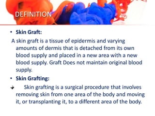
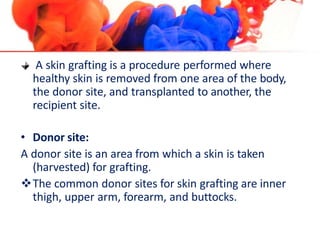
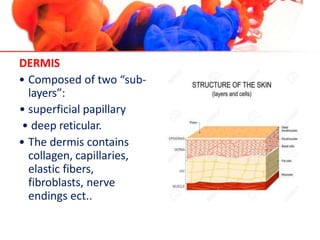
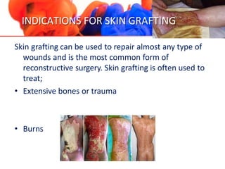
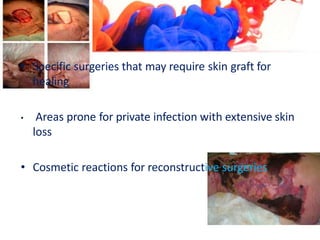
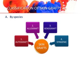
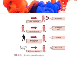
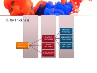
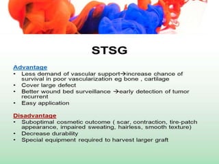
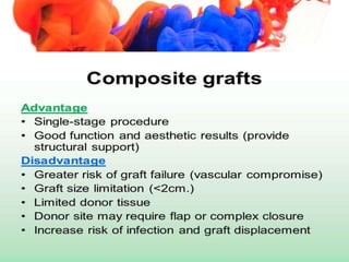
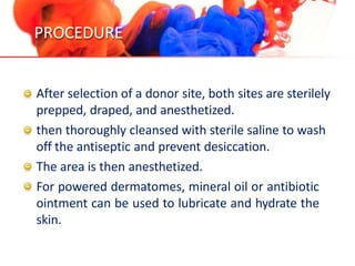
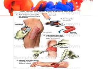
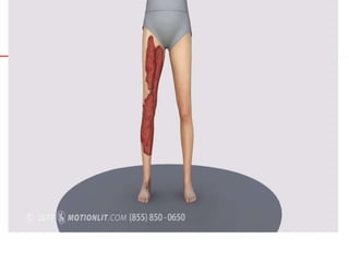
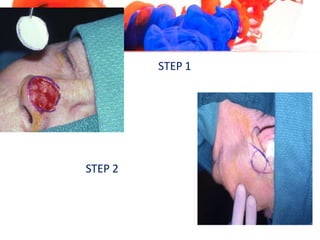
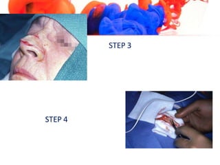
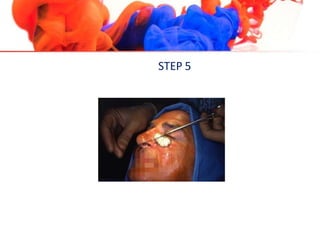
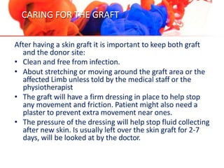
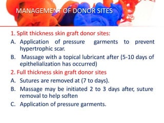
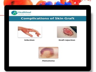
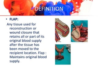
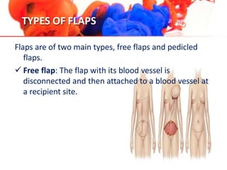
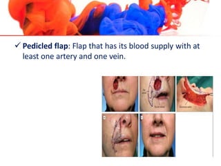
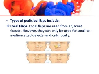
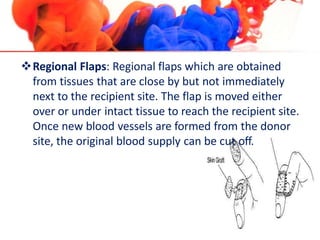
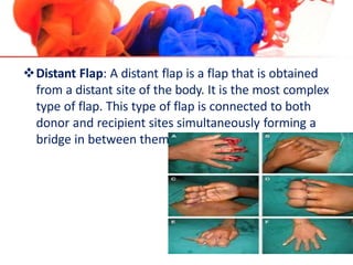
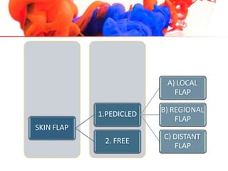
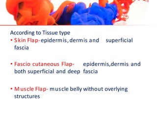
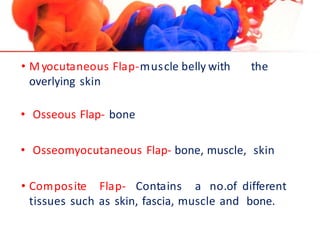
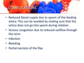
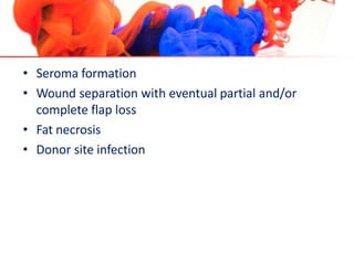
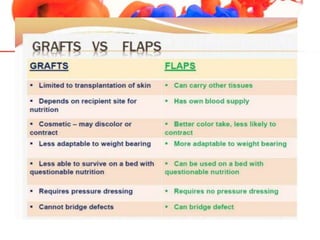
SKIN GRAFTING
INTRODUCTION
• Skin grafts are a common method of closing skin
defects and have been used since the early 1500s by
the Germans and Indians.
• Skin grafts are commonly used to promote healing of
shallow wounds and burns, and wound care nurses
play an important role in management of wounds
treated with grafting.
DEFINITION
• Skin Graft:
A skin graft is a tissue of epidermis and varying
amounts of dermis that is detached from its own
blood supply and placed in a new area with a new
blood supply. Graft Does not maintain original blood
supply.
• Skin Grafting:
Skin grafting is a surgical procedure that involves
removing skin from one area of the body and moving
it, or transplanting it, to a different area of the body.
A skin grafting is a procedure performed where
healthy skin is removed from one area of the body,
the donor site, and transplanted to another, the
recipient site.
• Donor site:
A donor site is an area from which a skin is taken
(harvested) for grafting.
The common donor sites for skin grafting are inner
thigh, upper arm, forearm, and buttocks.
• Recipient site:
A recipient site is an area to which a skin is attaching or
grafting.
HISTORICAL VIEW…
• Grafting of skin originated among the tilemaker caste
in India approximately 3000 years ago.
• 1817, Sir Astley Cooper grafted a FTS from a man’s
amputated thumb for stump coverage.
• Bunger in 1823 successfully reconstructed a nose
with a skin graft.
• Jonathan Warren in 1840 & Joseph Pancoast in 1844
grafted FTS from the arm to the nose & the earlobe,
respectively.
• In 1975 epithelial skin culture technology was
published by Rheinwald & Green.
REVIEW….
EPIDERMIS
• Stratified squamous
epithelium composed
primarily of keratinocytes.
• No blood vessels.
• Relies on diffusion from
underlying tissues.
• Separated from the dermis
by a basement membrane.
DERMIS
• Composed of two “sub-
layers”:
• superficial papillary
• deep reticular.
• The dermis contains
collagen, capillaries,
elastic fibers,
fibroblasts, nerve
endings ect..
INDICATIONS FOR SKIN GRAFTING
Skin grafting can be used to repair almost any type of
wounds and is the most common form of
reconstructive surgery. Skin grafting is often used to
treat;
• Extensive bones or trauma
• Burns
• Specific surgeries that may require skin graft for
healing
• Areas prone for private infection with extensive skin
loss
• Cosmetic reactions for reconstructive surgeries
CLASSIFICATION OF SKIN GRAFTS
A. By species
SKIN
GRAFTS
1.
AUTOGRAFT
2.
ALLOGRAFT
3.
XENOGRAFT
4.
SYNGENIC
SKINGRAFTS
1.SPLITH
THICKNESS
THIN SPLIT
THICKNESS
MEDIUM SPLIT
THICKNESS
THICK SPLIT
THICKNESS
2.FULL
THICKNESS
3.COMPOSITE
B. By Thickness
DONOR SITE SELECTION
• The selection of donor site is essential to achieve the
best possible outcome.
• Donor sites, from which the skin grafts are taken, can
be virtually anywhere in the body.
Criteria related with donor site selection are;
kind of skin graft is to be used.
Achieving the closest possible colour match.
Matching the texture and hair bearing qualities.
Obtaining the biggest possible skin graft without
jeopardizing the healing of the donor site.
Considering the cosmetic effects of the donor site
after healing so that it is in an inconspicuous location
PRE OPERATIVE PREPARATIONS
• No specific preoperative evaluation is unique to skin
grafting.
• NPO for 10-12 hrs
• Patients should wash or shower using soap and
water the evening before surgery.
• Prescribed medication should be reviewed pre-
operatively and only essential medicines given -
those taken orally should be swallowed with the
smallest amount of water possible.
• Medicines that will cause drowsiness should be
administered once the patient has been prepared for
theatre and the patient should be advised to stay on
the bed with a call bell.
• Hair around the incision site should be removed on
the day of surgery if necessary, using electric clippers
with a single-use disposable head.
• Patients’ comfort and dignity should be maintained
when they are changing into their theatre gown.
• Vital signs should be recorded and abnormal
readings reported.
• Allergies should be documented.
• The site of surgery should be marked on the ward or
day unit before patients go to theatre or receive
premeds; this should be checked by the nurse on the
ward or day unit who is completing the pre-operative
checklist.
• Consent should have been obtained
PROCEDURE
After selection of a donor site, both sites are sterilely
prepped, draped, and anesthetized.
then thoroughly cleansed with sterile saline to wash
off the antiseptic and prevent desiccation.
The area is then anesthetized.
For powered dermatomes, mineral oil or antibiotic
ointment can be used to lubricate and hydrate the
skin.
The dermatome applied firmly against the skin with
downward and forward pressure.
An assistant can use forceps to gently grasp and
apply traction to prevent the graft from folding in on
itself.
If desired, the graft can be subsequently
meshed; meshing in favored in larger grafts.
The graft is then applied to the defect and contoured
to fit the defect. The graft is then anchored in place
using sutures or staples depending on physician
preference.
A bolster is applied over the graft. The donor site
can be treated like an abrasion and covered with
petrolatum and a bandage.
• Full-thickness skin grafts:
FTSG is planned in the reconstruction of a surgical
defect, an appropriate donor site must be selected.
A template of the defect can be made using a gauze,
measurements or foil from the suture packaging.
FTSG harvesting does not require the use of additional
surgical instruments.
The donor graft tissue is placed in sterile sodium
chloride solution until it is used.
After harvesting the graft, the secondary defect should
be closed, and the FTSG defatted.
The yellow globular adipose is removed by using iris
scissors until the dermis is visualized.
Grafts are sutured with a quickly absorbing suture, such
as chromic gut or a non-absorbable suture such as
nylon.
STEP 1
STEP 2
STEP 3
STEP 4
STEP 5
HEALING OF GRAFT
• The healing of skin grafts occurs by three phases;
 first phase the phase of serum imbibition: it begins
immediately ,after placing the graft on the wound
bed. Because there are no vascular connections,
nutrients fluid supplied by diffusion of serum from
the bed. The graft is held in place only by weak fibrin
and fibronection bonds.
 Second phase: at 24-48 hours new capillaries start
invading the skin graft making the phase of
revascularization.
 Third phase: the phase of organization starts at 4-5
days when collagen linkages are made between the
wound bed and the graft to create firm attachments.
CARING FOR THE GRAFT
After having a skin graft it is important to keep both graft
and the donor site:
• Clean and free from infection.
• About stretching or moving around the graft area or the
affected Limb unless told by the medical staff or the
physiotherapist
• The graft will have a firm dressing in place to help stop
any movement and friction. Patient might also need a
plaster to prevent extra movement near ones.
• The pressure of the dressing will help stop fluid collecting
after new skin. Is usually left over the skin graft for 2-7
days, will be looked at by the doctor.
MANAGEMENT OF DONOR SITES
1. Split thickness skin graft donor sites:
A. Application of pressure garments to prevent
hypertrophic scar.
B. Massage with a topical lubricant after (5-10 days of
epithelialization has occurred)
2. Full thickness skin graft donor sites
A. Sutures are removed at (7 to days).
B. Massage may be initiated 2 to 3 days after, suture
removal to help soften
C. Application of pressure garments.
SKIN FLAP
INTRODUCTION
• Flap surgery is a technique
in plastic and reconstructive surgery where any type
of tissue is lifted from a donor site and moved to a
recipient site with an intact blood supply.
DEFINITION
• FLAP:
Any tissue used for
reconstruction or
wound closure that
retains all or part of its
original blood supply
after the tissue has
been moved to the
recipient location. Flap :
Maintains original blood
supply.
INDICATIONS
Use of skin flap for ground coverage when inadequate
vascularity of the wound bed prevents skin graft survival.
• Skin flaps are used to repair defects caused by congenital
deformity trauma or tumor ablation in an adjacent part
of the body.
• Skin flaps are also be used to heal extensive wants from
pressure ulcers and long-standing defects from
osteomyelitis.
• Use skin flaps for functional and cosmetic requirement
for wound coverage on the space particularly around the
eye nose and mouth.
TYPES OF FLAPS
Flaps are of two main types, free flaps and pedicled
flaps.
 Free flap: The flap with its blood vessel is
disconnected and then attached to a blood vessel at
a recipient site.
 Pedicled flap: Flap that has its blood supply with at
least one artery and one vein.
• Types of pedicled flaps include:
Local Flaps: Local flaps are used from adjacent
tissues. However, they can only be used for small to
medium sized defects, and only locally.
Regional Flaps: Regional flaps which are obtained
from tissues that are close by but not immediately
next to the recipient site. The flap is moved either
over or under intact tissue to reach the recipient site.
Once new blood vessels are formed from the donor
site, the original blood supply can be cut off.
Distant Flap: A distant flap is a flap that is obtained
from a distant site of the body. It is the most complex
type of flap. This type of flap is connected to both
donor and recipient sites simultaneously forming a
bridge in between them.
SKIN FLAP
1.PEDICLED
A) LOCAL
FLAP
B) REGIONAL
FLAP
C) DISTANT
FLAP
2. FREE
According to Blood supply;
• Random flaps, when the blood supply comes from
unrecognized blood vessels
• Axial flaps, when the blood supply comes from a
recognized and named artery or vein
• Perforator flaps, which have small blood vessels that
originate from a single large vessel.
According to Tissue type
• Skin Flap-epidermis,dermis and
fascia
superficial
• Fascio cutaneous Flap- epidermis,dermis and
both superficial and deep fascia
• Muscle Flap- muscle belly without overlying
structures
• Myocutaneous Flap-muscle belly with the
overlying skin
• Osseous Flap- bone
• Osseomyocutaneous Flap- bone, muscle, skin
• Composite Flap- Contains a no.of different
tissues such as skin, fascia, muscle and bone.
DONOR SITES
GENERAL PRINCIPLES FOR FLAP SURGERY
• Replace like for like.
• Think of reconstruction in terms of units
• Always have a pattern and a back up plan
• Steal from Peter to play Paul
• Do not forget the donor area
COMPLICATIONS
• Reduced blood supply due to spasm of the feeding
artery. This can be avoided by making sure that the
artery does not go into spasm during rotation.
• Venous congestion due to reduced outflow through
the veins
• Infection
• Bleeding
• Partial necrosis of the flap
• Seroma formation
• Wound separation with eventual partial and/or
complete flap loss
• Fat necrosis
• Donor site infection
skingrafting-200522091215.pptx

skingrafting-200522091215.pptx

  • 1.
  • 2.
    INTRODUCTION • Skin graftsare a common method of closing skin defects and have been used since the early 1500s by the Germans and Indians. • Skin grafts are commonly used to promote healing of shallow wounds and burns, and wound care nurses play an important role in management of wounds treated with grafting.
  • 3.
    DEFINITION • Skin Graft: Askin graft is a tissue of epidermis and varying amounts of dermis that is detached from its own blood supply and placed in a new area with a new blood supply. Graft Does not maintain original blood supply. • Skin Grafting: Skin grafting is a surgical procedure that involves removing skin from one area of the body and moving it, or transplanting it, to a different area of the body.
  • 4.
    A skin graftingis a procedure performed where healthy skin is removed from one area of the body, the donor site, and transplanted to another, the recipient site. • Donor site: A donor site is an area from which a skin is taken (harvested) for grafting. The common donor sites for skin grafting are inner thigh, upper arm, forearm, and buttocks.
  • 5.
    • Recipient site: Arecipient site is an area to which a skin is attaching or grafting.
  • 6.
    HISTORICAL VIEW… • Graftingof skin originated among the tilemaker caste in India approximately 3000 years ago. • 1817, Sir Astley Cooper grafted a FTS from a man’s amputated thumb for stump coverage. • Bunger in 1823 successfully reconstructed a nose with a skin graft. • Jonathan Warren in 1840 & Joseph Pancoast in 1844 grafted FTS from the arm to the nose & the earlobe, respectively.
  • 7.
    • In 1975epithelial skin culture technology was published by Rheinwald & Green.
  • 8.
    REVIEW…. EPIDERMIS • Stratified squamous epitheliumcomposed primarily of keratinocytes. • No blood vessels. • Relies on diffusion from underlying tissues. • Separated from the dermis by a basement membrane.
  • 9.
    DERMIS • Composed oftwo “sub- layers”: • superficial papillary • deep reticular. • The dermis contains collagen, capillaries, elastic fibers, fibroblasts, nerve endings ect..
  • 10.
    INDICATIONS FOR SKINGRAFTING Skin grafting can be used to repair almost any type of wounds and is the most common form of reconstructive surgery. Skin grafting is often used to treat; • Extensive bones or trauma • Burns
  • 11.
    • Specific surgeriesthat may require skin graft for healing • Areas prone for private infection with extensive skin loss • Cosmetic reactions for reconstructive surgeries
  • 12.
    CLASSIFICATION OF SKINGRAFTS A. By species SKIN GRAFTS 1. AUTOGRAFT 2. ALLOGRAFT 3. XENOGRAFT 4. SYNGENIC
  • 15.
    SKINGRAFTS 1.SPLITH THICKNESS THIN SPLIT THICKNESS MEDIUM SPLIT THICKNESS THICKSPLIT THICKNESS 2.FULL THICKNESS 3.COMPOSITE B. By Thickness
  • 19.
    DONOR SITE SELECTION •The selection of donor site is essential to achieve the best possible outcome. • Donor sites, from which the skin grafts are taken, can be virtually anywhere in the body.
  • 20.
    Criteria related withdonor site selection are; kind of skin graft is to be used. Achieving the closest possible colour match. Matching the texture and hair bearing qualities. Obtaining the biggest possible skin graft without jeopardizing the healing of the donor site. Considering the cosmetic effects of the donor site after healing so that it is in an inconspicuous location
  • 21.
    PRE OPERATIVE PREPARATIONS •No specific preoperative evaluation is unique to skin grafting. • NPO for 10-12 hrs • Patients should wash or shower using soap and water the evening before surgery. • Prescribed medication should be reviewed pre- operatively and only essential medicines given - those taken orally should be swallowed with the smallest amount of water possible.
  • 22.
    • Medicines thatwill cause drowsiness should be administered once the patient has been prepared for theatre and the patient should be advised to stay on the bed with a call bell. • Hair around the incision site should be removed on the day of surgery if necessary, using electric clippers with a single-use disposable head. • Patients’ comfort and dignity should be maintained when they are changing into their theatre gown.
  • 23.
    • Vital signsshould be recorded and abnormal readings reported. • Allergies should be documented. • The site of surgery should be marked on the ward or day unit before patients go to theatre or receive premeds; this should be checked by the nurse on the ward or day unit who is completing the pre-operative checklist. • Consent should have been obtained
  • 24.
    PROCEDURE After selection ofa donor site, both sites are sterilely prepped, draped, and anesthetized. then thoroughly cleansed with sterile saline to wash off the antiseptic and prevent desiccation. The area is then anesthetized. For powered dermatomes, mineral oil or antibiotic ointment can be used to lubricate and hydrate the skin.
  • 25.
    The dermatome appliedfirmly against the skin with downward and forward pressure. An assistant can use forceps to gently grasp and apply traction to prevent the graft from folding in on itself. If desired, the graft can be subsequently meshed; meshing in favored in larger grafts. The graft is then applied to the defect and contoured to fit the defect. The graft is then anchored in place using sutures or staples depending on physician preference.
  • 26.
    A bolster isapplied over the graft. The donor site can be treated like an abrasion and covered with petrolatum and a bandage.
  • 29.
    • Full-thickness skingrafts: FTSG is planned in the reconstruction of a surgical defect, an appropriate donor site must be selected. A template of the defect can be made using a gauze, measurements or foil from the suture packaging. FTSG harvesting does not require the use of additional surgical instruments. The donor graft tissue is placed in sterile sodium chloride solution until it is used.
  • 30.
    After harvesting thegraft, the secondary defect should be closed, and the FTSG defatted. The yellow globular adipose is removed by using iris scissors until the dermis is visualized. Grafts are sutured with a quickly absorbing suture, such as chromic gut or a non-absorbable suture such as nylon.
  • 31.
  • 32.
  • 33.
  • 35.
    HEALING OF GRAFT •The healing of skin grafts occurs by three phases;  first phase the phase of serum imbibition: it begins immediately ,after placing the graft on the wound bed. Because there are no vascular connections, nutrients fluid supplied by diffusion of serum from the bed. The graft is held in place only by weak fibrin and fibronection bonds.
  • 36.
     Second phase:at 24-48 hours new capillaries start invading the skin graft making the phase of revascularization.  Third phase: the phase of organization starts at 4-5 days when collagen linkages are made between the wound bed and the graft to create firm attachments.
  • 37.
    CARING FOR THEGRAFT After having a skin graft it is important to keep both graft and the donor site: • Clean and free from infection. • About stretching or moving around the graft area or the affected Limb unless told by the medical staff or the physiotherapist • The graft will have a firm dressing in place to help stop any movement and friction. Patient might also need a plaster to prevent extra movement near ones. • The pressure of the dressing will help stop fluid collecting after new skin. Is usually left over the skin graft for 2-7 days, will be looked at by the doctor.
  • 38.
    MANAGEMENT OF DONORSITES 1. Split thickness skin graft donor sites: A. Application of pressure garments to prevent hypertrophic scar. B. Massage with a topical lubricant after (5-10 days of epithelialization has occurred) 2. Full thickness skin graft donor sites A. Sutures are removed at (7 to days). B. Massage may be initiated 2 to 3 days after, suture removal to help soften C. Application of pressure garments.
  • 40.
  • 41.
    INTRODUCTION • Flap surgeryis a technique in plastic and reconstructive surgery where any type of tissue is lifted from a donor site and moved to a recipient site with an intact blood supply.
  • 42.
    DEFINITION • FLAP: Any tissueused for reconstruction or wound closure that retains all or part of its original blood supply after the tissue has been moved to the recipient location. Flap : Maintains original blood supply.
  • 43.
    INDICATIONS Use of skinflap for ground coverage when inadequate vascularity of the wound bed prevents skin graft survival. • Skin flaps are used to repair defects caused by congenital deformity trauma or tumor ablation in an adjacent part of the body. • Skin flaps are also be used to heal extensive wants from pressure ulcers and long-standing defects from osteomyelitis. • Use skin flaps for functional and cosmetic requirement for wound coverage on the space particularly around the eye nose and mouth.
  • 44.
    TYPES OF FLAPS Flapsare of two main types, free flaps and pedicled flaps.  Free flap: The flap with its blood vessel is disconnected and then attached to a blood vessel at a recipient site.
  • 45.
     Pedicled flap:Flap that has its blood supply with at least one artery and one vein.
  • 46.
    • Types ofpedicled flaps include: Local Flaps: Local flaps are used from adjacent tissues. However, they can only be used for small to medium sized defects, and only locally.
  • 47.
    Regional Flaps: Regionalflaps which are obtained from tissues that are close by but not immediately next to the recipient site. The flap is moved either over or under intact tissue to reach the recipient site. Once new blood vessels are formed from the donor site, the original blood supply can be cut off.
  • 48.
    Distant Flap: Adistant flap is a flap that is obtained from a distant site of the body. It is the most complex type of flap. This type of flap is connected to both donor and recipient sites simultaneously forming a bridge in between them.
  • 49.
    SKIN FLAP 1.PEDICLED A) LOCAL FLAP B)REGIONAL FLAP C) DISTANT FLAP 2. FREE
  • 50.
    According to Bloodsupply; • Random flaps, when the blood supply comes from unrecognized blood vessels • Axial flaps, when the blood supply comes from a recognized and named artery or vein
  • 51.
    • Perforator flaps,which have small blood vessels that originate from a single large vessel.
  • 52.
    According to Tissuetype • Skin Flap-epidermis,dermis and fascia superficial • Fascio cutaneous Flap- epidermis,dermis and both superficial and deep fascia • Muscle Flap- muscle belly without overlying structures
  • 53.
    • Myocutaneous Flap-musclebelly with the overlying skin • Osseous Flap- bone • Osseomyocutaneous Flap- bone, muscle, skin • Composite Flap- Contains a no.of different tissues such as skin, fascia, muscle and bone.
  • 54.
  • 56.
    GENERAL PRINCIPLES FORFLAP SURGERY • Replace like for like. • Think of reconstruction in terms of units • Always have a pattern and a back up plan • Steal from Peter to play Paul • Do not forget the donor area
  • 57.
    COMPLICATIONS • Reduced bloodsupply due to spasm of the feeding artery. This can be avoided by making sure that the artery does not go into spasm during rotation. • Venous congestion due to reduced outflow through the veins • Infection • Bleeding • Partial necrosis of the flap
  • 58.
    • Seroma formation •Wound separation with eventual partial and/or complete flap loss • Fat necrosis • Donor site infection