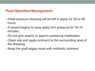
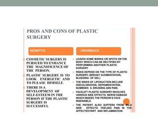
RECONSTRUCTIVE AND COSMETIC
SURGERIES
• The word "plastic derived from the Greek plastikos
meaning to mould or to shape.
• Plastic surgeon typically mold and reshape the following
tissues of the body; bone, cartilage, muscles, fat and skin.
• Plastic surgery is a medical specialty concerned with the
correction or restoration of form and function of the body
structures damaged by trauma, transformed by aging
process, changed by disease process and malformed as
a result of congenital defects.
• In plastic surgery the transfer of skin tissue (skin grafting)
is one of the most common procedures.
• Plastic surgery include closure of wounds, removal of
skin tumors, repair of soft tissue injuries or burns,
correction of deformities and repair of cosmetic defects.
During plastic surgery the following procedures are
common:
• tissue may be removed to fill depression to cover a wound
• or to improve appearance and tissue may be completely
removed to alter the shape.
• Plastic surgery is divided into two major areas:
• Reconstructive surgery and
• Cosmetic (aesthetic) surgery.
GOALS
The basic goals of the plastic surgery include following:
Correction of perceived disfigurement
Restoration of impaired function
Improvement of physical appearance
BENEFITS
The benefits of plastic surgery may include:
Correction of a congenital or acquired deformity
Correction of a perceived physical imperfection
Psychological benefits
Basic principles of plastic surgery:
Achieving minimal scarring
Careful planning of incisions
Appropriate choice of wound closure
 Use of best available suture materials
Early removal of exposed sutures
Documentation through photography
RECONSTRUCTIVE SURGERY
• Reconstructive surgery is the use of surgery to restore the
form and function of the body.
• It is performed to correct functional impairments caused
by: burns, traumatic injuries such as facial bone fractures,
congenital abnormalities such as cleft lip or palate,
developmental abnormalities, infection or disease and
removal of cancers or tumors such as mastectomy.
Common Reconstructive Surgical
Procedures
There are several operative and non-operative procedures
available to improve the shape and enhance the beauty of
various body parts.
• Most commonly performed procedures for various body
parts are as follows:
Breast Reconstruction
Cleft Lip and Palate
Ptosis/Drooping of Eyelid
Face Injury
Injuries to Limbs
 Defects of ears
Burn Contracture Surgery
Cranio-facial Defects
Pressure Sores
 Hand and finger Injuries
 Amputations
Spinal cord defects
RECONSTRUCTIVE MODALITIES
1. Skin grafting - split skin graft
- full thickness skin graft
- composite graft
2. Skin flap
a)Local flap - rotational flap
- advancement flap
- transposition flap
- interpolation flap
b)Distant flap - direct flap
- tubed flap
- microvascular free flap
Reconstructive Modalities
Skin Grafting
• Skin grafting is a type of medical grafting involving the
transplantation of skin.
• Here a section of skin is detached from its own blood
supply and transferred to a distant site .
• Skin grafts are commonly used to repair defects result
from excision of skin tumors, burn and to cover the
wound in which insufficient skin is available to permit
wound closure.
The grafting has two purposes:
• it can reduce course of treatment needed
• it can improve the function and appearance of the area of
the body which receives the skin graft .
Indications
• Extensive wounds or trauma
• Burns
• Specific surgeries
Classification
1.Autografts: An autografts is tissue obtained from the
patient's own skin.
2. Allografts: An allograft is tissue obtained from a donor of
the same species; these grafts are also called allogenic or
homografts.
3. Xenografts: A xenografts or heterograft is tissue from a
donor of a different species
Types of Skin Grafts
Split Skin Grafts
• A split-thickness graft can be cut at various thicknesses
and is commonly used to cover large wounds.
• It is taken by shaving the surface layers (epidermis and
dermis) of the skin with a large knife called a dermatome
• The shaved piece of skin is then applied to the wound.
This type of skin graft is often taken from the leg.
• It is often used after excision of a lesion on the lower leg.
Full Thickness Skin Grafts:
• Full-thickness graft consists of epidermis and the entire
dermis without the underlying fat
• This type of skin graft is taken by removing all the layers
of skin . It is done in a similar way to skin excision.
• The piece of skin is cut into the correct shape, and then
applied to the wound. This type of skin graft is often taken
from the arm, neck or behind the ear.
• It is often used after excisions on the hand or face.
Composite Graft:
• A composite skin graft is a combinations of skin and fat,
skin and cartilage.
• It is used in patients whose injuries require three-
dimensional reconstruction
• For example, a wedge of ear containing skin and cartilage
can be used to repair the nose.
Donor Site
The common areas which are used as donor sites are
• buttocks,
• Thighs
• upper arms
• the dressing will usually be left in place for 7-14 days
• Re- pad the dressing as the area often oozes/ bleeds after
the operation
• Donor site area is usually more painful than the grafted area as
the top layers of the skin are removed exposing nerve endings.
• Regular painkillers will need to be given to ease discomfort
• All dressing need to be kept dry
The donor site is selected with several criteria
The donor site is selected with several criteria :-
• Achieving the closest possible color match
• Matching the texture and hair bearing qualities
• Obtaining the thickest possible skin grafts without
affecting the healing of donor site
Graft Application
Wound preparation.
• The wound is cleaned and measured, and then a pattern
is traced for transfer over to the donor site.
Anesthesia is administered.
• Depending on the size, severity, and location of the
wound, as well as the type of graft, the procedure may
require local anesthesia, regional anesthesia, iv sedation,
general anesthesia, or a combination.
• The donor skin is harvested and prepared. The skin is
either removed with a scalpel, skin grafting knives or with
the help of a special harvesting machine called a
dermatome.
• The graft may also be "meshed," a process by which
multiple controlled incisions are placed in the graft.
This technique allows fluid to leak out from the underlying
tissue and the donor skin to spread out over a much
larger area.
• With full thickness or composite graft, the donor site is
then closed with sutures. With a split- thickness graft,
sutures are not needed at the donor site.
• The skin graft is taken from the donor or host site and
applied to the desired site called the recipient site or graft
bed. It is held in place by a few small stitches or surgical
staples.
• A pressure bandage is applied over the graft recipient
site. A special vacuum apparatus called a wound VAC
may be placed over the area for the first 3 to 5 days to
control drainage and increase the graft's chances of
survival.
• Healing begins. At first, the graft uses oxygen and
nutrients from the tissue at the recipient site to survive.
The graft is initially nourished by plasma. (plasmatic
imbibitions)
• New blood vessels begin to grow within the first 36 hours
by a process called capillary inosculation, followed by
new skin cells which then begin to grow from the graft to
cover the recipient area with new skin.
• The process of revascularization and reattachment of skin
graft to a recipient bed takesplace.
• After a skin graft is put in place, it may be left exposed or
covered with a light dressing or a pressure dressing
depending on the area.
Post-Op Care:
• Both the donor and recipient sites should be kept moist
and well- protected.
• Physician will instruct patient on the proper use of
medications and bandaging
For a graft to survive and be effective, certain
conditions must be met:
The recipient site must have an adequate blood supply
so that normal physiologic function can resume.
The graft must be in close contact with its bed to avoid
accumulation of blood or fluid.
The area must be free of infection
The graft must be fixed firmly (immobilized) so that it
remain in place on the recipient sites.
Complications of Skin Grafts and Donor Areas
1.Infection
• Smelly discharge from dressing
• High temperature
• Increased pain
• Redness and swelling around the skin graft and donor
area
2.Bleeding through dressing due to trauma or infection
which may cause clots and lift graftt
3. Inadequate excision of the wound bed
4. Inadequate vascular supply to the wound bed
5. Hematomas and seromas
6.Graft rejection
Rejection may occur in heterologous graft. To prevent this,
the patient usually must be treated with long term
immunosuppressant drugs
.
Graft Care
• Clean and free from infection.
• Avoid stretching or moving around the graft area or the
affected limb unless advised.
• The graft have firm dressing in place to help stop any
movement and friction.
• Patient might also need a plaster to prevent extra
movement near joints.
• The pressure of the dressing will help to stop fluid
collecting under the new skin.
• Dressing is usually kept over the skin graft for 2-7 days
After Care
• Once a skin graft has been put in place, it must be
maintained carefully even after it has healed.
• Patients who have grafts on their legs should remain in
bed for 7 to 10 days with their legs elevated.
• For several months, the patient should support the graft
with bandage or stocking.
• Grafted skin does not contain sweat or oil glands, and
should be lubricated daily for two to three months with
mineral oil to prevent drying and cracking
• Try to avoid scratching the wounds
• Administer anti-histamine.
• Antibiotics is given to fight or prevent an infection caused
by bacteria
• Clean and wash regularly on areas where there are no
dressings.
• Make sure patient wears cotton clothes to help stop
getting too hot.
Keep wounds clean and dry
• When allowed to bathe, carefully wash the graft and
donor sites with soap and water.
• Dry the area and put on clean, new bandages as (Change
bandages every time when they get wet or dirty.
• When the wounds have healed patient will need to apply
cream on to them, gently massaging them 2-3 times a
day, to prevent them getting dry and flaky.
• The graft and donor site will need to be protected from the
sun as they will burn more easily than rest of patient's
skin.
• As the wound heals scarring may occur.
• Patient may be given pressure garments to wear, or a
dressing or gel to be put on the scar(help flatten the
scarring)
Nursing Care
1.Circulation:
Assess the graft area for signs of adequate blood supply
Inspects the color of the graft area.
Make sure the graft area is warm(indicates sufficient
blood supply to the area)
2. Drainage:
 Checks the patency of drains placed in the graft area.
 Makes sure they are not blocked, so drainage can flow
out of the graft site instead of accumulating in it and
potentially causing an infection
3.Positioning:
• The nurse ensures blood circulation to the graft area by
positioning the patient off the graft
• Taking pressure off the graft and skin surrounding it
reduces the risk of decreased blood supply to the area.
4. Low Pressure:
 Place the patient on a low pressure bed when lying down
or low pressure cushion for sitting down.(adequate
perfusion)
5. Trapeze:
Provide an over the bed trapeze
Makes sure that the patient is aware of how to use it for
moving around in bed.
This reduces the amount of shearing and friction that
could occur during movement and possibly displace the
graft
Skin Flaps
It is a segment of tissue that remains attached at one end
(base or pedicle) while the other end is moved to a
recipient area.
Its survival depends on functioning arterial and venous
blood supplies and lymphatic drainage in its pedicle or
base.
 A flap differs from a graft in that a portion of the tissue
is attached to its original site and retains its blood
supply
Flaps may consist of skin, mucosa, muscle, adipose
tissue, omentum and bone based on its own blood supply.
They are used for wound coverage and provide bulk,
especially when bone, tendon, blood vessels or nerve
tissue is exposed.
Flap offer an aesthetic solution because a flap retains the
color and textures of the donor area.
The major complication is necrosis of the pedicle or base
as a result of failure of the blood supply.
Indications
To repair defects caused by congenital deformity, trauma,
or tumor ablation in an adjacent part of the body.
To heal extensive wounds from pressure ulcers.
 For functional and cosmetic requirements for wound
coverage on the face, particularly around the eye, nose
and mouth.
FREE FLAP
• A striking advance in reconstructive surgery is the use of
free flaps or free tissue transfer achieved by
microvascular techniques .
• Free flaps are harvested from one area of the body to
reconstruct a defect in a distant area.
• The donor tissue (skin, muscle, bone or a combination of
these) is detached from its blood supply at the donor site
and reattached by microvascular anastomosis to arteries
and veins at the recipient site.
• Microvascular surgery - uses a variety of donor sites for
tissue reconstruction
Methods of Flap Movement
• Skin flaps can be moved to a local or distant site.
Local Flaps:
• Used for defects that are adjacent to the donor site.
• There are 4 major types of local flaps based on the
predominant type of movement
1 . ADVANCEMENT FLAP
An advancement flap moves directly forward without
lateral movement.
A triangle of skin is excised from the base of the flap to
aid in closure.
2.ROTATIONAL FLAP
A rotational flap is a semicircular flap that rotates about a
pivot point into an adjacent defect.
3. TRANSPOSITION FLAP
• A transposition flap moves laterally about a pivot point
into an adjacent defect.
• Usually, it is designed as a rectangle.
• Design the flap to be longer than defect, since
transposition decreases the length.
• The donor site can be closed directly or closed with a skin
graft or second skin flap.
4.INTERPOLATION FLAP
The interpolation flap rotates about a pivot point into a
nearby defect, with the pedicle passing skin bridge.
Distant Flaps:
Use distant flaps to cover nonadjacent defects.
They may be transferred directly or transferred by
microvascular techniques.
DIRECT FLAP
• A direct flap is transferred to a distant site directly so that
the donor site and recipient sites are approximated
TUBED FLAP
• The tubed flap is transferred to a recipient site with the
lateral flap edges sewn together, while the new blood
supply is incorporated from the distant end of the flap.
• Sewing the edges together result in decreased risk of
infection and contraction of the flap.
MICROVASCULAR FREE FLAP
• A microvascular free flap is a type of distant flap in
which the flap, with its vascular pedicle, is detached
completely from its donor vessels and anastomosed to
the recipient vessels at the recipient site using a
microvascular surgical technique.
Pre-Operative Management
History and physical examination should be done for
(latex sensitivity, cardiovascular problems requiring
endocarditis antibiotics prophylaxis, bleeding problems
and high BP)
To enhance wound healing instruct to avoid smoking,
alcoholism and proper nutrition to be encouraged.
.
Aspirin, NSAID and vitamin E are discontinued 14 days
before the procedure
Prothrombin time and international normalized ratio
should be measured before the procedure
The operative site should be free of makeup.
Post-Operative Management
Initial pressure dressing will be left in place for 24 to 48
hours.
If wound begins to ooze apply firm pressure for 10-15
minutes.
Do not give aspirin or aspirin-containing medication.
Clean site and apply ointment to the surrounding area of
the dressing.
Keep the graft edges moist with antibiotic ointment
Protect the graft from the sun. The sun will cause
pigmentation changes in the graft
Inspect the dressing daily. Report unusual drainage or
signs of any inflammatory reaction
Avoid strenuous exercise.Anything that causes face to
flush raise BP and impair healing
RECONSTRUCTIVE SURGERIES & Cosmetic surgeries.pptx

RECONSTRUCTIVE SURGERIES & Cosmetic surgeries.pptx

  • 1.
  • 2.
    • The word"plastic derived from the Greek plastikos meaning to mould or to shape. • Plastic surgeon typically mold and reshape the following tissues of the body; bone, cartilage, muscles, fat and skin. • Plastic surgery is a medical specialty concerned with the correction or restoration of form and function of the body structures damaged by trauma, transformed by aging process, changed by disease process and malformed as a result of congenital defects.
  • 3.
    • In plasticsurgery the transfer of skin tissue (skin grafting) is one of the most common procedures. • Plastic surgery include closure of wounds, removal of skin tumors, repair of soft tissue injuries or burns, correction of deformities and repair of cosmetic defects.
  • 4.
    During plastic surgerythe following procedures are common: • tissue may be removed to fill depression to cover a wound • or to improve appearance and tissue may be completely removed to alter the shape. • Plastic surgery is divided into two major areas: • Reconstructive surgery and • Cosmetic (aesthetic) surgery.
  • 5.
    GOALS The basic goalsof the plastic surgery include following: Correction of perceived disfigurement Restoration of impaired function Improvement of physical appearance
  • 6.
    BENEFITS The benefits ofplastic surgery may include: Correction of a congenital or acquired deformity Correction of a perceived physical imperfection Psychological benefits
  • 7.
    Basic principles ofplastic surgery: Achieving minimal scarring Careful planning of incisions Appropriate choice of wound closure  Use of best available suture materials Early removal of exposed sutures Documentation through photography
  • 8.
    RECONSTRUCTIVE SURGERY • Reconstructivesurgery is the use of surgery to restore the form and function of the body. • It is performed to correct functional impairments caused by: burns, traumatic injuries such as facial bone fractures, congenital abnormalities such as cleft lip or palate, developmental abnormalities, infection or disease and removal of cancers or tumors such as mastectomy.
  • 9.
    Common Reconstructive Surgical Procedures Thereare several operative and non-operative procedures available to improve the shape and enhance the beauty of various body parts. • Most commonly performed procedures for various body parts are as follows: Breast Reconstruction Cleft Lip and Palate Ptosis/Drooping of Eyelid Face Injury
  • 10.
    Injuries to Limbs Defects of ears Burn Contracture Surgery Cranio-facial Defects Pressure Sores  Hand and finger Injuries  Amputations Spinal cord defects
  • 11.
    RECONSTRUCTIVE MODALITIES 1. Skingrafting - split skin graft - full thickness skin graft - composite graft 2. Skin flap a)Local flap - rotational flap - advancement flap - transposition flap - interpolation flap b)Distant flap - direct flap - tubed flap - microvascular free flap
  • 12.
    Reconstructive Modalities Skin Grafting •Skin grafting is a type of medical grafting involving the transplantation of skin. • Here a section of skin is detached from its own blood supply and transferred to a distant site . • Skin grafts are commonly used to repair defects result from excision of skin tumors, burn and to cover the wound in which insufficient skin is available to permit wound closure.
  • 13.
    The grafting hastwo purposes: • it can reduce course of treatment needed • it can improve the function and appearance of the area of the body which receives the skin graft . Indications • Extensive wounds or trauma • Burns • Specific surgeries
  • 14.
    Classification 1.Autografts: An autograftsis tissue obtained from the patient's own skin. 2. Allografts: An allograft is tissue obtained from a donor of the same species; these grafts are also called allogenic or homografts. 3. Xenografts: A xenografts or heterograft is tissue from a donor of a different species
  • 15.
    Types of SkinGrafts Split Skin Grafts • A split-thickness graft can be cut at various thicknesses and is commonly used to cover large wounds. • It is taken by shaving the surface layers (epidermis and dermis) of the skin with a large knife called a dermatome • The shaved piece of skin is then applied to the wound. This type of skin graft is often taken from the leg. • It is often used after excision of a lesion on the lower leg.
  • 17.
    Full Thickness SkinGrafts: • Full-thickness graft consists of epidermis and the entire dermis without the underlying fat • This type of skin graft is taken by removing all the layers of skin . It is done in a similar way to skin excision. • The piece of skin is cut into the correct shape, and then applied to the wound. This type of skin graft is often taken from the arm, neck or behind the ear. • It is often used after excisions on the hand or face.
  • 18.
    Composite Graft: • Acomposite skin graft is a combinations of skin and fat, skin and cartilage. • It is used in patients whose injuries require three- dimensional reconstruction • For example, a wedge of ear containing skin and cartilage can be used to repair the nose.
  • 20.
    Donor Site The commonareas which are used as donor sites are • buttocks, • Thighs • upper arms • the dressing will usually be left in place for 7-14 days • Re- pad the dressing as the area often oozes/ bleeds after the operation • Donor site area is usually more painful than the grafted area as the top layers of the skin are removed exposing nerve endings. • Regular painkillers will need to be given to ease discomfort • All dressing need to be kept dry
  • 21.
    The donor siteis selected with several criteria The donor site is selected with several criteria :- • Achieving the closest possible color match • Matching the texture and hair bearing qualities • Obtaining the thickest possible skin grafts without affecting the healing of donor site
  • 22.
    Graft Application Wound preparation. •The wound is cleaned and measured, and then a pattern is traced for transfer over to the donor site. Anesthesia is administered. • Depending on the size, severity, and location of the wound, as well as the type of graft, the procedure may require local anesthesia, regional anesthesia, iv sedation, general anesthesia, or a combination.
  • 23.
    • The donorskin is harvested and prepared. The skin is either removed with a scalpel, skin grafting knives or with the help of a special harvesting machine called a dermatome. • The graft may also be "meshed," a process by which multiple controlled incisions are placed in the graft. This technique allows fluid to leak out from the underlying tissue and the donor skin to spread out over a much larger area.
  • 24.
    • With fullthickness or composite graft, the donor site is then closed with sutures. With a split- thickness graft, sutures are not needed at the donor site. • The skin graft is taken from the donor or host site and applied to the desired site called the recipient site or graft bed. It is held in place by a few small stitches or surgical staples. • A pressure bandage is applied over the graft recipient site. A special vacuum apparatus called a wound VAC may be placed over the area for the first 3 to 5 days to control drainage and increase the graft's chances of survival.
  • 25.
    • Healing begins.At first, the graft uses oxygen and nutrients from the tissue at the recipient site to survive. The graft is initially nourished by plasma. (plasmatic imbibitions) • New blood vessels begin to grow within the first 36 hours by a process called capillary inosculation, followed by new skin cells which then begin to grow from the graft to cover the recipient area with new skin.
  • 26.
    • The processof revascularization and reattachment of skin graft to a recipient bed takesplace. • After a skin graft is put in place, it may be left exposed or covered with a light dressing or a pressure dressing depending on the area. Post-Op Care: • Both the donor and recipient sites should be kept moist and well- protected. • Physician will instruct patient on the proper use of medications and bandaging
  • 27.
    For a graftto survive and be effective, certain conditions must be met: The recipient site must have an adequate blood supply so that normal physiologic function can resume. The graft must be in close contact with its bed to avoid accumulation of blood or fluid. The area must be free of infection The graft must be fixed firmly (immobilized) so that it remain in place on the recipient sites.
  • 28.
    Complications of SkinGrafts and Donor Areas 1.Infection • Smelly discharge from dressing • High temperature • Increased pain • Redness and swelling around the skin graft and donor area 2.Bleeding through dressing due to trauma or infection which may cause clots and lift graftt
  • 29.
    3. Inadequate excisionof the wound bed 4. Inadequate vascular supply to the wound bed 5. Hematomas and seromas 6.Graft rejection Rejection may occur in heterologous graft. To prevent this, the patient usually must be treated with long term immunosuppressant drugs .
  • 30.
    Graft Care • Cleanand free from infection. • Avoid stretching or moving around the graft area or the affected limb unless advised. • The graft have firm dressing in place to help stop any movement and friction. • Patient might also need a plaster to prevent extra movement near joints. • The pressure of the dressing will help to stop fluid collecting under the new skin. • Dressing is usually kept over the skin graft for 2-7 days
  • 31.
    After Care • Oncea skin graft has been put in place, it must be maintained carefully even after it has healed. • Patients who have grafts on their legs should remain in bed for 7 to 10 days with their legs elevated. • For several months, the patient should support the graft with bandage or stocking.
  • 32.
    • Grafted skindoes not contain sweat or oil glands, and should be lubricated daily for two to three months with mineral oil to prevent drying and cracking
  • 33.
    • Try toavoid scratching the wounds • Administer anti-histamine. • Antibiotics is given to fight or prevent an infection caused by bacteria • Clean and wash regularly on areas where there are no dressings. • Make sure patient wears cotton clothes to help stop getting too hot.
  • 34.
    Keep wounds cleanand dry • When allowed to bathe, carefully wash the graft and donor sites with soap and water. • Dry the area and put on clean, new bandages as (Change bandages every time when they get wet or dirty. • When the wounds have healed patient will need to apply cream on to them, gently massaging them 2-3 times a day, to prevent them getting dry and flaky.
  • 35.
    • The graftand donor site will need to be protected from the sun as they will burn more easily than rest of patient's skin. • As the wound heals scarring may occur. • Patient may be given pressure garments to wear, or a dressing or gel to be put on the scar(help flatten the scarring)
  • 36.
    Nursing Care 1.Circulation: Assess thegraft area for signs of adequate blood supply Inspects the color of the graft area. Make sure the graft area is warm(indicates sufficient blood supply to the area) 2. Drainage:  Checks the patency of drains placed in the graft area.  Makes sure they are not blocked, so drainage can flow out of the graft site instead of accumulating in it and potentially causing an infection
  • 37.
    3.Positioning: • The nurseensures blood circulation to the graft area by positioning the patient off the graft • Taking pressure off the graft and skin surrounding it reduces the risk of decreased blood supply to the area.
  • 38.
    4. Low Pressure: Place the patient on a low pressure bed when lying down or low pressure cushion for sitting down.(adequate perfusion) 5. Trapeze: Provide an over the bed trapeze Makes sure that the patient is aware of how to use it for moving around in bed. This reduces the amount of shearing and friction that could occur during movement and possibly displace the graft
  • 39.
    Skin Flaps It isa segment of tissue that remains attached at one end (base or pedicle) while the other end is moved to a recipient area. Its survival depends on functioning arterial and venous blood supplies and lymphatic drainage in its pedicle or base.  A flap differs from a graft in that a portion of the tissue is attached to its original site and retains its blood supply
  • 40.
    Flaps may consistof skin, mucosa, muscle, adipose tissue, omentum and bone based on its own blood supply. They are used for wound coverage and provide bulk, especially when bone, tendon, blood vessels or nerve tissue is exposed. Flap offer an aesthetic solution because a flap retains the color and textures of the donor area. The major complication is necrosis of the pedicle or base as a result of failure of the blood supply.
  • 41.
    Indications To repair defectscaused by congenital deformity, trauma, or tumor ablation in an adjacent part of the body. To heal extensive wounds from pressure ulcers.  For functional and cosmetic requirements for wound coverage on the face, particularly around the eye, nose and mouth.
  • 42.
    FREE FLAP • Astriking advance in reconstructive surgery is the use of free flaps or free tissue transfer achieved by microvascular techniques . • Free flaps are harvested from one area of the body to reconstruct a defect in a distant area.
  • 43.
    • The donortissue (skin, muscle, bone or a combination of these) is detached from its blood supply at the donor site and reattached by microvascular anastomosis to arteries and veins at the recipient site. • Microvascular surgery - uses a variety of donor sites for tissue reconstruction
  • 44.
    Methods of FlapMovement • Skin flaps can be moved to a local or distant site. Local Flaps: • Used for defects that are adjacent to the donor site. • There are 4 major types of local flaps based on the predominant type of movement
  • 45.
    1 . ADVANCEMENTFLAP An advancement flap moves directly forward without lateral movement. A triangle of skin is excised from the base of the flap to aid in closure.
  • 46.
    2.ROTATIONAL FLAP A rotationalflap is a semicircular flap that rotates about a pivot point into an adjacent defect.
  • 47.
    3. TRANSPOSITION FLAP •A transposition flap moves laterally about a pivot point into an adjacent defect. • Usually, it is designed as a rectangle. • Design the flap to be longer than defect, since transposition decreases the length. • The donor site can be closed directly or closed with a skin graft or second skin flap.
  • 48.
    4.INTERPOLATION FLAP The interpolationflap rotates about a pivot point into a nearby defect, with the pedicle passing skin bridge.
  • 50.
    Distant Flaps: Use distantflaps to cover nonadjacent defects. They may be transferred directly or transferred by microvascular techniques.
  • 51.
    DIRECT FLAP • Adirect flap is transferred to a distant site directly so that the donor site and recipient sites are approximated
  • 52.
    TUBED FLAP • Thetubed flap is transferred to a recipient site with the lateral flap edges sewn together, while the new blood supply is incorporated from the distant end of the flap. • Sewing the edges together result in decreased risk of infection and contraction of the flap.
  • 53.
    MICROVASCULAR FREE FLAP •A microvascular free flap is a type of distant flap in which the flap, with its vascular pedicle, is detached completely from its donor vessels and anastomosed to the recipient vessels at the recipient site using a microvascular surgical technique.
  • 54.
    Pre-Operative Management History andphysical examination should be done for (latex sensitivity, cardiovascular problems requiring endocarditis antibiotics prophylaxis, bleeding problems and high BP) To enhance wound healing instruct to avoid smoking, alcoholism and proper nutrition to be encouraged. .
  • 55.
    Aspirin, NSAID andvitamin E are discontinued 14 days before the procedure Prothrombin time and international normalized ratio should be measured before the procedure The operative site should be free of makeup.
  • 56.
    Post-Operative Management Initial pressuredressing will be left in place for 24 to 48 hours. If wound begins to ooze apply firm pressure for 10-15 minutes. Do not give aspirin or aspirin-containing medication. Clean site and apply ointment to the surrounding area of the dressing. Keep the graft edges moist with antibiotic ointment
  • 57.
    Protect the graftfrom the sun. The sun will cause pigmentation changes in the graft Inspect the dressing daily. Report unusual drainage or signs of any inflammatory reaction Avoid strenuous exercise.Anything that causes face to flush raise BP and impair healing