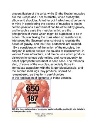
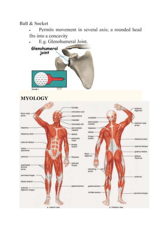
LOCOMOTOR SYSTEM 
The locomotor system is simply the system of 
movement of an organism. Adaptation by movement is 
the principal property distinguishing an animal from a 
plant. The human organism is born, develops and exists 
under the earth's gravitational pull. Each movement of 
the body is concerned with overcoming this gravitation 
and the weight-bearing and locomotor system therefore 
performs an anti gravitation function too. That is why it 
can also be called the antigravitation system.The weight 
bearing and locomotor system constitutes the greater 
part of the total body mass. This explains the fact that 
the weight-bearing and locomotor system is the main 
determinant of the shape of the body and its separation 
into the trunk, head and limbs. The weight-bearing and 
locomotor system is made up of three systems of 
organs: (1) Osteology, (2) Arthrology and (3) 
Myology. Osteology and arthrology form 
the passive part of the locomotor system and myology 
forms the active part of the locomotor system. 
The passive part of this system can also be called 
theosteoarthrosyndesmology, which is the 
science of the bones and their articulations. 
GENERAL OSTEOLOGY 
This branch of anatomy studies the skeleton. The 
skeleton is a complex of hard structures that originate in 
the mesenchyme and possesses mechanical importance. 
It consists of separate bones joined to each other by 
means of connective cartilaginous or bony tissue, which 
together make up the passive locomotor apparatus or 
system. The skeleton is composed of Axial(the skull,
thorax and vertebral column) 
and appendicular(skeleton of upper and lower limbs) 
skeleton. 
The skeleton consists of about 206 separate bones, out 
of which 24-40 are unpaired and the rest of them are 
paired.Each bone, with the exception of the 
cartilaginous joint surfaces and areas where flat tendons 
are attached, is enclosed in a connective tissue sheath, 
the periosteum.The structural and functional unit of 
the bone are the osteon and the trabecular 
pack. 
. 
22 bones in 
skull 
6 in middle ears 
1 hyoid bone 
26 in vertebral column 
25 in thoracic cage 
4 in pectoral girdle 
60 in upper limbs 
60 in lower limbs 
2 in pelvic girdle 
206 bones in 
all.
ARTHROLOGY 
This branch of anatomy studies joints and 
articulations.Site where rigid elements of the skeleton meet 
are called articulations. Joints are places of union between 
two or more skeletal elements.They can be classified by 
the type of tissue present. Using this method, we can 
split the joints of the body 
into fibrous, cartilaginous and synovial joints. 
Fibrous Joints 
A fibrous joint is where the bones concerned are bound 
by fibrous tissue. Fibrous joints can further be 
subclassified into sutures, gomphoses and 
syndesmoses. 
Sutures: 
 Immovable joint (called a synarthrosis) 
 Only found between the bones of the skull. 
 There is limited movement until about 20 years of 
age, after which they become fixed. 
Fig 1.0 – Lateral view of the skull, showing the suture joints joining the skull 
bones together. 
Gomphoses: 
 Immovable joint (called a synarthrosis)
 Where the teeth articulate with their sockets in 
the maxillae (upper jaw) or the mandible(lower jaw) 
 The fibrous connection that binds the tooth and 
socket is the periodontal ligament 
Syndesmoses: 
 Slightly movable joins (called an amphiarthrosis) 
 Usual structure is bones held together by 
an interosseous membrane 
 Middle radio-ulnar and middle tibiofibular joint are 
key examples. 
DonNMU. 
Cartilaginous: 
In cartilaginous joints, the bones are attached 
by fibrocartilage or hyaline cartilage. They can be split 
into synchondroses and symphysis cartilgainous 
joints.
Synchondroses: 
 Also known as primary cartilaginous joints, they 
only involve hyaline cartilage. 
 The joints can be immovable (synarthroses) or 
slightly movable (amphiarthroses) 
 The joint between the diaphysis and epiphysis of a 
growing long bone is a synchondrosis. This is 
interesting in that it is a temporary joint with no 
movement. 
DonNMU. 
Symphyses: 
 Also known as a secondary cartilaginous joint, can 
involve fibrocartilage or hyaline cartilage 
 The joints are slightly movable (amphiarthroses) 
 An example of a symphysis is the pubic symphysis. 
DonNMU. 
The pubic symphysis, formed by a junction of the two pubic bones. It is an
example of a symphysis. 
Synovial: 
A synovial joint is a joint filled with synovial fluid. These 
joints tend to be fully moveable (known as diarthroses), 
and are the main type of joint found around the body. 
They are defined by the arrangement of their articular 
surfaces, and types of movement they allow. 
Hinge: 
 Permits flexion and extension 
 Elbow joint is a notable example 
Saddle 
 Concave and convex joint surfaces 
 E.g. Metatarsophalangeal joint 
Plane 
 Permit gliding or sliding movements
 E.g. Acromioclavicular joint 
Pivot 
 Allow rotation; a round bony process fits into a 
bony ligamentous socket 
 E.g. Atlantoaxial joint & proximal radio-ulnar joint. 
Condyloid: 
 Permits flexion & extension, adduction, abduction 
& circumduction 
 E.g. Metacarpophalangeal joint.
Ball & Socket 
 Permits movement in several axis; a rounded head 
fits into a concavity 
 E.g. Glenohumeral Joint. 
MYOLOGY
Myology is the study of the muscular 
system.This includes the study of the structure, 
function,and diseases of muscle.The muscular system 
consists of skeletal muscles which contract to move or 
position parts of the body (e.g., the bones that articulate 
at joints),smooth and cardiac muscles that propel, 
expel,or control the flow of fluids and contained 
substances. 
Muscles are connected with the 
bones,cartilages,ligaments, and skin, either directly, or 
through the intervention of fibrous structures called 
tendons or aponeuroses. Where a muscle is attached to 
bone or cartilage, the fibers end in blunt extremities 
upon the periosteum or perichondrium, and do not come 
into direct relation with the osseous or cartilaginous 
tissue. Where muscles are connected with its skin, they 
lie as a flattened layer beneath it, and are connected 
with its areolar tissue by larger or smaller bundles of 
fibers, as in the muscles of the face. 
The muscles vary extremely in their form.In the limbs, 
they are of considerable length, especially the more 
superficial ones; they surround the bones, and constitute 
an important protection to the various joints. In the trunk, 
they are broad, flattened, and expanded, and assist in 
forming the walls of the trunk cavities. Hence the reason 
of the terms, long, broad, short, etc., used in the 
description of a muscle. 
There is considerable variation in the arrangement of 
the fibers of certain muscles with reference to the 
tendons to which they are attached. In some muscles 
the fibers are parallel and run directly from their origin to 
their insertion; these arequadrilateral muscles, such as
the Thyrohyoideus. A modification of these is found in 
the fusiform muscles, in which the fibers are not quite 
parallel, but slightly curved, so that the muscle tapers at 
either end; in their actions, however, they resemble the 
quadrilateral muscles. Secondly, in other muscles the 
fibers are convergent; arising by a broad origin, they 
converge to a narrow or pointed insertion. This 
arrangement of these fibers is found in the triangular 
muscles—e. g., the Temporalis. In some muscles, 
which otherwise would belong to the quadrilateral or 
triangular type, the origin and insertion are not in the 
same plane, but the plane of the line of origin intersects 
that of the line of insertion; such is the case in the 
Pectineus. Thirdly, in some muscles (e. g., the Peronei) 
the fibers are oblique and converge, like the plumes of a 
quill pen, to one side of a tendon which runs the entire 
length of the muscle; such muscles are 
termed unipennate. A modification of this condition is 
found where oblique fibers converge to both sides of a 
central tendon; these are called bipennate, and an 
example is afforded in the Rectus femoris. Finally, there 
are muscles in which the fibers are arranged in curved 
bundles in one or more planes, as in the Sphincters. The 
arrangement of the fibers is of considerable importance 
in respect to the relative strength and range of 
movement of the muscle. Those muscles where the 
fibers are long and few in number have great range, but 
diminished strength; where, on the other hand, the fibers 
are short and more numerous, there is great power, but 
lessened range.
The names applied to the various muscles have been 
derived: (1) from their situation, as the Tibialis, Radialis, 
Ulnaris, Peronæus; (2) from their direction, as the 
Rectus abdominis, Obliqui capitis, Transversus 
abdominis; (3) from their uses, as Flexors, Extensors, 
Abductors, etc.; (4) from their shape, as the 
Deltoideus, Rhomboideus; (5) from the number of their 
divisions, as the Biceps and Triceps; (6) from their 
points of attachment, as the Sternocleidomastoideus, 
Sternohyoideus, Sternothyreoideus. 
In the description of a muscle, the term origin is meant 
to imply its more fixed or central attachment; and the 
term insertion the movable point on which the force of 
the muscle is applied; but the origin is absolutely fixed in 
only a small number of muscles, such as those of the 
face which are attached by one extremity to immovable 
bones, and by the other to the movable integument; in 
the greater number, the muscle can be made to act from
either extremity. 
In the dissection of the muscles, attention should be 
directed to the exact origin, insertion,actions and 
innervation of each, and to its more 
important relations with surrounding parts. While 
accurate knowledge of the points of attachment of the 
muscles is of great importance in the determination of 
their actions, it is not to be regarded as conclusive. The 
action of the muscle deduced from its attachments, or 
even by pulling on it in the dead subject, is not 
necessarily its action in the living. By pulling, for 
example, on the Brachioradialis in the cadaver the hand 
may be slightly supinated when in the prone position 
and slightly pronated when in the supine position, but 
there is no evidence that these actions are performed by 
the muscle during life. It is impossible for an individual to 
throw into action any one muscle; in other words, 
movements, not muscles, are represented in the central 
nervous system. To carry out a movement a definite 
combination of muscles is called into play, and the 
individual has no power either to leave out a muscle 
from this combination or to add one to it. One (or more) 
muscle of the combination is the chief moving force; 
when this muscle passes over more than one joint other 
muscles (synergic muscles) come into play to inhibit 
the movements not required; a third set of muscles 
(fixation muscles) fix the limb—i. e., in the case of the 
limb-movements—and also prevent disturbances of the 
equilibrium of the body generally. As an example, the 
movement of the closing of the fist may be considered: 
(1) the prime movers are the Flexores digitorum, Flexor 
pollicis longus, and the small muscles of the thumb; (2) 
the synergic muscles are the Extensores carpi, which
prevent flexion of the wrist; while (3) the fixation muscles 
are the Biceps and Triceps brachii, which steady the 
elbow and shoulder. A further point which must be borne 
in mind in considering the actions of muscles is that in 
certain positions a movement can be effected by gravity, 
and in such a case the muscles acting are the 
antagonists of those which might be supposed to be in 
action. Thus in flexing the trunk when no resistance is 
interposed the Sacrospinales contract to regulate the 
action of gravity, and the Recti abdominis are relaxed. 
By a consideration of the action of the muscles, the 
surgeon is able to explain the causes of displacement in 
various forms of fracture, and the causes which produce 
distortion in various deformities, and, consequently, to 
adopt appropriate treatment in each case. The relations, 
also, of some of the muscles, especially those in 
immediate apposition with the larger bloodvessels, and 
the surface markings they produce, should be 
remembered, as they form useful guides 
in the application of ligatures to those vessels. 
NB: the three categories of locomotor system shall be dealt with into details in 
subsequent lectures. 
Kamal Umar Labaran 
DonNMU.

Locomotor system

  • 1.
    LOCOMOTOR SYSTEM Thelocomotor system is simply the system of movement of an organism. Adaptation by movement is the principal property distinguishing an animal from a plant. The human organism is born, develops and exists under the earth's gravitational pull. Each movement of the body is concerned with overcoming this gravitation and the weight-bearing and locomotor system therefore performs an anti gravitation function too. That is why it can also be called the antigravitation system.The weight bearing and locomotor system constitutes the greater part of the total body mass. This explains the fact that the weight-bearing and locomotor system is the main determinant of the shape of the body and its separation into the trunk, head and limbs. The weight-bearing and locomotor system is made up of three systems of organs: (1) Osteology, (2) Arthrology and (3) Myology. Osteology and arthrology form the passive part of the locomotor system and myology forms the active part of the locomotor system. The passive part of this system can also be called theosteoarthrosyndesmology, which is the science of the bones and their articulations. GENERAL OSTEOLOGY This branch of anatomy studies the skeleton. The skeleton is a complex of hard structures that originate in the mesenchyme and possesses mechanical importance. It consists of separate bones joined to each other by means of connective cartilaginous or bony tissue, which together make up the passive locomotor apparatus or system. The skeleton is composed of Axial(the skull,
  • 2.
    thorax and vertebralcolumn) and appendicular(skeleton of upper and lower limbs) skeleton. The skeleton consists of about 206 separate bones, out of which 24-40 are unpaired and the rest of them are paired.Each bone, with the exception of the cartilaginous joint surfaces and areas where flat tendons are attached, is enclosed in a connective tissue sheath, the periosteum.The structural and functional unit of the bone are the osteon and the trabecular pack. . 22 bones in skull 6 in middle ears 1 hyoid bone 26 in vertebral column 25 in thoracic cage 4 in pectoral girdle 60 in upper limbs 60 in lower limbs 2 in pelvic girdle 206 bones in all.
  • 3.
    ARTHROLOGY This branchof anatomy studies joints and articulations.Site where rigid elements of the skeleton meet are called articulations. Joints are places of union between two or more skeletal elements.They can be classified by the type of tissue present. Using this method, we can split the joints of the body into fibrous, cartilaginous and synovial joints. Fibrous Joints A fibrous joint is where the bones concerned are bound by fibrous tissue. Fibrous joints can further be subclassified into sutures, gomphoses and syndesmoses. Sutures:  Immovable joint (called a synarthrosis)  Only found between the bones of the skull.  There is limited movement until about 20 years of age, after which they become fixed. Fig 1.0 – Lateral view of the skull, showing the suture joints joining the skull bones together. Gomphoses:  Immovable joint (called a synarthrosis)
  • 4.
     Where theteeth articulate with their sockets in the maxillae (upper jaw) or the mandible(lower jaw)  The fibrous connection that binds the tooth and socket is the periodontal ligament Syndesmoses:  Slightly movable joins (called an amphiarthrosis)  Usual structure is bones held together by an interosseous membrane  Middle radio-ulnar and middle tibiofibular joint are key examples. DonNMU. Cartilaginous: In cartilaginous joints, the bones are attached by fibrocartilage or hyaline cartilage. They can be split into synchondroses and symphysis cartilgainous joints.
  • 5.
    Synchondroses:  Alsoknown as primary cartilaginous joints, they only involve hyaline cartilage.  The joints can be immovable (synarthroses) or slightly movable (amphiarthroses)  The joint between the diaphysis and epiphysis of a growing long bone is a synchondrosis. This is interesting in that it is a temporary joint with no movement. DonNMU. Symphyses:  Also known as a secondary cartilaginous joint, can involve fibrocartilage or hyaline cartilage  The joints are slightly movable (amphiarthroses)  An example of a symphysis is the pubic symphysis. DonNMU. The pubic symphysis, formed by a junction of the two pubic bones. It is an
  • 6.
    example of asymphysis. Synovial: A synovial joint is a joint filled with synovial fluid. These joints tend to be fully moveable (known as diarthroses), and are the main type of joint found around the body. They are defined by the arrangement of their articular surfaces, and types of movement they allow. Hinge:  Permits flexion and extension  Elbow joint is a notable example Saddle  Concave and convex joint surfaces  E.g. Metatarsophalangeal joint Plane  Permit gliding or sliding movements
  • 7.
     E.g. Acromioclavicularjoint Pivot  Allow rotation; a round bony process fits into a bony ligamentous socket  E.g. Atlantoaxial joint & proximal radio-ulnar joint. Condyloid:  Permits flexion & extension, adduction, abduction & circumduction  E.g. Metacarpophalangeal joint.
  • 8.
    Ball & Socket  Permits movement in several axis; a rounded head fits into a concavity  E.g. Glenohumeral Joint. MYOLOGY
  • 9.
    Myology is thestudy of the muscular system.This includes the study of the structure, function,and diseases of muscle.The muscular system consists of skeletal muscles which contract to move or position parts of the body (e.g., the bones that articulate at joints),smooth and cardiac muscles that propel, expel,or control the flow of fluids and contained substances. Muscles are connected with the bones,cartilages,ligaments, and skin, either directly, or through the intervention of fibrous structures called tendons or aponeuroses. Where a muscle is attached to bone or cartilage, the fibers end in blunt extremities upon the periosteum or perichondrium, and do not come into direct relation with the osseous or cartilaginous tissue. Where muscles are connected with its skin, they lie as a flattened layer beneath it, and are connected with its areolar tissue by larger or smaller bundles of fibers, as in the muscles of the face. The muscles vary extremely in their form.In the limbs, they are of considerable length, especially the more superficial ones; they surround the bones, and constitute an important protection to the various joints. In the trunk, they are broad, flattened, and expanded, and assist in forming the walls of the trunk cavities. Hence the reason of the terms, long, broad, short, etc., used in the description of a muscle. There is considerable variation in the arrangement of the fibers of certain muscles with reference to the tendons to which they are attached. In some muscles the fibers are parallel and run directly from their origin to their insertion; these arequadrilateral muscles, such as
  • 10.
    the Thyrohyoideus. Amodification of these is found in the fusiform muscles, in which the fibers are not quite parallel, but slightly curved, so that the muscle tapers at either end; in their actions, however, they resemble the quadrilateral muscles. Secondly, in other muscles the fibers are convergent; arising by a broad origin, they converge to a narrow or pointed insertion. This arrangement of these fibers is found in the triangular muscles—e. g., the Temporalis. In some muscles, which otherwise would belong to the quadrilateral or triangular type, the origin and insertion are not in the same plane, but the plane of the line of origin intersects that of the line of insertion; such is the case in the Pectineus. Thirdly, in some muscles (e. g., the Peronei) the fibers are oblique and converge, like the plumes of a quill pen, to one side of a tendon which runs the entire length of the muscle; such muscles are termed unipennate. A modification of this condition is found where oblique fibers converge to both sides of a central tendon; these are called bipennate, and an example is afforded in the Rectus femoris. Finally, there are muscles in which the fibers are arranged in curved bundles in one or more planes, as in the Sphincters. The arrangement of the fibers is of considerable importance in respect to the relative strength and range of movement of the muscle. Those muscles where the fibers are long and few in number have great range, but diminished strength; where, on the other hand, the fibers are short and more numerous, there is great power, but lessened range.
  • 11.
    The names appliedto the various muscles have been derived: (1) from their situation, as the Tibialis, Radialis, Ulnaris, Peronæus; (2) from their direction, as the Rectus abdominis, Obliqui capitis, Transversus abdominis; (3) from their uses, as Flexors, Extensors, Abductors, etc.; (4) from their shape, as the Deltoideus, Rhomboideus; (5) from the number of their divisions, as the Biceps and Triceps; (6) from their points of attachment, as the Sternocleidomastoideus, Sternohyoideus, Sternothyreoideus. In the description of a muscle, the term origin is meant to imply its more fixed or central attachment; and the term insertion the movable point on which the force of the muscle is applied; but the origin is absolutely fixed in only a small number of muscles, such as those of the face which are attached by one extremity to immovable bones, and by the other to the movable integument; in the greater number, the muscle can be made to act from
  • 12.
    either extremity. Inthe dissection of the muscles, attention should be directed to the exact origin, insertion,actions and innervation of each, and to its more important relations with surrounding parts. While accurate knowledge of the points of attachment of the muscles is of great importance in the determination of their actions, it is not to be regarded as conclusive. The action of the muscle deduced from its attachments, or even by pulling on it in the dead subject, is not necessarily its action in the living. By pulling, for example, on the Brachioradialis in the cadaver the hand may be slightly supinated when in the prone position and slightly pronated when in the supine position, but there is no evidence that these actions are performed by the muscle during life. It is impossible for an individual to throw into action any one muscle; in other words, movements, not muscles, are represented in the central nervous system. To carry out a movement a definite combination of muscles is called into play, and the individual has no power either to leave out a muscle from this combination or to add one to it. One (or more) muscle of the combination is the chief moving force; when this muscle passes over more than one joint other muscles (synergic muscles) come into play to inhibit the movements not required; a third set of muscles (fixation muscles) fix the limb—i. e., in the case of the limb-movements—and also prevent disturbances of the equilibrium of the body generally. As an example, the movement of the closing of the fist may be considered: (1) the prime movers are the Flexores digitorum, Flexor pollicis longus, and the small muscles of the thumb; (2) the synergic muscles are the Extensores carpi, which
  • 13.
    prevent flexion ofthe wrist; while (3) the fixation muscles are the Biceps and Triceps brachii, which steady the elbow and shoulder. A further point which must be borne in mind in considering the actions of muscles is that in certain positions a movement can be effected by gravity, and in such a case the muscles acting are the antagonists of those which might be supposed to be in action. Thus in flexing the trunk when no resistance is interposed the Sacrospinales contract to regulate the action of gravity, and the Recti abdominis are relaxed. By a consideration of the action of the muscles, the surgeon is able to explain the causes of displacement in various forms of fracture, and the causes which produce distortion in various deformities, and, consequently, to adopt appropriate treatment in each case. The relations, also, of some of the muscles, especially those in immediate apposition with the larger bloodvessels, and the surface markings they produce, should be remembered, as they form useful guides in the application of ligatures to those vessels. NB: the three categories of locomotor system shall be dealt with into details in subsequent lectures. Kamal Umar Labaran DonNMU.