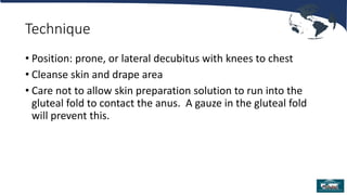
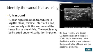
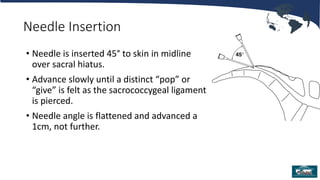
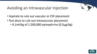
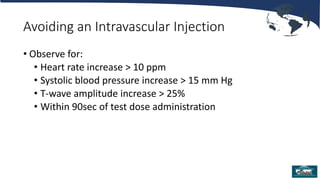
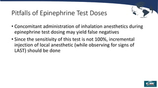
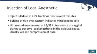
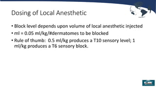
1. Single Injection (non-
catheter) Caudal Blocks
for Pediatric Anesthesia
James Ko, M.D., M.P.H.
Elliot Krane, M.D.
Stanford Children’s Health, Stanford
University
Stanford, CA
Updated 7/2017
2. Overview
• Indications
• Special considerations
• Technique
• Local anesthesia options
• Adjuncts to local anesthetics
• Possible complications
• Summary
3. Scope
• Single “shot” caudal epidural injection is a frequently utilized
regional anesthetic technique in children.
• There is over 50 years of experience and safety data available
for review
4. Indications for a Single “Shot” Caudal
• Surgery involving dermatomes below T10:
• Urologic procedures
• Lower abdominal surgery
• Pelvic and lower extremity surgery
• If performed prior to incision, caudal block will
• decrease maintenance anesthetic requirement
• facilitate earlier extubation
• provide early postoperative pain control
5. Age Parameters
• There is no literature definitively establishing upper or lower
age limits for caudal blocks.
• The decision is based upon a risk/benefit analysis and ease
of use analysis individualized to each patient, situation, and
anesthesia provider.
6. Special Considerations:
Contraindications
• Sacral deformities
• Unclear landmarks
• Sacral dimple
• May overlie spinal dysraphism, spina bifida occulta or tethered cord
• If MR imaging is available, it may rule out anomalies
• Ultrasound evaluation of the sacrum in skilled hands will also rule
out anomalies and allow visualization of the thecal sac and spinal
cord.
• Sacral dimples are common and themselves are not an indication
for MR scanning unless unusually deep or associated with
neurologic abnormalities
7. Special Considerations: Contraindications
• Neurologic abnormalities of the spinal cord or peripheral nervous system
• Postoperative progression of neuropathy often occurs after a surgical
procedure, and may implicate the caudal block if performed
• Local anesthetics are neurotoxic, and neuropathic peripheral nerves are at
increased risk of expression of neurotoxicity
• New postoperative neurologic deficits will require imaging to rule out
complications of an epidural injection or needle insertion
10. Non-styleted Needle: A Warning
• The reason for styling spinal and epidural needles is to avoid introducing skin cells into
the CSF, where they may grow into a dermoid tumor mass. Styleted needles prevent a
core of skin filling the needle tip.
• The risk of dermoid tumors in the epidural space is negligible. The risk is in entering
the thecae sac.
• Some anesthesiologists recommend making an entry hole in the skin with a first
needle (e.g. 21g hypodermic needle) then introducing the caudal needle into the
previously broken skin if a styleted needle is not being used.
11. Supples
• Sterile gloves
• Skin preparation solution
• Chlorhexidine
• Povidone iodine if allergic to chlorhexidine
• Sterile towels to drape area
• Sterile gauze
• Syringe containing injectate
• Sterile dressing (“Band-Aid”)
12. Technique
• Position: prone, or lateral decubitus with knees to chest
• Cleanse skin and drape area
• Care not to allow skin preparation solution to run into the
gluteal fold to contact the anus. A gauze in the gluteal fold
will prevent this.
13. Identify the sacral hiatus using
• Palpation and Surface Anatomy
•Start by palpating the posterior superior iliac spines
(PSIS), visualize an equilateral triangle defined by
the two PSIS’s. The 3rd angle will overlie the sacral
hiatus in most children. Palpate the two sacral
cornuae at this location; the indentation between
them will be the sacral ligament and sacral hiatus.
14. D: Dura (ventral and dorsal)
ES: Termination of thecae sac
SCM: Sacral membrane. Note
the hiatus as the space between
the ventral table of bone and the
posterior elements.
Identify the sacral hiatus using
• Ultrasound
•Linear high resolution transducer in
sagittal plane, midline. Start at L5 and
scan caudally until the sacrum and the
sacral hiatus are visible. The needle may
be inserted under visualization in-plane.
15. Needle Insertion
• Needle is inserted 45° to skin in midline
over sacral hiatus.
• Advance slowly until a distinct “pop” or
“give” is felt as the sacrococcygeal ligament
is pierced.
• Needle angle is flattened and advanced a
1cm, not further.
16. Avoiding an Intravascular Injection
• Aspirate to rule out vascular or CSF placement
• Test dose to rule out intravascular placement
• 0.1ml/kg of 1:200,000 epinephrine (0.5µg/kg)
17. Avoiding an Intravascular Injection
• Observe for:
• Heart rate increase > 10 ppm
• Systolic blood pressure increase > 15 mm Hg
• T-wave amplitude increase > 25%
• Within 90sec of test dose administration
18. Pitfalls of Epinephrine Test Doses
• Concomitant administration of inhalation anesthetics during
epinephrine test dosing may yield false negatives
• Since the sensitivity of this test is not 100%, incremental
injection of local anesthetic (while observing for signs of
LAST) should be done
19. Injection of Local Anesthetic
• Inject full dose in 25% fractions over several minutes
• Bulging of skin over sacrum indicates misplaced needle
• Ultrasound may be used at L5/S1 in transverse or saggital
planes to observe local anesthetic in the epidural space.
Usually will see compression of dura.
20. Dosing of Local Anesthetic
• Block level depends upon volume of local anesthetic injected
• ml = 0.05 ml/kg/#dermatomes to be blocked
• Rule of thumb: 0.5 ml/kg produces a T10 sensory level; 1
ml/kg produces a T6 sensory block.
21. Choice of Local Anesthetic
• Bupivacaine
• Levo-Bupivacaine (in Europe)
• Ropivacaine
• Lidocaine
22. Dosing of Local Anesthetic
• Reduce dose 50% if newborn < 6 weeks
• Reduce dose by 25% – 50% in presence of hepatic
dysfunction, cardiac dysfunction, volume contraction,
hypoproteinemia
23. Bupivacaine
• The most cardiotoxic of choices
• Maximum dose = 3 mg/kg in an otherwise healthy child
• Duration of action 2 – 6 hours (with or without epinephrine)
24. Levo-bupivacaine
• Maximum dose = 3 mg/kg
• Levo enantiomer has similar duration as bupivacaine
• Levo enantiomer reduces cardio and CNS toxicity
• Not marketed in the U.S. or Canada
25. Ropivacaine
• Maximum dose = 3 - 4 mg/kg
• Provides similar duration as bupivacaine
• Compared with bupivacaine:
• Reduced cardio and CNS toxicity
• Reduced motor block
26. Lidocaine
• Maximum dose = 5 mg/kg
• Provides very short duration of action
• Advantage: ability to monitor blood levels during continuous
infusions
28. Adjuvants: Opioids
• Fentanyl exerts most or all of its effects via systemic
absorption from epidural space and not through selective
spinal analgesia
• Hydromorphone, more lipophilic than morphine, causes less
nausea and vomiting than morphine
• Risk of delayed respiratory depression mandates observation
& monitoring for >12 hours
29. Adjuvants: Clonidine
• Enhanced analgesia by direct stimulation of alpha-2 receptors
and mu opioid receptors in spinal cord
• Benefits: lengthens duration of opioid analgesia
• No respiratory depression, pruritus, urinary effect
• Dose 1 – 2 µg/kg
30. Adjuvants: Other
• There have been other adjuvants reported (ketamine, diazepam,
neostigmine, dexamethasone, dexmedetomidine for example) but none
have been proven to not be neurotoxic in vitro or in vivo and should not
be used.
• In fact, ketamine is indeed neurotoxic in the rabbit epidural model.
• We do not recommend clinically using these in children at this time.
31. Postoperative Management
• A clear plan should address pain once caudal block recedes
and should be communicated to caregivers and parents
• Some patients may have distress over lower extremity
numbness and weakness. This is usually alleviated with small
doses of benzodiazepines.
33. Keys to Success
• Equilateral triangle method is most reliable method of
locating the sacral hiatus. Most technique failures are due to
needle placement that is too caudad.
• Test dose
• Clonidine to lengthen duration of analgesia
34. Keys to Success
• Equilateral triangle method is most reliable method of
locating the sacral hiatus. Most technique failures are due to
needle placement that is too caudad.
• Test dose
• Clonidine to lengthen duration of analgesia
35. Keys to Success
• If surgery or pain is located above the umbilicus, a lumbar
epidural (with or without catheter) will provide more reliable
and effective analgesia.
• The success rate will be enhanced with the use of ultrasound
to confirm epidural drug injection if there is any doubt about
the anatomy
36. Summary
• Caudal analgesia/anesthesia is safe and effective for pediatric
patients
• Complications and failures are minimized by close attention
to anatomic landmarks, sterile technique, and ultrasound
guidance in some cases
• Medication choice should be individualized to patient and
surgery
37. Bibliography
Baird R, et al. A systematic review and meta-analysis of caudal blockade versus alternative analgesic strategies for pediatric
inguinal hernia repair. J Ped Surg 2013;48:1077
Cook B, et al. Comparison of adrenaline, clonidine and ketamine on the duration of caudal analgesia produced by bupivacaine in
children. BJA 1995;75:698
Dobereiner E, et al: Evidence-based clinical update: which local anesthetic drug for pediatric caudal block provides optimal
efficacy with the fewest side effects? Can J Anesth 2010;57:1102
Krane E. Spinal epidermoid tumors: will a forgotten complication rise again? [editorial]. Reg Anes Pain Med 1999;24:494
Menzies R, et al. A survey of pediatric caudal extradural anesthesia practice Ped Anesth 2009;19:829
Polaner DM, et al. Pediatric Regional Anesthesia Network (PRAN): a multi-institutional study of the use and incidence of
complications of pediatric regional anesthesia. Anes Analg 2012;115:1353
Roberts S. Ultrasonographic guidance in pediatric regional anesthesia. Part 2: techniques. Pet Anes 2006;16:1112
Tobias J Caudal epidural block: a review of test dosing and recognition of systemic injection in children. Anes Analg
2001;93:1156
Tsui B, et al. Color flow Doppler ultrasonography can distinguish caudal epidural injection from intrathecal injection. Anes Analg
2013;116:1376