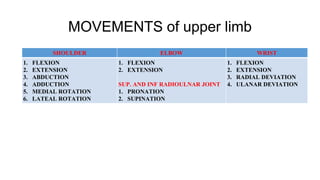
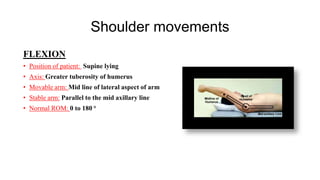
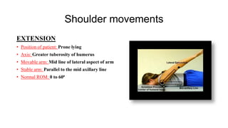
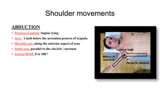
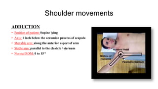
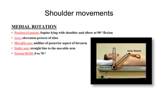
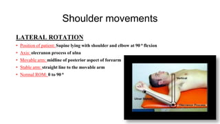
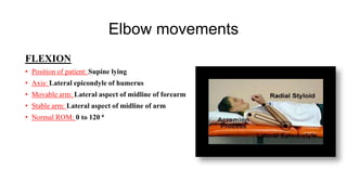
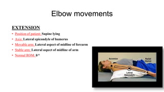
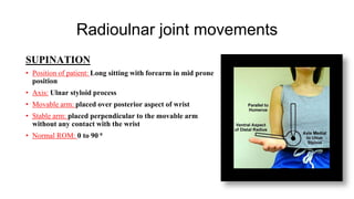
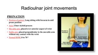
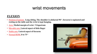
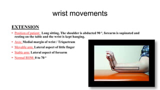
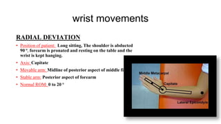
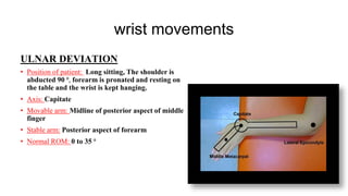
Dr. Sairam PT
GONIOMETRY
UPPER LIMB
RANGE measurement
Dr. Peteti Sairam, MPT(Neuro),
Assistant Professor
Introduction
• A goniometer is an instrument that measures the available range of motion at a joint. The art and
science of measuring the joint ranges in each plane of the joint are called goniometry.
• To measure the range of motion physical therapists most commonly use a goniometer.
• If a patient is suffering from decreased range of motion in a particular joint, the physiotherapist
can use a goniometer to assess the range of motion is at the initial assessment, and then make sure
the intervention is working by using the goniometer in subsequent sessions.
Types of Goniometer
Universal Goniometer - comes in two forms: short arm and
long arm.
• The short arm goniometer is used for smaller joints like the
wrist, elbow, or ankle,
• The long arm goniometers are more accurate for joints with
long levers like the knee and hip joints.
Gravity Goniometer/Inclinometer:
• One arm has a weighted pointer that
remains vertical under the influence
of gravity.
Software/Smartphone-based Goniometer:
• A smartphone as a digital goniometer has several
benefits like availability, ease of measurement,
application-based tracking of measurements, and
one-hand use. These applications use the
accelerometers in phones to calculate the joint
angles.
Arthrodial Goniometer:
• Ideal for measuring cervical rotation,
anteroposterior flexion, and lateral
flexion of the cervical spine.
Twin Axis Electrogoniometer
• The inter-rater and intra-rater reliability
of the electrogoniometer is higher than
the universal goniometer but challenging
to apply in patients' clinical evaluation,
hence used more often for research
purposes
PROCEDURE
• Position the patient in a relaxed manner and the joint to be measured should be free from
obstructions like pillows, couch.
• Patient has to wear loose clothes, to allow smoot movement of the measuring joint.
• Measuring joint has to be in neural position or it has to be at 0⁰ position.
• Total procedure has to explain to the patient.
• Therapist has to stand near to the patient and facing the joint, which has to be measured.
• Axis or the fulcrum of the goniometer is placed over the axis of the joint to be measured.
• Stable arm is fixed to the proximal segment of the joint.
• Movable arm is fixed with the distal segment of the joint.
• Therapist has to move the distal segment of the joint with the movable arm of the
goniometer to measure the joint range.
Selection of the Goniometer
• An important factor for measuring ROM.
• Size of the joint
-Bigger the joint = Size of goniometer is big, movable arm and stable
arm bigger in length.
-Smaller the joint = Size of goniometer is small, movable arm and
stable arm smaller in length.
• An inclinometer also can be used for the measuring the joint ROM,
specifically used for measuring pelvic movements.
MOVEMENTS of upper limb
SHOULDER ELBOW WRIST
1. FLEXION
2. EXTENSION
3. ABDUCTION
4. ADDUCTION
5. MEDIAL ROTATION
6. LATEAL ROTATION
1. FLEXION
2. EXTENSION
SUP. AND INF RADIOULNAR JOINT
1. PRONATION
2. SUPINATION
1. FLEXION
2. EXTENSION
3. RADIAL DEVIATION
4. ULANAR DEVIATION
Shoulder movements
FLEXION
• Position of patient: Supine lying
• Axis: Greater tuberosity of humerus
• Movable arm: Mid line of lateral aspect of arm
• Stable arm: Parallel to the mid axillary line
• Normal ROM: 0 to 180 ⁰
Shoulder movements
EXTENSION
• Position of patient: Prone lying
• Axis: Greater tuberosity of humerus
• Movable arm: Mid line of lateral aspect of arm
• Stable arm: Parallel to the mid axillary line
• Normal ROM: 0 to 60⁰
Shoulder movements
ABDUCTION
• Position of patient: Supine lying
• Axis: 1 inch below the acromion process of scapula
• Movable arm: along the anterior aspect of arm
• Stable arm: parallel to the clavicle / sternum
• Normal ROM: 0 to 180 ⁰
Shoulder movements
ADDUCTION
• Position of patient: Supine lying
• Axis: 1 inch below the acromion process of scapula
• Movable arm: along the anterior aspect of arm
• Stable arm: parallel to the clavicle / sternum
• Normal ROM: 0 to 15 ⁰
Shoulder movements
MEDIAL ROTATION
• Position of patient: Supine lying with shoulder and elbow at 90 ⁰ flexion
• Axis: olecranon process of ulna
• Movable arm: midline of posterior aspect of forearm
• Stable arm: straight line to the movable arm
• Normal ROM: 0 to 70 ⁰
Shoulder movements
LATERAL ROTATION
• Position of patient: Supine lying with shoulder and elbow at 90 ⁰ flexion
• Axis: olecranon process of ulna
• Movable arm: midline of posterior aspect of forearm
• Stable arm: straight line to the movable arm
• Normal ROM: 0 to 90 ⁰
Elbow movements
FLEXION
• Position of patient: Supine lying
• Axis: Lateral epicondyle of humerus
• Movable arm: Lateral aspect of midline of forearm
• Stable arm: Lateral aspect of midline of arm
• Normal ROM: 0 to 120 ⁰
Elbow movements
EXTENSION
• Position of patient: Supine lying
• Axis: Lateral epicondyle of humerus
• Movable arm: Lateral aspect of midline of forearm
• Stable arm: Lateral aspect of midline of arm
• Normal ROM: 0 ⁰
Radioulnar joint movements
SUPINATION
• Position of patient: Long sitting with forearm in mid prone
position
• Axis: Ulnar styloid process
• Movable arm: placed over posterior aspect of wrist
• Stable arm: placed perpendicular to the movable arm
without any contact with the wrist
• Normal ROM: 0 to 90 ⁰
Radioulnar joint movements
PRONATION
• Position of patient: Long sitting with forearm in mid
prone position
• Axis: Ulnar styloid process
• Movable arm: placed over anterior aspect of wrist
• Stable arm: placed perpendicular to the movable arm
without any contact with the wrist
• Normal ROM: 0 to 70 ⁰
wrist movements
FLEXION
• Position of patient: Long sitting, The shoulder is abducted 90 ⁰, forearm is supinated and
resting on the table and the wrist is kept hanging.
• Axis: Medial margin of wrist / Triquetrum
• Movable arm: Lateral aspect of little finger
• Stable arm: Lateral aspect of forearm
• Normal ROM: 0 to 75 ⁰
wrist movements
EXTENSION
• Position of patient: Long sitting, The shoulder is abducted 90 ⁰, forearm is supinated and
resting on the table and the wrist is kept hanging.
• Axis: Medial margin of wrist / Triquetrum
• Movable arm: Lateral aspect of little finger
• Stable arm: Lateral aspect of forearm
• Normal ROM: 0 to 70 ⁰
wrist movements
RADIAL DEVIATION
• Position of patient: Long sitting, The shoulder is abducted
90 ⁰, forearm is pronated and resting on the table and the
wrist is kept hanging.
• Axis: Capitate
• Movable arm: Midline of posterior aspect of middle finger
• Stable arm: Posterior aspect of forearm
• Normal ROM: 0 to 20 ⁰
wrist movements
ULNAR DEVIATION
• Position of patient: Long sitting, The shoulder is
abducted 90 ⁰, forearm is pronated and resting on
the table and the wrist is kept hanging.
• Axis: Capitate
• Movable arm: Midline of posterior aspect of middle
finger
• Stable arm: Posterior aspect of forearm
• Normal ROM: 0 to 35 ⁰
ANY DOUBTS ????????????????????????

GONIOMETRY UPPER LIMB PPT.pptx

  • 1.
    Dr. Sairam PT GONIOMETRY UPPERLIMB RANGE measurement Dr. Peteti Sairam, MPT(Neuro), Assistant Professor
  • 2.
    Introduction • A goniometeris an instrument that measures the available range of motion at a joint. The art and science of measuring the joint ranges in each plane of the joint are called goniometry. • To measure the range of motion physical therapists most commonly use a goniometer. • If a patient is suffering from decreased range of motion in a particular joint, the physiotherapist can use a goniometer to assess the range of motion is at the initial assessment, and then make sure the intervention is working by using the goniometer in subsequent sessions.
  • 3.
    Types of Goniometer UniversalGoniometer - comes in two forms: short arm and long arm. • The short arm goniometer is used for smaller joints like the wrist, elbow, or ankle, • The long arm goniometers are more accurate for joints with long levers like the knee and hip joints.
  • 5.
    Gravity Goniometer/Inclinometer: • Onearm has a weighted pointer that remains vertical under the influence of gravity.
  • 6.
    Software/Smartphone-based Goniometer: • Asmartphone as a digital goniometer has several benefits like availability, ease of measurement, application-based tracking of measurements, and one-hand use. These applications use the accelerometers in phones to calculate the joint angles.
  • 7.
    Arthrodial Goniometer: • Idealfor measuring cervical rotation, anteroposterior flexion, and lateral flexion of the cervical spine.
  • 8.
    Twin Axis Electrogoniometer •The inter-rater and intra-rater reliability of the electrogoniometer is higher than the universal goniometer but challenging to apply in patients' clinical evaluation, hence used more often for research purposes
  • 9.
    PROCEDURE • Position thepatient in a relaxed manner and the joint to be measured should be free from obstructions like pillows, couch. • Patient has to wear loose clothes, to allow smoot movement of the measuring joint. • Measuring joint has to be in neural position or it has to be at 0⁰ position. • Total procedure has to explain to the patient. • Therapist has to stand near to the patient and facing the joint, which has to be measured. • Axis or the fulcrum of the goniometer is placed over the axis of the joint to be measured. • Stable arm is fixed to the proximal segment of the joint. • Movable arm is fixed with the distal segment of the joint. • Therapist has to move the distal segment of the joint with the movable arm of the goniometer to measure the joint range.
  • 10.
    Selection of theGoniometer • An important factor for measuring ROM. • Size of the joint -Bigger the joint = Size of goniometer is big, movable arm and stable arm bigger in length. -Smaller the joint = Size of goniometer is small, movable arm and stable arm smaller in length. • An inclinometer also can be used for the measuring the joint ROM, specifically used for measuring pelvic movements.
  • 11.
    MOVEMENTS of upperlimb SHOULDER ELBOW WRIST 1. FLEXION 2. EXTENSION 3. ABDUCTION 4. ADDUCTION 5. MEDIAL ROTATION 6. LATEAL ROTATION 1. FLEXION 2. EXTENSION SUP. AND INF RADIOULNAR JOINT 1. PRONATION 2. SUPINATION 1. FLEXION 2. EXTENSION 3. RADIAL DEVIATION 4. ULANAR DEVIATION
  • 12.
    Shoulder movements FLEXION • Positionof patient: Supine lying • Axis: Greater tuberosity of humerus • Movable arm: Mid line of lateral aspect of arm • Stable arm: Parallel to the mid axillary line • Normal ROM: 0 to 180 ⁰
  • 13.
    Shoulder movements EXTENSION • Positionof patient: Prone lying • Axis: Greater tuberosity of humerus • Movable arm: Mid line of lateral aspect of arm • Stable arm: Parallel to the mid axillary line • Normal ROM: 0 to 60⁰
  • 14.
    Shoulder movements ABDUCTION • Positionof patient: Supine lying • Axis: 1 inch below the acromion process of scapula • Movable arm: along the anterior aspect of arm • Stable arm: parallel to the clavicle / sternum • Normal ROM: 0 to 180 ⁰
  • 15.
    Shoulder movements ADDUCTION • Positionof patient: Supine lying • Axis: 1 inch below the acromion process of scapula • Movable arm: along the anterior aspect of arm • Stable arm: parallel to the clavicle / sternum • Normal ROM: 0 to 15 ⁰
  • 16.
    Shoulder movements MEDIAL ROTATION •Position of patient: Supine lying with shoulder and elbow at 90 ⁰ flexion • Axis: olecranon process of ulna • Movable arm: midline of posterior aspect of forearm • Stable arm: straight line to the movable arm • Normal ROM: 0 to 70 ⁰
  • 17.
    Shoulder movements LATERAL ROTATION •Position of patient: Supine lying with shoulder and elbow at 90 ⁰ flexion • Axis: olecranon process of ulna • Movable arm: midline of posterior aspect of forearm • Stable arm: straight line to the movable arm • Normal ROM: 0 to 90 ⁰
  • 18.
    Elbow movements FLEXION • Positionof patient: Supine lying • Axis: Lateral epicondyle of humerus • Movable arm: Lateral aspect of midline of forearm • Stable arm: Lateral aspect of midline of arm • Normal ROM: 0 to 120 ⁰
  • 19.
    Elbow movements EXTENSION • Positionof patient: Supine lying • Axis: Lateral epicondyle of humerus • Movable arm: Lateral aspect of midline of forearm • Stable arm: Lateral aspect of midline of arm • Normal ROM: 0 ⁰
  • 20.
    Radioulnar joint movements SUPINATION •Position of patient: Long sitting with forearm in mid prone position • Axis: Ulnar styloid process • Movable arm: placed over posterior aspect of wrist • Stable arm: placed perpendicular to the movable arm without any contact with the wrist • Normal ROM: 0 to 90 ⁰
  • 21.
    Radioulnar joint movements PRONATION •Position of patient: Long sitting with forearm in mid prone position • Axis: Ulnar styloid process • Movable arm: placed over anterior aspect of wrist • Stable arm: placed perpendicular to the movable arm without any contact with the wrist • Normal ROM: 0 to 70 ⁰
  • 22.
    wrist movements FLEXION • Positionof patient: Long sitting, The shoulder is abducted 90 ⁰, forearm is supinated and resting on the table and the wrist is kept hanging. • Axis: Medial margin of wrist / Triquetrum • Movable arm: Lateral aspect of little finger • Stable arm: Lateral aspect of forearm • Normal ROM: 0 to 75 ⁰
  • 23.
    wrist movements EXTENSION • Positionof patient: Long sitting, The shoulder is abducted 90 ⁰, forearm is supinated and resting on the table and the wrist is kept hanging. • Axis: Medial margin of wrist / Triquetrum • Movable arm: Lateral aspect of little finger • Stable arm: Lateral aspect of forearm • Normal ROM: 0 to 70 ⁰
  • 24.
    wrist movements RADIAL DEVIATION •Position of patient: Long sitting, The shoulder is abducted 90 ⁰, forearm is pronated and resting on the table and the wrist is kept hanging. • Axis: Capitate • Movable arm: Midline of posterior aspect of middle finger • Stable arm: Posterior aspect of forearm • Normal ROM: 0 to 20 ⁰
  • 25.
    wrist movements ULNAR DEVIATION •Position of patient: Long sitting, The shoulder is abducted 90 ⁰, forearm is pronated and resting on the table and the wrist is kept hanging. • Axis: Capitate • Movable arm: Midline of posterior aspect of middle finger • Stable arm: Posterior aspect of forearm • Normal ROM: 0 to 35 ⁰
  • 26.