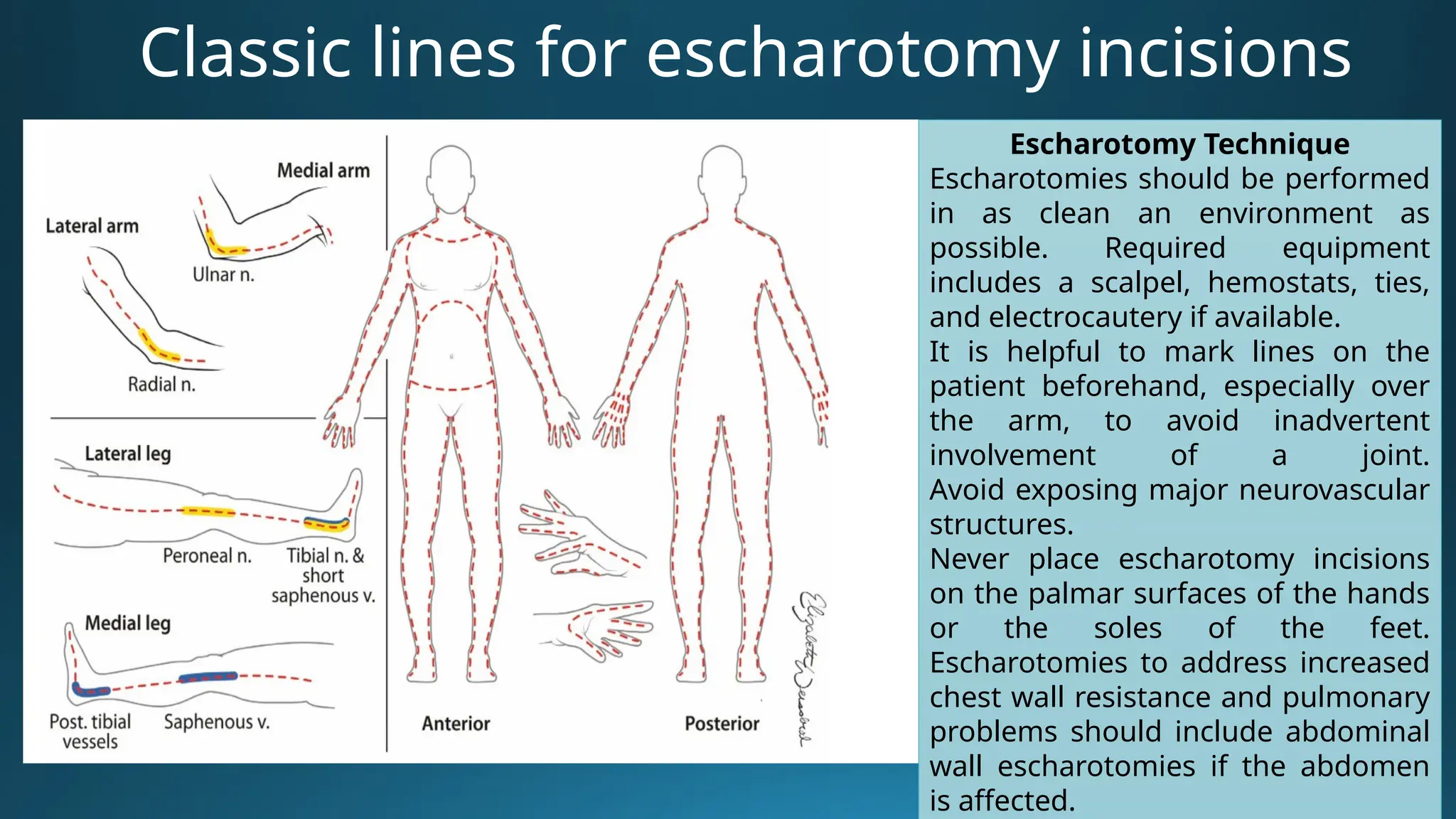
Ivano-Frankivsk National Medical University
Department of disaster and military medicine
Premedical care in emergency situation
LECTURE
“Shock in trauma, their clinical characteristics.
Combat thermal injury. Diagnosis of burns.
Inhalation injuries. Emergency medical care in the
tactical field care, at the stages of evacuation.
Effect of low temperatures”
Ivano-Frankivsk - 2025
lecturer
Anna Ovchar
Plan of lecture
• Shock in trauma.
• Combat thermal injury.
• Inhalation injuries.
• Effect of low temperatures.
Purpose of lecture
The management of shock has been the subject of intensive research for
decades. As a result, changes continue to be made in the recommendations
for prehospital treatment of the patient with shock and burns.
Shock
The first step in the initial management of shock is to recognize its presence.
Shock — abnormality of the circulatory system that
results in inadequate organ perfusion and tissue
oxygenation.
Normal Perfusion
• Blood Pressure = Cardiac Output х Peripheral Vascular
Resistance
• Cardiac Output = Heart Rate х Stroke Volume
Types of Traumatic Shock
Hypovolemic shock
The term hypovolemic means
low fluid volume. Hypovolemic
shock includes all types of shock
caused by fluid loss, such as
bleeding, burns, vomiting,
diarrhea, and severe
dehydration. There must be an
adequate amount of fluid within
the body and circulatory system
at all times. If there is loss of
fluid volume, blood pressure will
drop and shock will result.
Types of Traumatic Shock
Cardiogenic shock
The heart (pump) is at the center of the
circulatory system and must be
functioning properly to maintain a
proper blood pressure to circulate blood
adequately. If the heart fails to pump an
adequate volume of blood, the result can
be an abnormally low blood pressure
(hypotension). This category of shock is
sometimes referred to as pump failure.
Types of Traumatic Shock
Distributive shock
Blood circulates throughout the body
by way of a closed system of vessels.
The vessels are made up of smooth
muscle and can dilate and constrict,
depending on the needs of the body.
Certain conditions can cause the
vessels to dilate excessively, resulting
in a much larger space than the
available blood supply can fill. When
there is more space within the
system than blood to fill it, blood
pressure drops and shock results.
Types of Traumatic Shock
Obstructive shock
The adequate flow of blood can be disrupted due to a variety of
obstructions to the heart, lungs, and great vessels. Damage to the
heart from trauma and blockages in the vessels that connect the
heart and lungs are causes of obstructive shock. The obstruction of
blood flow reduces perfusion, which can lead to shock
The Body’s Response During Shock
Compensated shock the condition in
which the body is using specific
mechanisms, such as increased pulse
rate and increased breathing rate, to
compensate for a lack of adequate
perfusion.
Decompensated
shock the
condition in which
the body is no
longer able to
compensate for a
lack of adequate
If the root cause of
the shock is not cared
for soon enough, the
patient will eventually
enter a condition
called irreversible
shock and will die.
HEMORRAGIC SHOCK
SIGNS of shock:
• Mental confusion or altered
mental status in the absence of a
head injury
• Weak or absent radial pulses
• Tachycardia
• Tachypnea
• Excessive thirst
• Cyanosis with pale, grey, or
blotchy blue skin
• Nausea and/or vomiting
• Diaphoresis with sweaty, cool,
clammy skin
One of the key signs for shock is Altered Mental
Status in the absence of head injury. Check
casualty every 15 minutes for AVPU.
Alertness: Knows who, where they are.
Verbal: Orally responds to verbal commands.
Pain: Level of pain felt when the sternum is briskly
rubbed with the knuckle (if needed)
Unconscious: Unresponsive
Decreasing: AVPU could indicate condition
SIGNS and SYMPTHOMS of
hemorrhagic shock
ATLS classification of hemorrhagic shock
CLASS I CLASS II CLASS III CLASS IV
BloodLoss (ml)
%
<750
15%
750-1500
15%-30%
1500-2000
30-40%
>2000
>40%
HR <100 >100 >120 >140
BP normal normal decrease decrease
PP normal decrease decrease decrease
RR 14-20 20-30 30-40 >35
UOP >30 20-30 5-15 negligible
CNS slightly
anxious
mildly
anxious
anxious
confused
confused
lethargic
Progression of symptoms
• Usually, up to 500 ml of blood loss is well-tolerated, often with no effects
except perhaps a slight tachycardia.
• 1,000 ml of blood loss will usually produce tachycardia greater than 100,
but otherwise, the casualty may appear normal.
• 1,500 ml of blood loss may be associated with changes in mental status, a
weak radial pulse, persistent tachycardia, and tachypnea.
• 2,000 ml of blood loss is accompanied by confusion and lethargy, a weak
radial pulse often greater than 120, and tachypnea greater than 35. This
amount of blood loss is possibly fatal if not managed quickly.
• And 2,500 ml of blood loss will usually present with the casualty
unconscious, no radial pulse, a carotid pulse greater than 140, and
tachypnea greater than 35. This amount of blood loss will be fatal without
immediate and rapid intervention.
PROGRESIVE CHANGES IN
SHOCK
Lethal triad of hemorrhagic shock
Acidosis is most commonly
the result of lactic acid
buildup as cells revert to
anaerobic processes in the
absence of adequate
cellular oxygenation (or
shock). Sometimes there is
a respiratory contribution if
the respiratory drive is
blunted and there is a
buildup of carbon dioxide,
but the metabolic acidosis
from lactic acid buildup in
trauma tends to be the
predominant issue.
Coagulopathy can result from
direct losses of clotting factors
and platelets due to massive
hemorrhage but can be
accentuated by hemodilution
if fluids are replaced by
crystalloid or colloid fluids that
do not replace clotting factors
and platelets like you would
get from using whole blood
transfusions as fluid
replacement therapy.
Additionally, the function of
clotting factors is
temperature-dependent and
hypothermia leads to clotting
factor dysfunction, even if
there is an adequate quantity.
Hypothermia results from a
combination of
environmental factors
(exposure, even in warmer
climates) and physiologic
responses to blood loss. As
mentioned previously, the
functions of the clotting
cascade are impaired in
hypothermia, and further
blood loss from coagulopathy
leads to increasing
hypothermia. This vicious
cycle of acidosis,
hypothermia, and
coagulation (or the lethal
triad) requires prevention
Steps in the management of shock are as
follows: Management:
– Control bleeding
–Shock position
– High-flow oxygen
– Rapid safe transport
– Large-bore IV access
– Fluid bolus (500–1,00
mL for adult and repeat
if necessary
– Cardiac monitor,
SpO2, ETCO2
–Ongoing exam
The initial fluid bolus elicits three
possible responses:
• Rapid response. The vital signs return to and remain normal. This
typically indicates that the patient has lost less than 20% of blood
volume and that the hemorrhage has stopped.
• Transient response. The vital signs initially improve (pulse slows and
blood pressure increases); however, during reassessment, these
patients show deterioration with recurrent signs of shock. These
patients have typically lost between 20% and 40% of their blood
volume.
• Minimal or no response. These patients show virtually no change in
the profound signs of shock after a 1- to 2-liter bolus.
Volume Resuscitation
There are two general categories of fluid resuscitation products that have been
for the management of trauma patients-blood and IV solutions.
Isotonic Crystalloid Solutions
Isotonic crystalloids are balanced salt solutions
comprised of electrolytes (substances that
separate into charged ions when dissolved in
solutions). They act as effective volume expanders
for a short time, but they possess no oxygen-
carrying capacity.
Immediately after infusion, crystalloids fill the
vascular space that was depleted by blood loss,
improving preload and cardiac output.
Lactated Ringer's
Lactated Ringer's solution remains the isotonic
crystalloid solution of choice for the management
of shock because its composition is most similar to
the electrolyte composition of blood plasma. It
contains specific amounts of sodium, potassium,
calcium, chloride, and lactate ions.
The order of preference for blood
products is:
- Cold-stored low-titer O whole
blood
- Pre-screened low-titer O fresh
whole blood
- Plasma, red blood cells (RBCs) and
platelets in a 1:1:1 ratio
- Plasma and RBCs in a 1:1 ratio
- Plasma or RBCs alone
If possible, IV fluids should be warmed to about
39°C before infusion. Infusion of large amounts of
room temperature or cold IV fluid contributes to
hypothermia and increased hemorrhage.
Distributive (Vasogenic) Shock
Distributive shock, or vasogenic shock, occurs when the vascular
container enlarges without a proportional increase in fluid volume.
After trauma, this is typically found in patients who have sustained
a spinal cord injury.
Spinal shock is a neurologic phenomenon
that occurs for an unpredictable and variable
amount of time after spinal cord injury,
resulting in temporary loss of all sensory and
motor function, muscle flaccidity and
paralysis, and loss of reflexes below the level
of the spinal cord injury.
Cord contusion is usually caused by a
penetrating type of injury or movement of
bony fragments. The severity of injury
resulting from the contusion is related to the
amount of bleeding into the spinal cord
tissue.
Damage to or disruption of the spinal blood
Neurogenic "shock" secondary to spinal cord injury
represents a significant additional finding. When the
spinal cord is disrupted, the body's sympathetic control
mechanism is interrupted and can no longer maintain
constriction of the muscles in the walls of the blood
vessels below the point of disruption. These arteries and
arterioles dilate, enlarging the size of the vascular
container, which leads to a partial loss of systemic
vascular resistance.
Signs and symptoms:
– Hypotension
– Heart rate normal or slow
– Skin warm, dry, pink
– Paralysis or deficit (No chest movement, simple
Cardiogenic Shock
Pumping strength is reduced, cardiac output falls, and blood pressure goes
down. Such a condition is called cardiogenic shock.
Myocardial contusion
Direct trauma to the heart can injure it,
damaging the muscle and reducing
pumping strength, a condition called
myocardial contusion.
Myocardial contusion can result in
diminished cardiac output because of
this heart muscle damage, as well as
possibly causing cardiac dysrhythmia.
Myocardial contusion often cannot be
differentiated from cardiac tamponade
in the field.
Myocardial infarction (MI)
Though this is a medical cause of loss
of heart muscle
strength, the emergency care provider
must remember that cardiogenic
shock must be considered in the
hypotensive and usually tachycardic
patient who presents with chest pain.
Especially in older patients, an MI may
be the precipitating cause of the
traumatic event.
In the trauma patient, the significant causes of cardiogenic shock are the
following:
Myocardial contusion may cause ventricular
ectopy
Mechanical (or Obstructive)
Shock
Traumatic conditions that can cause mechanical shock are tension
pneumothorax and cardiac tamponade:
Physical findings of tension pneumothorax Pathophysiology and physical findings of cardiac
tamponade
Algorithm MARCH-PAWS
• MARCH PAWS
Burns
Categories of burns based on source include:
• Heat (thermal) burns, which may be caused
by fire, steam, or hot objects.
•Chemical burns, which may be caused by
caustics, such as acids and alkalis.
•Electrical burns, which originate from
outlets, frayed wires, and faulty circuits.
•Lightning burns, which occur during
electrical storms.
•Light burns, which occur with intense light.
Light from the arc welder or industrial laser will
damage unprotected eyes. Also, ultraviolet light
(including sunlight) can burn the eyes and skin.
•Radiation burns, which usually result from
nuclear sources.
Most often burns are categorized according
to the depth of the burn
Most often burns are categorized according
to the depth of the burn
Burn Depth Characteristics
Total burns surface area
• The Lund-Browder chart is a
diagram that takes into account
age-related changes in children.
Using these charts, a prehospital
care provider maps the bum and
then determines bum size based
on an accompanying reference
table.
• This method requires drawing a
map of the bums and then
converting the map to a calculated
burned surface area. The
complexity of this method makes it
difficult to use in a prehospital
situation.
Burns greater than 15% surface area (adult), greater than 10% (child)
or any burn occurring in the extremes of age are considered serious
Serious burns requiring hospitalization include the following:
•Adult: greater than 15% burn
•Pediatric: greater than 10% burn
•Any burn in the very young, elderly or the infirm
•Any full thickness burn
•Specific regions: face, ears, eyes, hands, feet, perineum
•Circumferential burns
•High-voltage electrical burns
•Inhalational injury
•Associated trauma or significant pre-burn illness, e.g. diabetes
Burns management
1. Stop the burning process immediately.
2. Flush superficial burns with water (or saline) for several min.
3. Remove smoldering clothing and jewelry. Do not remove
any clothing that is melted onto the skin.
4. Continually monitor the airway. Any burns to the face or exposure to smoke
may cause airway problems.
5. Cover partial- and full-thickness burns with dry, clean dressings.
6. If the eyes or eyelids have been burned, place clean dressings or pads over
them. Moisten these pads with sterile water if possible.
7. If a serious burn involves the hands or feet, always place a clean pad
between toes or fingers before completing the dressing.
8. Provide oxygen and care for shock.
9. Painkillers. Narcotic analgesics such as fentanyl (1 mcg per kg body weight) or
morphine (0.1 mg per kg body weight) in adequate dosages will be required to
control pain.
11. Maintain body temperature. Prevent hypothermia, by remove wet clothing
Burns management
11. Fluids should be given to all patients over one year
with burns covering 15% or more of TBSA and to all
infants under one year with burns covering 10% or
more of TBSA.
Parkland formula to calculate the patient’s fluid
requirement over the first 24 hours. The formula tells
what volume of Ringer’s lactate to give, in milliliters.
Parkland formula
Volume (ml) = 4 x body weight (kg) x % TBSA
burned
Circumferential Burns
Circumferential burns of the trunk or limbs are
capable of producing a life- or limb-threatening
condition as a result of the thick, inelastic eschar that is
formed.
Circumferential bums of the chest can constrict the
chest wall (inability to inhale)
Circumferential burns of the extremities create a
tourniquet-like effect that can render an arm or leg
pulseless.
Management:
• Monitor respiration and chest expansion
• Monitor distal PMS
• Escharotomies (surgical incisions made through the
burn eschar to allow expansion of the deeper tissues
and decompression of previously compressed and
often occluded vascular structures)
Classic lines for escharotomy incisions
Escharotomy Technique
Escharotomies should be performed
in as clean an environment as
possible. Required equipment
includes a scalpel, hemostats, ties,
and electrocautery if available.
It is helpful to mark lines on the
patient beforehand, especially over
the arm, to avoid inadvertent
involvement of a joint.
Avoid exposing major neurovascular
structures.
Never place escharotomy incisions
on the palmar surfaces of the hands
or the soles of the feet.
Escharotomies to address increased
chest wall resistance and pulmonary
problems should include abdominal
wall escharotomies if the abdomen
is affected.
Chemical burns
Chemical injury can result from exposure to acids, alkalizes, and
petroleum products.
Acidic burns cause a coagulation necrosis of the surrounding tissue, which
impedes the penetration of the acid to some extent (pH between 7
(neutral) and 0 (strong acid)).
Alkali burns are generally more serious than acid burns, as the alkali
penetrates more deeply by liquefaction necrosis of the tissue (pH between
7 and 14 (strong base)).
The severity of chemical injury is
determined by four factors:
• nature of the chemical,
• concentration of the chemical,
• duration of contact,
• mechanism of action of the
chemical.
Prehospital Management
•Wear appropriate protective gloves, eyewear, and respiratory
protection if needed. In some situations you will need to wear a
chemical protective suit.
•Remove all the patient’s clothing. Place in plastic bags to limit
further contact.
•If dry powder is still present on the skin, brush it away before
irrigating with water.
•Immediately lush away the chemical with large amounts of
water, for at least 20 to 30 minutes, using a shower or hose (alkali
burns require longer irrigation).
•Neutralizing agents offer no advantage over water lavage
(reaction with the neutralizing agent can itself produce heat and
cause further tissue damage).
•Alkali burns to the eye require continuous irrigation during
the first 8 hours after the burn, If possible, ocular
decontamination with continuous irrigation using a Morgan lens
Electrical burns
The factors that determine severity of
electrical injury:
• Type and amount of current (alternating
versus direct current and also the voltage)
• Path of the current through the body
• Duration of contact with the current
source
Electrical injuries cause skin burns at the entrance and exit sites because of high
temperatures generated by the electric arc 2,500°C at the skin surface.
Fractures and dislocations may be present due to the violent muscle contractions
that electrical injuries cause.
The most serious and immediate injury that results from electrical contact is
cardiac arrhythmia (premature ventricular contractions, ventricular tachycardia,
and ventricular fibrillation).
In cases of electrical burns, damage is caused by electricity entering the body and traveling
through the tissues.
Lightning injury:
injury by the
multiple effects of
very short duration,
very high voltage
direct current on the
body. The most
serious effect is
cardiac and
respiratory arrest.
Prehospital Management
• At the scene of an electrical injury, your first priority is scene safety.
• Determine if the patient is still in contact with the electrical current.
• If so, you must remove the patient from contact without becoming a victim yourself
(connect with power company personnel are turning off the electricity).
• Due to the potential for arrhythmia development, routine IV access should be
initiated in the ambulance, along with continuous cardiac monitoring (be ready for CPR).
Follow standard guidelines for CPR and advanced cardiac life support (ACLS).
• Electrical burn patients are at risk for developing rhabdomyolysis. In those cases,
adequate fluid resuscitation is indicated by maintaining a urine output of 0.5 to 1 cc/kg
body weight per hour.
• Also with breakdown of muscle cells, potassium is released into the circulation and
can lead to hyperkalemia, which can result in cardiac arrhythmias and death. Tall peaked
T-waves on the ECG may be a sign of hyperkalemia. Intravenous calcium (gluconate or
chloride) along with intravenous sodium bicarbonate, 50% dextrose, and insulin can
temporarily reduce the effects of hyperkalemia on the heart.
Inhalation Injuries
There are three elements of smoke inhalation:
• thermal injury (injury to the skin caused by heat from
flame, hot liquids, hot gases, or hot solids)
• asphyxiation (two gaseous products that are clinically
important as asphyxiants are carbon monoxide and
cyanide gas)
• delayed toxin-induced lung injury (injury to the lungs or
other body organs from inhalation of toxic gases found
in smoke). Danger signs of upper
airway burns:
• Burns of the face
• Singed eyebrows or nasal
hair
• Burns in the mouth
• Carbonaceous (sooty)
sputum
• History of being confined in
a
Heat inhalation can cause complete
airway
obstruction by swelling of the
hypopharynx: left side-normal
Frostbite
• Damage from frostbite can be due to freezing of tissue,
ice crystal formation causing cell membrane injury,
microvascular occlusion, and subsequent tissue anoxia.
Some of the tissue damage also can result from
reperfusion injury that occurs on rewarming.
Frostbite is classified into (according to depth):
• first-degree,
• second-degree,
• third-degree,
• fourth-degree.
Classification of frostbites
• 1. First-degree frostbite: Hyperemia and edema are present
without skin necrosis.
• 2. Second-degree frostbite: Large, clear vesicle formation
accompanies the hyperemia and edema with partial-thickness
skin necrosis.
• 3. Third-degree frostbite: Full-thickness and subcutaneous tissue
necrosis occurs, commonly with hemorrhagic vesicle formation.
• 4. Fourth-degree frostbite: Full-thickness skin necrosis occurs,
including muscle and bone with later necrosis.
Prehospital Management
Patients with superficial frostbite should be placed with the affected area against a warm body
surface (covering the patient's ears with warm hands or placing affected fingers into armpits, axillae,
or groin regions).
Management of deep frostbite in the prehospital setting includes:
• Assessing and treating the patient for hypothermia, if present. Get the patient out of the cold. Take
the patient indoors (depending on circumstances). Remove wet clothing.
• Do not rub or massage the frostbitten area (massage will cause further damage to injured tissues).
• Transport the patient to the hospital with the injured area elevated.
• Administer analgesia as needed.
• Cover blisters with a dry, sterile dressing.
• Consider rewarming only if the potential to refreeze does not exist.
IV opiate analgesics are usually required for pain relief and should be initiated before the tissues have
thawed. Initiate IV NaCl with a 250-ml bolus to treat dehydration and reduce blood viscosity and capillary
sludging.
Attempts to begin rewarming of deep frostbite patients in the field can be hazardous to the patient's eventual
recovery and are not recommended unless prolonged transport times (over 2 hours) are involved. If prolonged
transport is involved, thaw the affected part in a warm water bath at a temperature no greater than 37°C to 38.9°C on
the affected area until the area becomes soft and pliable to the touch (-30 min).
Administer ibuprofen (12 mg/kg up to 800 mg) if available. NSAIDs such as ibuprofen help decrease
Hypothermia
• Hypothermia is defined as a decrease in BT generally
35°C, owing to inadequate thermogenesis and/or
excess environmental cold stress. Extreme cold
weather does not need to be present for a person to
become hypothermic.
• Hypothermia is sometimes also called accidental
hypothermia to distinguish it from therapeutic or
induced hypothermia, which is a key step in the last
link in the chain of survival for comatose patients
with return of spontaneous circulation.
Mechanism of heat loss of the body
Signs and symptoms of hypothermia
Decreasing mental status:
Amnesia, memory lapses, and incoherence
Mood changes
Impaired judgment
Reduced ability to communicate
Dizziness
Vague, slow, slurred, or thick speech
Drowsiness progressing even to
unresponsiveness
Decreasing motor and sensory
function:
Stiffness, rigidity
Lack of coordination
Exhaustion
Shivering at first,
little or no shivering later
Loss of sensation
Changing vital signs:
Breathing rapid at first; shallow,
slow later; absent near end
Pulse rapid at first; slow and barely
palpable later; irregular or absent
near end
Skin red in early stages, changing
to pale, to cyanotic, to gray, waxen,
and hard; cold to the touch
Slowly responding pupils
Low to absent blood pressure
Stage Clinical Findings
Estimated Core
Temperature ( °C)
Hypothermia I (mild) Conscious, shivering * 35–32 °C
Hypothermia II
(moderate)
Impaired consciousness
*; may or may not be
shivering
<32–28 °C
Hypothermia III
(severe)
Unconscious *; vital
signs present
<28 °C
Hypothermia IV
(severe)
Apparent death; vital
signs absent
Classically < 24 °C **
Classical staging of
accidental hypothermia
based on clinical signs
Copyright 2021 European
Resuscitation Council.
Principles of pre-hospital management of hypothermia
Treatment of hypothermia
Mild hypothermia
Passive rewarming:
 removing the patient
from the cold
environment,
 optimizing insulation,
 offering food and warm
drinks,
 and promoting active
movements,
Moderate and Severe Hypothermia
 Patients with moderate or severe hypothermia require active rewarming. The whole body
should be insulated to reduce the risk of further cooling. Protection should be provided
against cold, wind, and moisture. A hypothermic patient should be packaged with several
layers.
Attempts to rewarm should not delay transport. They should receive adequate oxygenation and
be placed on a cardiac monitor. When IV or IO fluids are required, they should be warmed to
38–42 °C and should be given in boluses guided by vital signs . Use of heated fluids helps to
limit secondary cooling and may protect lines from freezing but has little direct effect on
rewarming.
L7. Shock. Trauma _ emergency care in Premedical care

L7. Shock. Trauma _ emergency care in Premedical care

  • 1.
    Ivano-Frankivsk National MedicalUniversity Department of disaster and military medicine Premedical care in emergency situation LECTURE “Shock in trauma, their clinical characteristics. Combat thermal injury. Diagnosis of burns. Inhalation injuries. Emergency medical care in the tactical field care, at the stages of evacuation. Effect of low temperatures” Ivano-Frankivsk - 2025 lecturer Anna Ovchar
  • 2.
    Plan of lecture •Shock in trauma. • Combat thermal injury. • Inhalation injuries. • Effect of low temperatures.
  • 3.
    Purpose of lecture Themanagement of shock has been the subject of intensive research for decades. As a result, changes continue to be made in the recommendations for prehospital treatment of the patient with shock and burns.
  • 4.
    Shock The first stepin the initial management of shock is to recognize its presence. Shock — abnormality of the circulatory system that results in inadequate organ perfusion and tissue oxygenation.
  • 5.
    Normal Perfusion • BloodPressure = Cardiac Output х Peripheral Vascular Resistance • Cardiac Output = Heart Rate х Stroke Volume
  • 6.
    Types of TraumaticShock Hypovolemic shock The term hypovolemic means low fluid volume. Hypovolemic shock includes all types of shock caused by fluid loss, such as bleeding, burns, vomiting, diarrhea, and severe dehydration. There must be an adequate amount of fluid within the body and circulatory system at all times. If there is loss of fluid volume, blood pressure will drop and shock will result.
  • 7.
    Types of TraumaticShock Cardiogenic shock The heart (pump) is at the center of the circulatory system and must be functioning properly to maintain a proper blood pressure to circulate blood adequately. If the heart fails to pump an adequate volume of blood, the result can be an abnormally low blood pressure (hypotension). This category of shock is sometimes referred to as pump failure.
  • 8.
    Types of TraumaticShock Distributive shock Blood circulates throughout the body by way of a closed system of vessels. The vessels are made up of smooth muscle and can dilate and constrict, depending on the needs of the body. Certain conditions can cause the vessels to dilate excessively, resulting in a much larger space than the available blood supply can fill. When there is more space within the system than blood to fill it, blood pressure drops and shock results.
  • 9.
    Types of TraumaticShock Obstructive shock The adequate flow of blood can be disrupted due to a variety of obstructions to the heart, lungs, and great vessels. Damage to the heart from trauma and blockages in the vessels that connect the heart and lungs are causes of obstructive shock. The obstruction of blood flow reduces perfusion, which can lead to shock
  • 10.
    The Body’s ResponseDuring Shock Compensated shock the condition in which the body is using specific mechanisms, such as increased pulse rate and increased breathing rate, to compensate for a lack of adequate perfusion. Decompensated shock the condition in which the body is no longer able to compensate for a lack of adequate If the root cause of the shock is not cared for soon enough, the patient will eventually enter a condition called irreversible shock and will die.
  • 11.
  • 12.
    SIGNS of shock: •Mental confusion or altered mental status in the absence of a head injury • Weak or absent radial pulses • Tachycardia • Tachypnea • Excessive thirst • Cyanosis with pale, grey, or blotchy blue skin • Nausea and/or vomiting • Diaphoresis with sweaty, cool, clammy skin One of the key signs for shock is Altered Mental Status in the absence of head injury. Check casualty every 15 minutes for AVPU. Alertness: Knows who, where they are. Verbal: Orally responds to verbal commands. Pain: Level of pain felt when the sternum is briskly rubbed with the knuckle (if needed) Unconscious: Unresponsive Decreasing: AVPU could indicate condition
  • 13.
    SIGNS and SYMPTHOMSof hemorrhagic shock
  • 14.
    ATLS classification ofhemorrhagic shock CLASS I CLASS II CLASS III CLASS IV BloodLoss (ml) % <750 15% 750-1500 15%-30% 1500-2000 30-40% >2000 >40% HR <100 >100 >120 >140 BP normal normal decrease decrease PP normal decrease decrease decrease RR 14-20 20-30 30-40 >35 UOP >30 20-30 5-15 negligible CNS slightly anxious mildly anxious anxious confused confused lethargic
  • 15.
    Progression of symptoms •Usually, up to 500 ml of blood loss is well-tolerated, often with no effects except perhaps a slight tachycardia. • 1,000 ml of blood loss will usually produce tachycardia greater than 100, but otherwise, the casualty may appear normal. • 1,500 ml of blood loss may be associated with changes in mental status, a weak radial pulse, persistent tachycardia, and tachypnea. • 2,000 ml of blood loss is accompanied by confusion and lethargy, a weak radial pulse often greater than 120, and tachypnea greater than 35. This amount of blood loss is possibly fatal if not managed quickly. • And 2,500 ml of blood loss will usually present with the casualty unconscious, no radial pulse, a carotid pulse greater than 140, and tachypnea greater than 35. This amount of blood loss will be fatal without immediate and rapid intervention.
  • 16.
  • 17.
    Lethal triad ofhemorrhagic shock
  • 18.
    Lethal triad ofhemorrhagic shock Acidosis is most commonly the result of lactic acid buildup as cells revert to anaerobic processes in the absence of adequate cellular oxygenation (or shock). Sometimes there is a respiratory contribution if the respiratory drive is blunted and there is a buildup of carbon dioxide, but the metabolic acidosis from lactic acid buildup in trauma tends to be the predominant issue. Coagulopathy can result from direct losses of clotting factors and platelets due to massive hemorrhage but can be accentuated by hemodilution if fluids are replaced by crystalloid or colloid fluids that do not replace clotting factors and platelets like you would get from using whole blood transfusions as fluid replacement therapy. Additionally, the function of clotting factors is temperature-dependent and hypothermia leads to clotting factor dysfunction, even if there is an adequate quantity. Hypothermia results from a combination of environmental factors (exposure, even in warmer climates) and physiologic responses to blood loss. As mentioned previously, the functions of the clotting cascade are impaired in hypothermia, and further blood loss from coagulopathy leads to increasing hypothermia. This vicious cycle of acidosis, hypothermia, and coagulation (or the lethal triad) requires prevention
  • 19.
    Steps in themanagement of shock are as follows: Management: – Control bleeding –Shock position – High-flow oxygen – Rapid safe transport – Large-bore IV access – Fluid bolus (500–1,00 mL for adult and repeat if necessary – Cardiac monitor, SpO2, ETCO2 –Ongoing exam
  • 20.
    The initial fluidbolus elicits three possible responses: • Rapid response. The vital signs return to and remain normal. This typically indicates that the patient has lost less than 20% of blood volume and that the hemorrhage has stopped. • Transient response. The vital signs initially improve (pulse slows and blood pressure increases); however, during reassessment, these patients show deterioration with recurrent signs of shock. These patients have typically lost between 20% and 40% of their blood volume. • Minimal or no response. These patients show virtually no change in the profound signs of shock after a 1- to 2-liter bolus.
  • 21.
    Volume Resuscitation There aretwo general categories of fluid resuscitation products that have been for the management of trauma patients-blood and IV solutions. Isotonic Crystalloid Solutions Isotonic crystalloids are balanced salt solutions comprised of electrolytes (substances that separate into charged ions when dissolved in solutions). They act as effective volume expanders for a short time, but they possess no oxygen- carrying capacity. Immediately after infusion, crystalloids fill the vascular space that was depleted by blood loss, improving preload and cardiac output. Lactated Ringer's Lactated Ringer's solution remains the isotonic crystalloid solution of choice for the management of shock because its composition is most similar to the electrolyte composition of blood plasma. It contains specific amounts of sodium, potassium, calcium, chloride, and lactate ions. The order of preference for blood products is: - Cold-stored low-titer O whole blood - Pre-screened low-titer O fresh whole blood - Plasma, red blood cells (RBCs) and platelets in a 1:1:1 ratio - Plasma and RBCs in a 1:1 ratio - Plasma or RBCs alone If possible, IV fluids should be warmed to about 39°C before infusion. Infusion of large amounts of room temperature or cold IV fluid contributes to hypothermia and increased hemorrhage.
  • 23.
    Distributive (Vasogenic) Shock Distributiveshock, or vasogenic shock, occurs when the vascular container enlarges without a proportional increase in fluid volume. After trauma, this is typically found in patients who have sustained a spinal cord injury. Spinal shock is a neurologic phenomenon that occurs for an unpredictable and variable amount of time after spinal cord injury, resulting in temporary loss of all sensory and motor function, muscle flaccidity and paralysis, and loss of reflexes below the level of the spinal cord injury. Cord contusion is usually caused by a penetrating type of injury or movement of bony fragments. The severity of injury resulting from the contusion is related to the amount of bleeding into the spinal cord tissue. Damage to or disruption of the spinal blood Neurogenic "shock" secondary to spinal cord injury represents a significant additional finding. When the spinal cord is disrupted, the body's sympathetic control mechanism is interrupted and can no longer maintain constriction of the muscles in the walls of the blood vessels below the point of disruption. These arteries and arterioles dilate, enlarging the size of the vascular container, which leads to a partial loss of systemic vascular resistance. Signs and symptoms: – Hypotension – Heart rate normal or slow – Skin warm, dry, pink – Paralysis or deficit (No chest movement, simple
  • 24.
    Cardiogenic Shock Pumping strengthis reduced, cardiac output falls, and blood pressure goes down. Such a condition is called cardiogenic shock. Myocardial contusion Direct trauma to the heart can injure it, damaging the muscle and reducing pumping strength, a condition called myocardial contusion. Myocardial contusion can result in diminished cardiac output because of this heart muscle damage, as well as possibly causing cardiac dysrhythmia. Myocardial contusion often cannot be differentiated from cardiac tamponade in the field. Myocardial infarction (MI) Though this is a medical cause of loss of heart muscle strength, the emergency care provider must remember that cardiogenic shock must be considered in the hypotensive and usually tachycardic patient who presents with chest pain. Especially in older patients, an MI may be the precipitating cause of the traumatic event. In the trauma patient, the significant causes of cardiogenic shock are the following: Myocardial contusion may cause ventricular ectopy
  • 25.
    Mechanical (or Obstructive) Shock Traumaticconditions that can cause mechanical shock are tension pneumothorax and cardiac tamponade: Physical findings of tension pneumothorax Pathophysiology and physical findings of cardiac tamponade
  • 26.
  • 27.
    Burns Categories of burnsbased on source include: • Heat (thermal) burns, which may be caused by fire, steam, or hot objects. •Chemical burns, which may be caused by caustics, such as acids and alkalis. •Electrical burns, which originate from outlets, frayed wires, and faulty circuits. •Lightning burns, which occur during electrical storms. •Light burns, which occur with intense light. Light from the arc welder or industrial laser will damage unprotected eyes. Also, ultraviolet light (including sunlight) can burn the eyes and skin. •Radiation burns, which usually result from nuclear sources.
  • 28.
    Most often burnsare categorized according to the depth of the burn
  • 29.
    Most often burnsare categorized according to the depth of the burn
  • 30.
  • 31.
  • 32.
  • 33.
  • 34.
    Total burns surfacearea • The Lund-Browder chart is a diagram that takes into account age-related changes in children. Using these charts, a prehospital care provider maps the bum and then determines bum size based on an accompanying reference table. • This method requires drawing a map of the bums and then converting the map to a calculated burned surface area. The complexity of this method makes it difficult to use in a prehospital situation.
  • 35.
    Burns greater than15% surface area (adult), greater than 10% (child) or any burn occurring in the extremes of age are considered serious Serious burns requiring hospitalization include the following: •Adult: greater than 15% burn •Pediatric: greater than 10% burn •Any burn in the very young, elderly or the infirm •Any full thickness burn •Specific regions: face, ears, eyes, hands, feet, perineum •Circumferential burns •High-voltage electrical burns •Inhalational injury •Associated trauma or significant pre-burn illness, e.g. diabetes
  • 36.
    Burns management 1. Stopthe burning process immediately. 2. Flush superficial burns with water (or saline) for several min. 3. Remove smoldering clothing and jewelry. Do not remove any clothing that is melted onto the skin. 4. Continually monitor the airway. Any burns to the face or exposure to smoke may cause airway problems. 5. Cover partial- and full-thickness burns with dry, clean dressings. 6. If the eyes or eyelids have been burned, place clean dressings or pads over them. Moisten these pads with sterile water if possible. 7. If a serious burn involves the hands or feet, always place a clean pad between toes or fingers before completing the dressing. 8. Provide oxygen and care for shock. 9. Painkillers. Narcotic analgesics such as fentanyl (1 mcg per kg body weight) or morphine (0.1 mg per kg body weight) in adequate dosages will be required to control pain. 11. Maintain body temperature. Prevent hypothermia, by remove wet clothing
  • 37.
    Burns management 11. Fluidsshould be given to all patients over one year with burns covering 15% or more of TBSA and to all infants under one year with burns covering 10% or more of TBSA. Parkland formula to calculate the patient’s fluid requirement over the first 24 hours. The formula tells what volume of Ringer’s lactate to give, in milliliters. Parkland formula Volume (ml) = 4 x body weight (kg) x % TBSA burned
  • 38.
    Circumferential Burns Circumferential burnsof the trunk or limbs are capable of producing a life- or limb-threatening condition as a result of the thick, inelastic eschar that is formed. Circumferential bums of the chest can constrict the chest wall (inability to inhale) Circumferential burns of the extremities create a tourniquet-like effect that can render an arm or leg pulseless. Management: • Monitor respiration and chest expansion • Monitor distal PMS • Escharotomies (surgical incisions made through the burn eschar to allow expansion of the deeper tissues and decompression of previously compressed and often occluded vascular structures)
  • 39.
    Classic lines forescharotomy incisions Escharotomy Technique Escharotomies should be performed in as clean an environment as possible. Required equipment includes a scalpel, hemostats, ties, and electrocautery if available. It is helpful to mark lines on the patient beforehand, especially over the arm, to avoid inadvertent involvement of a joint. Avoid exposing major neurovascular structures. Never place escharotomy incisions on the palmar surfaces of the hands or the soles of the feet. Escharotomies to address increased chest wall resistance and pulmonary problems should include abdominal wall escharotomies if the abdomen is affected.
  • 40.
    Chemical burns Chemical injurycan result from exposure to acids, alkalizes, and petroleum products. Acidic burns cause a coagulation necrosis of the surrounding tissue, which impedes the penetration of the acid to some extent (pH between 7 (neutral) and 0 (strong acid)). Alkali burns are generally more serious than acid burns, as the alkali penetrates more deeply by liquefaction necrosis of the tissue (pH between 7 and 14 (strong base)). The severity of chemical injury is determined by four factors: • nature of the chemical, • concentration of the chemical, • duration of contact, • mechanism of action of the chemical.
  • 41.
    Prehospital Management •Wear appropriateprotective gloves, eyewear, and respiratory protection if needed. In some situations you will need to wear a chemical protective suit. •Remove all the patient’s clothing. Place in plastic bags to limit further contact. •If dry powder is still present on the skin, brush it away before irrigating with water. •Immediately lush away the chemical with large amounts of water, for at least 20 to 30 minutes, using a shower or hose (alkali burns require longer irrigation). •Neutralizing agents offer no advantage over water lavage (reaction with the neutralizing agent can itself produce heat and cause further tissue damage). •Alkali burns to the eye require continuous irrigation during the first 8 hours after the burn, If possible, ocular decontamination with continuous irrigation using a Morgan lens
  • 42.
    Electrical burns The factorsthat determine severity of electrical injury: • Type and amount of current (alternating versus direct current and also the voltage) • Path of the current through the body • Duration of contact with the current source Electrical injuries cause skin burns at the entrance and exit sites because of high temperatures generated by the electric arc 2,500°C at the skin surface. Fractures and dislocations may be present due to the violent muscle contractions that electrical injuries cause. The most serious and immediate injury that results from electrical contact is cardiac arrhythmia (premature ventricular contractions, ventricular tachycardia, and ventricular fibrillation). In cases of electrical burns, damage is caused by electricity entering the body and traveling through the tissues. Lightning injury: injury by the multiple effects of very short duration, very high voltage direct current on the body. The most serious effect is cardiac and respiratory arrest.
  • 43.
    Prehospital Management • Atthe scene of an electrical injury, your first priority is scene safety. • Determine if the patient is still in contact with the electrical current. • If so, you must remove the patient from contact without becoming a victim yourself (connect with power company personnel are turning off the electricity). • Due to the potential for arrhythmia development, routine IV access should be initiated in the ambulance, along with continuous cardiac monitoring (be ready for CPR). Follow standard guidelines for CPR and advanced cardiac life support (ACLS). • Electrical burn patients are at risk for developing rhabdomyolysis. In those cases, adequate fluid resuscitation is indicated by maintaining a urine output of 0.5 to 1 cc/kg body weight per hour. • Also with breakdown of muscle cells, potassium is released into the circulation and can lead to hyperkalemia, which can result in cardiac arrhythmias and death. Tall peaked T-waves on the ECG may be a sign of hyperkalemia. Intravenous calcium (gluconate or chloride) along with intravenous sodium bicarbonate, 50% dextrose, and insulin can temporarily reduce the effects of hyperkalemia on the heart.
  • 44.
    Inhalation Injuries There arethree elements of smoke inhalation: • thermal injury (injury to the skin caused by heat from flame, hot liquids, hot gases, or hot solids) • asphyxiation (two gaseous products that are clinically important as asphyxiants are carbon monoxide and cyanide gas) • delayed toxin-induced lung injury (injury to the lungs or other body organs from inhalation of toxic gases found in smoke). Danger signs of upper airway burns: • Burns of the face • Singed eyebrows or nasal hair • Burns in the mouth • Carbonaceous (sooty) sputum • History of being confined in a Heat inhalation can cause complete airway obstruction by swelling of the hypopharynx: left side-normal
  • 45.
    Frostbite • Damage fromfrostbite can be due to freezing of tissue, ice crystal formation causing cell membrane injury, microvascular occlusion, and subsequent tissue anoxia. Some of the tissue damage also can result from reperfusion injury that occurs on rewarming. Frostbite is classified into (according to depth): • first-degree, • second-degree, • third-degree, • fourth-degree.
  • 46.
    Classification of frostbites •1. First-degree frostbite: Hyperemia and edema are present without skin necrosis. • 2. Second-degree frostbite: Large, clear vesicle formation accompanies the hyperemia and edema with partial-thickness skin necrosis. • 3. Third-degree frostbite: Full-thickness and subcutaneous tissue necrosis occurs, commonly with hemorrhagic vesicle formation. • 4. Fourth-degree frostbite: Full-thickness skin necrosis occurs, including muscle and bone with later necrosis.
  • 47.
    Prehospital Management Patients withsuperficial frostbite should be placed with the affected area against a warm body surface (covering the patient's ears with warm hands or placing affected fingers into armpits, axillae, or groin regions). Management of deep frostbite in the prehospital setting includes: • Assessing and treating the patient for hypothermia, if present. Get the patient out of the cold. Take the patient indoors (depending on circumstances). Remove wet clothing. • Do not rub or massage the frostbitten area (massage will cause further damage to injured tissues). • Transport the patient to the hospital with the injured area elevated. • Administer analgesia as needed. • Cover blisters with a dry, sterile dressing. • Consider rewarming only if the potential to refreeze does not exist. IV opiate analgesics are usually required for pain relief and should be initiated before the tissues have thawed. Initiate IV NaCl with a 250-ml bolus to treat dehydration and reduce blood viscosity and capillary sludging. Attempts to begin rewarming of deep frostbite patients in the field can be hazardous to the patient's eventual recovery and are not recommended unless prolonged transport times (over 2 hours) are involved. If prolonged transport is involved, thaw the affected part in a warm water bath at a temperature no greater than 37°C to 38.9°C on the affected area until the area becomes soft and pliable to the touch (-30 min). Administer ibuprofen (12 mg/kg up to 800 mg) if available. NSAIDs such as ibuprofen help decrease
  • 48.
    Hypothermia • Hypothermia isdefined as a decrease in BT generally 35°C, owing to inadequate thermogenesis and/or excess environmental cold stress. Extreme cold weather does not need to be present for a person to become hypothermic. • Hypothermia is sometimes also called accidental hypothermia to distinguish it from therapeutic or induced hypothermia, which is a key step in the last link in the chain of survival for comatose patients with return of spontaneous circulation.
  • 49.
    Mechanism of heatloss of the body
  • 50.
    Signs and symptomsof hypothermia Decreasing mental status: Amnesia, memory lapses, and incoherence Mood changes Impaired judgment Reduced ability to communicate Dizziness Vague, slow, slurred, or thick speech Drowsiness progressing even to unresponsiveness Decreasing motor and sensory function: Stiffness, rigidity Lack of coordination Exhaustion Shivering at first, little or no shivering later Loss of sensation Changing vital signs: Breathing rapid at first; shallow, slow later; absent near end Pulse rapid at first; slow and barely palpable later; irregular or absent near end Skin red in early stages, changing to pale, to cyanotic, to gray, waxen, and hard; cold to the touch Slowly responding pupils Low to absent blood pressure Stage Clinical Findings Estimated Core Temperature ( °C) Hypothermia I (mild) Conscious, shivering * 35–32 °C Hypothermia II (moderate) Impaired consciousness *; may or may not be shivering <32–28 °C Hypothermia III (severe) Unconscious *; vital signs present <28 °C Hypothermia IV (severe) Apparent death; vital signs absent Classically < 24 °C ** Classical staging of accidental hypothermia based on clinical signs Copyright 2021 European Resuscitation Council.
  • 51.
    Principles of pre-hospitalmanagement of hypothermia
  • 52.
    Treatment of hypothermia Mildhypothermia Passive rewarming:  removing the patient from the cold environment,  optimizing insulation,  offering food and warm drinks,  and promoting active movements,
  • 53.
    Moderate and SevereHypothermia  Patients with moderate or severe hypothermia require active rewarming. The whole body should be insulated to reduce the risk of further cooling. Protection should be provided against cold, wind, and moisture. A hypothermic patient should be packaged with several layers. Attempts to rewarm should not delay transport. They should receive adequate oxygenation and be placed on a cardiac monitor. When IV or IO fluids are required, they should be warmed to 38–42 °C and should be given in boluses guided by vital signs . Use of heated fluids helps to limit secondary cooling and may protect lines from freezing but has little direct effect on rewarming.

Editor's Notes

  • #4 Shock can kill a patient in the field, the emergency department, the operating room, or the intensive care unit. Although actual death may be delayed for several hours to several days or even weeks, the most common cause of that death is the failure of early resuscitation. The lack of perfusion of cells by oxygenated blood results in anaerobic metabolism and decreased function of cells needed for organ survival. Even when some cells are initially spared, death can occur later, because the remaining cells are unable to adequately carry out the function of that organ indefinitely.
  • #5 The normal perfusion of body tissues requires four intact components. They are as follows: • Intact vascular system to deliver oxygenated blood throughout the body: the blood vessels • Adequate air exchange in the lungs to allow oxygen to enter the blood: oxygenation • Adequate volume of fluid in the vascular system: red blood cells and plasma • Functioning pump: the heart It is important to remember that blood pressure requires a “steady state” activity of all the preceding factors. The heart must be pumping, the blood volume must be adequate, the blood vessels must be intact, and the lungs must be oxygenating the blood. An important formula regarding the maintenance of blood pressure should be fresh in the mind of every emergency care provider:
  • #21 Normal saline (0.9% sodium chloride [NaCl] solution) remains an acceptable alternative, although hyperchloremia (a marked increase in the blood chloride level) may occur with massive volume resuscitation with normal saline administration. Solutions of dextrose in water (e.g., D5 W) are not effective volume expanders and have no place in the resuscitation of trauma patients. In fact, administration of glucose-containing fluids only serves to increase the patient's blood glucose level, which then has a diuretic effect and will actually increase fluid loss via the kidneys.
  • #23 Spinal shock . This condition is different than spinal shock. Although many people use the terms "neurogenic shock" and "spinal shock“ interchangeably, in fact, they represent different entities.
  • #25 Tension pneumothorax is so named because of the high air tension (pressure) that can develop in the pleural space (between the lung and chest wall) due to a lung or chest-wall injury. This very high positive pressure collapses the low-pressure superior and inferior vena cava, preventing the return of venous blood to the heart. The resulting “backup” of blood presents is seen as distended neck veins. Shifting of mediastinal structures also may lower venous return by impinging on the superior and inferior vena cava, also causing a deviation of the trachea away from the affected side (rarely seen clinically). Decreased venous return results in lower cardiac output and the development of shock. Also, decreased blood flow through the lungs can result in hypoxia, ultimately causing cyanosis of the patient’s skin. (See Figure 8-3 and Chapters 6 and 7 for a complete description of the signs, symptoms, and treatment of tension pneumothorax.) • Cardiac tamponade, or “pericardial tamponade,” of a traumatic nature occurs when blood fills the “potential” space between the heart and the pericardium, squeezing the heart and preventing the heart from filling (Figure 8-4). Just as the “squeezing” of the superior and inferior vena cava present as distended neck veins in the patient who has tension pneumothorax, the “squeezing” of the heart during tamponade produces the same sign. This decreased filling of the heart causes cardiac output to fall, resulting in the development of shock. As with tension pneumothorax, cyanosis may occur during cardiac tamponade also due to decreased blood flow through the lungs from this form of mechanical shock. Pericardial tamponade may occur in more than 75% of cases of penetrating cardiac injury. The signs of tamponade have been labeled “Beck’s triad,” consisting of hypotension, distended neck veins, and muffled heart tones. A “pulsus paradoxus” may be present, which is an abnormal reduction in the patient’s pulse pressure during inspiration. (When a normal patient inhales, the pulse weakens slightly during inspiration, usually less than 10 mm Hg; a pulsus paradoxus is the marked dropping off of the strength of the pulse during inspiration.) Scene interventions should be avoided if the diagnosis of cardiac tamponade is suspected because time wasted on scene increases the chances of death for the patient. If it is within your scope of practice, perform a pericardiocentesis. Otherwise, immediate transport to a facility that has the capability to perform a pericardiocentesis or pericardial decompression may be the only life-saving measure available. Using intravenous fluids to increase filling pressure of the heart may possibly be of some value in tamponade, but IV fluids might also worsen the condition if there is an additional internal exsanguinating injury. Use of IV fluids in this situation should be during transport and only at the order of medical direction
  • #28 Historically, burns have been described by degree (first, second, and third) of injury, with second degree further divided into superficial and deep, and burns to deep muscle and bone described as “fourth” degree.
  • #38 When attempting to ventilate patients with circumferential chest wall burns, the bag-mask device may become difficult or impossible to compress. In such cases, prompt escharotomies of the chest wall will allow re-establishment of ventilation.
  • #42 Any patient who receives an electric current injury, regardless of how stable he looks, should have a careful immediate evaluation of his cardiac status and continuous monitoring of cardiac activity.
  • #50 * Shivering or consciousness may be impaired by comorbid conditions such as trauma, central nervous system conditions, toxins or drugs, such as sedative-hypnotic drugs or opioids, independent of core temperature. ** Cardiac arrest can occur at earlier or later stages of hypothermia. Some patients may have vital signs with core temperatures < 24 °C.
  • #53 Hypothermia without vital signs (HT-IV) Diagnosis of cardiac arrest It can be difficult to diagnose CA in an unconscious patient in a cold setting. The vital signs may be minimal and extremely difficult to detect. Rescuers should attempt to find vital signs for 60 s. The use of electrocardiography, ETCO2 (any detectable expiratory CO2 correlates with the presence of vital signs) or point-of-care ultrasound (POCUS) may help detect organised cardiac activity and significant cardiac output. Cardiopulmonary resuscitation (CPR) Chest compressions and ventilation of a hypothermic patient in CA should be performed as for a normothermic patient in CA [34]. If ventricular fibrillation persists after three shocks and the core temperature is <30 °C, further attempts to defibrillate should be delayed until core temperature is >30 °C [34]. Epinephrine and amiodarone should not be given if the core temperature is <30 °C. The administration interval for epinephrine should be doubled from every 3–5 min to every 6–10 min if core temperature is <30 °C. Standard protocols should be resumed once normothermia (≥35 °C) is achieved