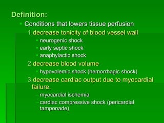
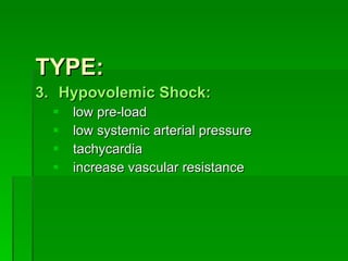
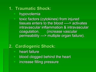
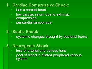
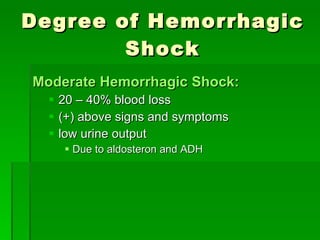
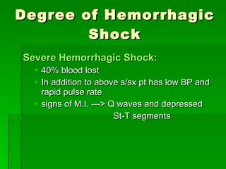
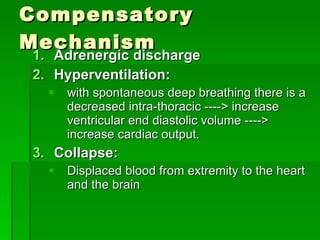
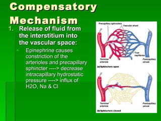
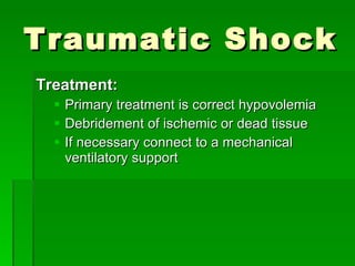
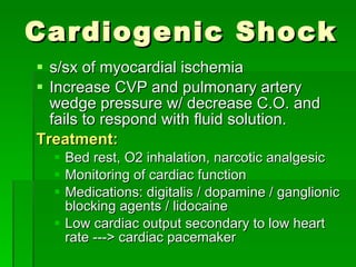
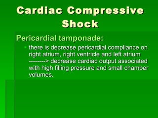
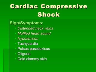
SHOCK James Taclin C. Banez, M.D.
Definition: Conditions that lowers tissue perfusion decrease tonicity of blood vessel wall   neurogenic shock early septic shock anaphylactic shock decrease blood volume hypovolemic shock (hemorrhagic shock) decrease cardiac output due to myocardial failure. myocardial ischemia cardiac compressive shock (pericardial tamponade)
TYPE: Hypovolemic Shock: low pre-load low systemic arterial pressure tachycardia increase vascular resistance
Traumatic Shock: hypovolemia toxic factors (cytokines) from injured tissues enters to the blood ----> activates intravascular inflammation & intravascular coagulation.  (increase vascular permeability ---> multiple organ failure). Cardiogenic Shock: heart failure blood clogged behind the heart increase filling pressure
Cardiac Compressive Shock: has a normal heart low cardiac return due to extrinsic compression pericardial tamponade Septic Shock systemic changes brought by bacterial toxins Neurogenic Shock loss of arterial and venous tone pool of blood in dilated peripheral venous system
Cellular Changes: decrease tissue perfusion ----> decrease oxygen delivery ----> decrease ATP -----> Na/K membrane pump fails ----> Na & Cl enters the cell ---> isotonic cellular swelling ----> cell death.
Renal Response: Normal kidneys can tolerate renal ischemia from 15 mins to a maximum of 90 mins. Can be prolonged w/ hypothermia and/or steroid) Prolonged hypoperfusion leads to functional and anatomic changes presenting as azotemia. Renal failure index : ( Urine Na x Plasma creatinine )  Urine creatinine < 1 usually indicates pre-renal oliguria > 1 indicates acute renal failure
Pulmonary Response: Damage Alveolar – Capillary interface  in case of shock ----> acute diffuse lung injury. Leakage of proteinaceous fluid into the interstitium and alveolar space. Leads to  ARDS :  Hypoxemia unresponsive to elevation of FiO2 Decreased pulmonary compliance Needs high airway pressure to attain adequate tidal volume
Pathophysiology Hypovolemic Shock: most common Most of the blood is lost from systemic and small veins (50%) ----> decrease cardiac return ----> low cardiac output ----> decrease blood pressure
Degree of Hemorrhagic Shock Mild Hemorrhagic Shock: < 20% blood lost adrenergic constriction of blood vessels in the skin thirsty, feels cold normal BP, PR and urine output
Degree of Hemorrhagic Shock Moderate Hemorrhagic Shock: 20 – 40% blood loss (+) above signs and symptoms low urine output Due to aldosteron and ADH
Degree of Hemorrhagic Shock Severe Hemorrhagic Shock: 40% blood lost In addition to above s/sx pt has low BP and rapid pulse rate signs of M.I. ---> Q waves and depressed  St-T segments
Compensatory Mechanism Adrenergic discharge Hyperventilation: with spontaneous deep breathing there is a decreased intra-thoracic ----> increase ventricular end diastolic volume ----> increase cardiac output. Collapse: Displaced blood from extremity to the heart and the brain
Compensatory Mechanism Release of fluid from the interstitium into the vascular space: Epinephrine causes constriction of the arterioles and precapillary sphincter ----> decrease intracapillary hydrostatic pressure ----> influx of H2O, Na & Cl
Compensatory Mechanism Release of vasoactive hormones: Resorption of fluid from intracellular to extracellular space: Epinephrine, cortisol and glucagon increases concentration of interstitial glucose  ---->   extracellular hyperosmolality  ---->  draws H2O from cell  ---->   hydrostatic pressure in the interstitium  ---->  forced H2O and CHON into the lymphatics  ------------->  oncotic pressure intravascular  ---->  attracts H2O intravascularly further
Compensatory Mechanism Renal conservation of body H2O and electrolyte aldosterone anti-diuretic hormone (vasopressin)
Decompensation in Hypovolemic Shock Relaxation of arteriolar and precapillary spasm forced H20 and electrolyte from vascular space into the interstitium Deterioration of cell membrane function
Two Most Sensitive Signs of Hypovolemia are: Cutaneous vasoconstriction Oliguria Alcohol  can cause vasodilation and inhibits secretion of ADH
Monitoring: Management: resuscitate patient and control blood lost and correct dehydration give balance salt solution (crytalloid) disadvantage of giving colloid resuscitation. Post-resuscitation HPN Increase intravascular volume at the expense of necessary interstitial fluid Depression of albumin synthesis Depression of circulating immunoglobulin More expensive and less easier to titrate
Management: Supine position w/ elevated both lower   extremities :    preferred position for patient in shock Elderly patient ---> check condition of the heart (digitalis) ability to compensate. Steroid is not indicated O2 inhalation is of little value if vascular volume is not corrected
Causes of Refractory Shock: Continuing blood loss from primary injury or another source Inadequate replacement of fluids Massive trauma or other derangement Myocardial infarction Concomitant septic shock
Traumatic Shock Traumatized tissue activates coagulation system forming: Microthrombi: Occludes pulmonary vasculature ---> increase pulmonary vascular resistance ----> increase right arterial pressure Humoral products of microthrombi : cytoxines Increases vascular permeability ---> loss of plasma
Traumatic Shock Treatment: Primary treatment is correct hypovolemia Debridement of ischemic or dead tissue If necessary connect to a mechanical ventilatory support
Cardiogenic Shock s/sx of myocardial ischemia Increase CVP and pulmonary artery wedge pressure w/ decrease C.O. and fails to respond with fluid solution. Treatment: Bed rest, O2 inhalation, narcotic analgesic Monitoring of cardiac function Medications: digitalis / dopamine / ganglionic blocking agents / lidocaine Low cardiac output secondary to low heart rate ---> cardiac pacemaker
Cardiac Compressive Shock Pericardial tamponade: there is decrease pericardial compliance on right atrium, right ventricle and left atrium --------> decrease cardiac output associated with high filling pressure and small chamber volumes.
Cardiac Compressive Shock Sign/Symptoms: Distended neck veins Muffled heart sound Hypotension Tachycardia Pulsus paradoxicus Oliguria Cold clammy skin
Cardiac Compressive Shock Chest X-ray: Water bottle shape (erect film) Management: in  unstable pt .  – do anterolateral thoracotomy w/ decompression of the tamponade and surgical control of the abnormality In  stable pt.  – do pericardiocentesis, withdrawal of 50cc. Blood shd. Return cardiovascular dynamics to normal
Septic Shock Causes: Gram positive sepsis (exotoxin) Gram negative sepsis (endotoxin) More common Source of infection: genito-urinary respiratory alimentary integumentary
Septic Shock Early septic shock: warm extremity, pt is still normovolemic toxin increases core body temperature BP decreases due to vasodilatation  (decrease peripheral resistance) increase cardiac output due to minimal resistance increase heart rate and increase cardiac contractility hyperventilation  ---->  respiratory alkalosis
Septic Shock Late septic shock: cold extremity, patient is hypovolemic hypovolemia due to --> increase vascular permeability and decrease cardiac output Increase vascular permeability (endotoxin) Decrease cardiac output    due to increased pulmonary vascular resistance (endotoxin) Increase right ventricular pressure --> due to impairment of emptying. Increase peripheral resistance  (cold cyanotic extremities) Decrease CVP
Septic Shock Treatment: volume replacement look for the source of infection to give the proper antibiotic (culture and sensitivity) Earliest sign of gm(-) infection: hyperventilation respiratory alkalosis altered sensorium
Neurogenic Shock Seen in spinal cord injuries and spinal anesthesia. normovolemic / hypovolemic pt.: Blood pool in the systemic venules and small veins resulting to decrease right heart filling ----> decrease stroke volume ----> decrease cardiac output ----> (+) angiotensin and ADH
Neurogenic Shock hypervolemic pt.: cardiac output increase due to expanded blood volume and decrease vascular resistance. hypotension due to decrease vascular resistance Treatment: Give IVF, if BP does not respond ----> give vasoconstrictor to restore venous tone ----> restore right cardiac filling ----> increase cardiac output. Elevate the lower extremities
THANK YOU

Shock 2

  • 1.
    SHOCK James TaclinC. Banez, M.D.
  • 2.
    Definition: Conditions thatlowers tissue perfusion decrease tonicity of blood vessel wall neurogenic shock early septic shock anaphylactic shock decrease blood volume hypovolemic shock (hemorrhagic shock) decrease cardiac output due to myocardial failure. myocardial ischemia cardiac compressive shock (pericardial tamponade)
  • 3.
    TYPE: Hypovolemic Shock:low pre-load low systemic arterial pressure tachycardia increase vascular resistance
  • 4.
    Traumatic Shock: hypovolemiatoxic factors (cytokines) from injured tissues enters to the blood ----> activates intravascular inflammation & intravascular coagulation. (increase vascular permeability ---> multiple organ failure). Cardiogenic Shock: heart failure blood clogged behind the heart increase filling pressure
  • 5.
    Cardiac Compressive Shock:has a normal heart low cardiac return due to extrinsic compression pericardial tamponade Septic Shock systemic changes brought by bacterial toxins Neurogenic Shock loss of arterial and venous tone pool of blood in dilated peripheral venous system
  • 6.
    Cellular Changes: decreasetissue perfusion ----> decrease oxygen delivery ----> decrease ATP -----> Na/K membrane pump fails ----> Na & Cl enters the cell ---> isotonic cellular swelling ----> cell death.
  • 7.
    Renal Response: Normalkidneys can tolerate renal ischemia from 15 mins to a maximum of 90 mins. Can be prolonged w/ hypothermia and/or steroid) Prolonged hypoperfusion leads to functional and anatomic changes presenting as azotemia. Renal failure index : ( Urine Na x Plasma creatinine ) Urine creatinine < 1 usually indicates pre-renal oliguria > 1 indicates acute renal failure
  • 8.
    Pulmonary Response: DamageAlveolar – Capillary interface in case of shock ----> acute diffuse lung injury. Leakage of proteinaceous fluid into the interstitium and alveolar space. Leads to ARDS : Hypoxemia unresponsive to elevation of FiO2 Decreased pulmonary compliance Needs high airway pressure to attain adequate tidal volume
  • 9.
    Pathophysiology Hypovolemic Shock:most common Most of the blood is lost from systemic and small veins (50%) ----> decrease cardiac return ----> low cardiac output ----> decrease blood pressure
  • 10.
    Degree of HemorrhagicShock Mild Hemorrhagic Shock: < 20% blood lost adrenergic constriction of blood vessels in the skin thirsty, feels cold normal BP, PR and urine output
  • 11.
    Degree of HemorrhagicShock Moderate Hemorrhagic Shock: 20 – 40% blood loss (+) above signs and symptoms low urine output Due to aldosteron and ADH
  • 12.
    Degree of HemorrhagicShock Severe Hemorrhagic Shock: 40% blood lost In addition to above s/sx pt has low BP and rapid pulse rate signs of M.I. ---> Q waves and depressed St-T segments
  • 13.
    Compensatory Mechanism Adrenergicdischarge Hyperventilation: with spontaneous deep breathing there is a decreased intra-thoracic ----> increase ventricular end diastolic volume ----> increase cardiac output. Collapse: Displaced blood from extremity to the heart and the brain
  • 14.
    Compensatory Mechanism Releaseof fluid from the interstitium into the vascular space: Epinephrine causes constriction of the arterioles and precapillary sphincter ----> decrease intracapillary hydrostatic pressure ----> influx of H2O, Na & Cl
  • 15.
    Compensatory Mechanism Releaseof vasoactive hormones: Resorption of fluid from intracellular to extracellular space: Epinephrine, cortisol and glucagon increases concentration of interstitial glucose ----> extracellular hyperosmolality ----> draws H2O from cell ----> hydrostatic pressure in the interstitium ----> forced H2O and CHON into the lymphatics -------------> oncotic pressure intravascular ----> attracts H2O intravascularly further
  • 16.
    Compensatory Mechanism Renalconservation of body H2O and electrolyte aldosterone anti-diuretic hormone (vasopressin)
  • 17.
    Decompensation in HypovolemicShock Relaxation of arteriolar and precapillary spasm forced H20 and electrolyte from vascular space into the interstitium Deterioration of cell membrane function
  • 18.
    Two Most SensitiveSigns of Hypovolemia are: Cutaneous vasoconstriction Oliguria Alcohol can cause vasodilation and inhibits secretion of ADH
  • 19.
    Monitoring: Management: resuscitatepatient and control blood lost and correct dehydration give balance salt solution (crytalloid) disadvantage of giving colloid resuscitation. Post-resuscitation HPN Increase intravascular volume at the expense of necessary interstitial fluid Depression of albumin synthesis Depression of circulating immunoglobulin More expensive and less easier to titrate
  • 20.
    Management: Supine positionw/ elevated both lower extremities :  preferred position for patient in shock Elderly patient ---> check condition of the heart (digitalis) ability to compensate. Steroid is not indicated O2 inhalation is of little value if vascular volume is not corrected
  • 21.
    Causes of RefractoryShock: Continuing blood loss from primary injury or another source Inadequate replacement of fluids Massive trauma or other derangement Myocardial infarction Concomitant septic shock
  • 22.
    Traumatic Shock Traumatizedtissue activates coagulation system forming: Microthrombi: Occludes pulmonary vasculature ---> increase pulmonary vascular resistance ----> increase right arterial pressure Humoral products of microthrombi : cytoxines Increases vascular permeability ---> loss of plasma
  • 23.
    Traumatic Shock Treatment:Primary treatment is correct hypovolemia Debridement of ischemic or dead tissue If necessary connect to a mechanical ventilatory support
  • 24.
    Cardiogenic Shock s/sxof myocardial ischemia Increase CVP and pulmonary artery wedge pressure w/ decrease C.O. and fails to respond with fluid solution. Treatment: Bed rest, O2 inhalation, narcotic analgesic Monitoring of cardiac function Medications: digitalis / dopamine / ganglionic blocking agents / lidocaine Low cardiac output secondary to low heart rate ---> cardiac pacemaker
  • 25.
    Cardiac Compressive ShockPericardial tamponade: there is decrease pericardial compliance on right atrium, right ventricle and left atrium --------> decrease cardiac output associated with high filling pressure and small chamber volumes.
  • 26.
    Cardiac Compressive ShockSign/Symptoms: Distended neck veins Muffled heart sound Hypotension Tachycardia Pulsus paradoxicus Oliguria Cold clammy skin
  • 27.
    Cardiac Compressive ShockChest X-ray: Water bottle shape (erect film) Management: in unstable pt . – do anterolateral thoracotomy w/ decompression of the tamponade and surgical control of the abnormality In stable pt. – do pericardiocentesis, withdrawal of 50cc. Blood shd. Return cardiovascular dynamics to normal
  • 28.
    Septic Shock Causes:Gram positive sepsis (exotoxin) Gram negative sepsis (endotoxin) More common Source of infection: genito-urinary respiratory alimentary integumentary
  • 29.
    Septic Shock Earlyseptic shock: warm extremity, pt is still normovolemic toxin increases core body temperature BP decreases due to vasodilatation (decrease peripheral resistance) increase cardiac output due to minimal resistance increase heart rate and increase cardiac contractility hyperventilation ----> respiratory alkalosis
  • 30.
    Septic Shock Lateseptic shock: cold extremity, patient is hypovolemic hypovolemia due to --> increase vascular permeability and decrease cardiac output Increase vascular permeability (endotoxin) Decrease cardiac output  due to increased pulmonary vascular resistance (endotoxin) Increase right ventricular pressure --> due to impairment of emptying. Increase peripheral resistance (cold cyanotic extremities) Decrease CVP
  • 31.
    Septic Shock Treatment:volume replacement look for the source of infection to give the proper antibiotic (culture and sensitivity) Earliest sign of gm(-) infection: hyperventilation respiratory alkalosis altered sensorium
  • 32.
    Neurogenic Shock Seenin spinal cord injuries and spinal anesthesia. normovolemic / hypovolemic pt.: Blood pool in the systemic venules and small veins resulting to decrease right heart filling ----> decrease stroke volume ----> decrease cardiac output ----> (+) angiotensin and ADH
  • 33.
    Neurogenic Shock hypervolemicpt.: cardiac output increase due to expanded blood volume and decrease vascular resistance. hypotension due to decrease vascular resistance Treatment: Give IVF, if BP does not respond ----> give vasoconstrictor to restore venous tone ----> restore right cardiac filling ----> increase cardiac output. Elevate the lower extremities
  • 34.