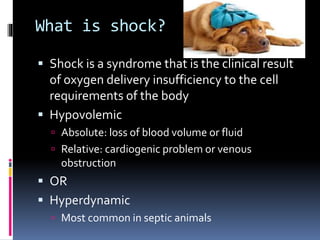
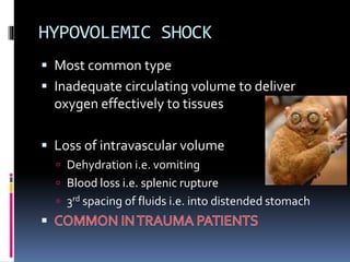
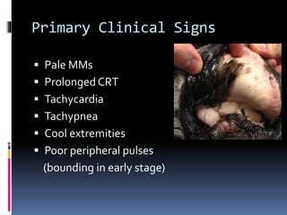
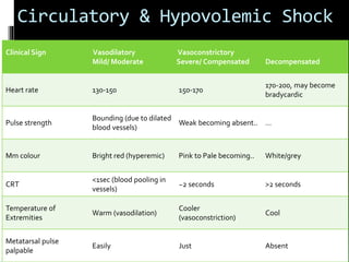
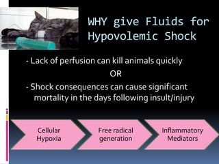
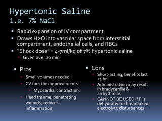
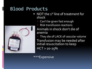
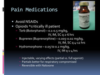
HYPOVOLEMIC SHOCK
By: Abigail Schmidt
What is shock?
 Shock is a syndrome that is the clinical result
of oxygen delivery insufficiency to the cell
requirements of the body
 Hypovolemic
 Absolute: loss of blood volume or fluid
 Relative: cardiogenic problem or venous
obstruction
 OR
 Hyperdynamic
 Most common in septic animals
HYPOVOLEMIC SHOCK
 Most common type
 Inadequate circulating volume to deliver
oxygen effectively to tissues
 Loss of intravascular volume
 Dehydration i.e. vomiting
 Blood loss i.e. splenic rupture
 3rd spacing of fluids i.e. into distended stomach
Signs of Hypovolemic Shock
 Compensatory events take place:
 Tachycardia (low preload = reduces cardiac output)
 Peripheral vasoconstriction
 Hypotension
Decreased perfusion to essential organs,
development of acidosis
Primary Clinical Signs
 Pale MMs
 Prolonged CRT
 Tachycardia
 Tachypnea
 Cool extremities
 Poor peripheral pulses
(bounding in early stage)
Triage Primary Assessment
ABCD:
 AB: Airway & Breathing
 Is P breathing? Is airway patent?
 Is P working too hard to breathe?
 C: Circulation
 Is heart beating effectively?
 ASSESS FOR SHOCK
 MM/CRT, BP, Pulse Assessment
 D: Debilitating Disease
 Neuro assessment
 Minimum BW: renal parameters, PCV/TP, ALT,
Glucose, E-lytes
Triage 2
 Secondary Survey
 Respiratory
 RR, RE, MM, URT (inspiratory) vs LRT (expiratory)
 LOCALIZE
 Cardiovascular
 Arrhythmias, tissue perfusion (BP, lactate, CRT, MM)
 SHOCK
 Neurological
 Abdominal
VITAL 1st STEPS
 Necessary to increase tissue perfusion via
fluids
 NEED to exclude:
 Heart disease
 Respiratory disease
 Intracranial disease
BEFORE loading patient with shock-dose rate fluids
Heart Disease
 Failure of pump, caused by:
 Heart disease
 Cardiac tamponade
 Arrhythmias
 Thoracic auscultation
 Murmurs
 Pulmonary edema heard as crackles
 Ascites/Jugular distension
Large volume fluid
administration
contraindicated!!!
Respiratory Disease
 Tachypnea/Dyspnea with increased sounds
heard on auscultation
 Crackles, wheezes, etc.
 ALTERNATIVELY: dullness indicates pleural
effusion, another contraindication to shock fluids
 Altered mentation/increased ICP
 Neurologic signs
 Seizures, Blindness, Absent PLR, Incoordination etc.
Intracranial Disease
 Low BP  activation of sympathetic nervous
system
 Tachycardia
 PeripheralVasoconstriction
 Fluid retention
 Protective response may make patients seem
more stable than what they truly are.
Pathological Consequences
In attempt to
maintain BP and
perfusion
What to do BEFORE referring
 Check important parameters:
 HR, BP, temperature
 BP may initially be normal due to sympathetic NS
 Then decline/drop low
 MM, CRT, Pulse rate/quality, RR
 Cats vs. Dogs
 Dogs: bradycardia
 worsening “shock” state = worse prognosis
 Cats often don’t present tachycardic (less scary)
 Cats more susceptible to fluid overload
 Monitor respiratory rate/effort!
 Cats more prone to hypothermia  active warming
Circulatory & Hypovolemic Shock
Clinical Sign Vasodilatory Vasoconstrictory
Mild/ Moderate Severe/ Compensated Decompensated
Heart rate 130-150 150-170
170-200, may become
bradycardic
Pulse strength
Bounding (due to dilated
blood vessels)
Weak becoming absent.. …
Mm colour Bright red (hyperemic) Pink to Pale becoming.. White/grey
CRT
<1sec (blood pooling in
vessels)
~2 seconds >2 seconds
Temperature of
Extremities
Warm (vasodilation)
Cooler
(vasoconstriction)
Cool
Metatarsal pulse
palpable
Easily Just Absent
WHY give Fluids for
Hypovolemic Shock
- Lack of perfusion can kill animals quickly
OR
- Shock consequences can cause significant
mortality in the days following insult/injury
Cellular
Hypoxia
Free radical
generation
Inflammatory
Mediators
SIRS  MODS  DIC
 Systemic Inflammatory Response Syndrome
 Inflammatory mediators cause disruption of
homeostasis
 Loss of vascular tone
 Endothelial permeability barrier disruption
 Stimulation of coagulation
 Microvascular thrombosis resulting in…
 Multiple Organ Dysfunction Syndrome
 Disseminated Intravascular Coagulation
 IV activation of coagulation with loss of localization
DIC aka
Shock Treatment Aims
 Provide oxygen support
 Connect to appropriate monitoring
 Vascular access and BP
 Shock fluid boluses to restore vascular perfusion
and oxygen delivery to tissues
 Pain medications if needed
 Stabilize the patient and send to IndyVet! 
Isotonic Crystalloids
i.e. Hartmann’s aka LRS,
0.9% Sodium Chloride
 Same concentration of solutes as blood; same
osmotic pressure
 Small molecules freely pass out of BVs, able
to enter all body compartments
 1/5 of total volume given = actually remain in BVs
1-2 hrs later
Crystalloids 2
 “Shock doses” = 60-90 ml/kg, but given in
incremental boluses delivered over 15-20 min
 Assess P after each bolus; repeat if necessary
**Rapid expansion of blood volume with
crystalloids may worsen blood loss
**Risk of interstitial edema, dilution of RBCs
and clotting factors with repeated boluses
Colloids
i.e. Hetastarch, Dextran 70
 Large molecules which do NOT pass out of BVs
 Expand IV space by increasing oncotic pressure
 “Shock doses” = 20 ml/kg
 Given as boluses of 5-10 ml/kg
 Cons
 Synthetic can cause
acquired coagulopathy
 Expensive, not multi-
purpose
 Pros
 Less volume needed
 Useful with large Ps
 IV expansion lasts longer
(up to 12 hrs)
Hypertonic Saline
i.e. 7% NaCl
 Rapid expansion of IV compartment
 Draws H2O into vascular space from interstitial
compartment, endothelial cells, and RBCs
 “Shock dose” = 4-7ml/kg of 7% hypertonic saline
 Given over 20 min
 Cons
 Short-acting, benefits last
<1 hr
 Administration may result
in bradycardia &
arrhythmias
 CANNOT BE USED if P is
dehydrated or has marked
electrolyte disturbances
 Pros
 Small volumes needed
 CV function improvements
 Myocardial contraction,
 Head trauma, penetrating
wounds, reduces
inflammation
Blood Products
 NOT the 1st line of treatment for
shock
 Can’t be given fast enough
 Risk transfusion reactions
 Animals in shock don’t die of
anemia
 They die of LACK of vascular volume
 Transfusion may be needed after
initial resuscitation to keep
HCT > 20-25%
***Expensive
Pain Medications
 Avoid NSAIDs
 Opioids *critically ill patient
 Torb (Butorphanol) – 0.1-0.5 mg/kg,
IV, IM, SC q 2-6 hrs
 Buprenex (Buprenorphine) – 0.005-0.02 mg/kg,
IV, IM, SC q 4-12 hrs
 Hydromorphone – 0.05 to 0.2 mg/kg,
IV, IM q 1-4 hrs
 Injectable, varying effects (partial vs. full agonist)
 Partials better for respiratory compromised
 Reversible with Naloxone
Questions?

Indy vet hypovolemic shock

  • 1.
  • 2.
    What is shock? Shock is a syndrome that is the clinical result of oxygen delivery insufficiency to the cell requirements of the body  Hypovolemic  Absolute: loss of blood volume or fluid  Relative: cardiogenic problem or venous obstruction  OR  Hyperdynamic  Most common in septic animals
  • 3.
    HYPOVOLEMIC SHOCK  Mostcommon type  Inadequate circulating volume to deliver oxygen effectively to tissues  Loss of intravascular volume  Dehydration i.e. vomiting  Blood loss i.e. splenic rupture  3rd spacing of fluids i.e. into distended stomach
  • 4.
    Signs of HypovolemicShock  Compensatory events take place:  Tachycardia (low preload = reduces cardiac output)  Peripheral vasoconstriction  Hypotension Decreased perfusion to essential organs, development of acidosis
  • 5.
    Primary Clinical Signs Pale MMs  Prolonged CRT  Tachycardia  Tachypnea  Cool extremities  Poor peripheral pulses (bounding in early stage)
  • 6.
    Triage Primary Assessment ABCD: AB: Airway & Breathing  Is P breathing? Is airway patent?  Is P working too hard to breathe?  C: Circulation  Is heart beating effectively?  ASSESS FOR SHOCK  MM/CRT, BP, Pulse Assessment  D: Debilitating Disease  Neuro assessment  Minimum BW: renal parameters, PCV/TP, ALT, Glucose, E-lytes
  • 7.
    Triage 2  SecondarySurvey  Respiratory  RR, RE, MM, URT (inspiratory) vs LRT (expiratory)  LOCALIZE  Cardiovascular  Arrhythmias, tissue perfusion (BP, lactate, CRT, MM)  SHOCK  Neurological  Abdominal
  • 8.
    VITAL 1st STEPS Necessary to increase tissue perfusion via fluids  NEED to exclude:  Heart disease  Respiratory disease  Intracranial disease BEFORE loading patient with shock-dose rate fluids
  • 9.
    Heart Disease  Failureof pump, caused by:  Heart disease  Cardiac tamponade  Arrhythmias  Thoracic auscultation  Murmurs  Pulmonary edema heard as crackles  Ascites/Jugular distension Large volume fluid administration contraindicated!!!
  • 10.
    Respiratory Disease  Tachypnea/Dyspneawith increased sounds heard on auscultation  Crackles, wheezes, etc.  ALTERNATIVELY: dullness indicates pleural effusion, another contraindication to shock fluids  Altered mentation/increased ICP  Neurologic signs  Seizures, Blindness, Absent PLR, Incoordination etc. Intracranial Disease
  • 11.
     Low BP activation of sympathetic nervous system  Tachycardia  PeripheralVasoconstriction  Fluid retention  Protective response may make patients seem more stable than what they truly are. Pathological Consequences In attempt to maintain BP and perfusion
  • 12.
    What to doBEFORE referring  Check important parameters:  HR, BP, temperature  BP may initially be normal due to sympathetic NS  Then decline/drop low  MM, CRT, Pulse rate/quality, RR  Cats vs. Dogs  Dogs: bradycardia  worsening “shock” state = worse prognosis  Cats often don’t present tachycardic (less scary)  Cats more susceptible to fluid overload  Monitor respiratory rate/effort!  Cats more prone to hypothermia  active warming
  • 13.
    Circulatory & HypovolemicShock Clinical Sign Vasodilatory Vasoconstrictory Mild/ Moderate Severe/ Compensated Decompensated Heart rate 130-150 150-170 170-200, may become bradycardic Pulse strength Bounding (due to dilated blood vessels) Weak becoming absent.. … Mm colour Bright red (hyperemic) Pink to Pale becoming.. White/grey CRT <1sec (blood pooling in vessels) ~2 seconds >2 seconds Temperature of Extremities Warm (vasodilation) Cooler (vasoconstriction) Cool Metatarsal pulse palpable Easily Just Absent
  • 14.
    WHY give Fluidsfor Hypovolemic Shock - Lack of perfusion can kill animals quickly OR - Shock consequences can cause significant mortality in the days following insult/injury Cellular Hypoxia Free radical generation Inflammatory Mediators
  • 15.
    SIRS  MODS DIC  Systemic Inflammatory Response Syndrome  Inflammatory mediators cause disruption of homeostasis  Loss of vascular tone  Endothelial permeability barrier disruption  Stimulation of coagulation  Microvascular thrombosis resulting in…  Multiple Organ Dysfunction Syndrome  Disseminated Intravascular Coagulation  IV activation of coagulation with loss of localization DIC aka
  • 16.
    Shock Treatment Aims Provide oxygen support  Connect to appropriate monitoring  Vascular access and BP  Shock fluid boluses to restore vascular perfusion and oxygen delivery to tissues  Pain medications if needed  Stabilize the patient and send to IndyVet! 
  • 17.
    Isotonic Crystalloids i.e. Hartmann’saka LRS, 0.9% Sodium Chloride  Same concentration of solutes as blood; same osmotic pressure  Small molecules freely pass out of BVs, able to enter all body compartments  1/5 of total volume given = actually remain in BVs 1-2 hrs later
  • 18.
    Crystalloids 2  “Shockdoses” = 60-90 ml/kg, but given in incremental boluses delivered over 15-20 min  Assess P after each bolus; repeat if necessary **Rapid expansion of blood volume with crystalloids may worsen blood loss **Risk of interstitial edema, dilution of RBCs and clotting factors with repeated boluses
  • 19.
    Colloids i.e. Hetastarch, Dextran70  Large molecules which do NOT pass out of BVs  Expand IV space by increasing oncotic pressure  “Shock doses” = 20 ml/kg  Given as boluses of 5-10 ml/kg  Cons  Synthetic can cause acquired coagulopathy  Expensive, not multi- purpose  Pros  Less volume needed  Useful with large Ps  IV expansion lasts longer (up to 12 hrs)
  • 20.
    Hypertonic Saline i.e. 7%NaCl  Rapid expansion of IV compartment  Draws H2O into vascular space from interstitial compartment, endothelial cells, and RBCs  “Shock dose” = 4-7ml/kg of 7% hypertonic saline  Given over 20 min  Cons  Short-acting, benefits last <1 hr  Administration may result in bradycardia & arrhythmias  CANNOT BE USED if P is dehydrated or has marked electrolyte disturbances  Pros  Small volumes needed  CV function improvements  Myocardial contraction,  Head trauma, penetrating wounds, reduces inflammation
  • 21.
    Blood Products  NOTthe 1st line of treatment for shock  Can’t be given fast enough  Risk transfusion reactions  Animals in shock don’t die of anemia  They die of LACK of vascular volume  Transfusion may be needed after initial resuscitation to keep HCT > 20-25% ***Expensive
  • 22.
    Pain Medications  AvoidNSAIDs  Opioids *critically ill patient  Torb (Butorphanol) – 0.1-0.5 mg/kg, IV, IM, SC q 2-6 hrs  Buprenex (Buprenorphine) – 0.005-0.02 mg/kg, IV, IM, SC q 4-12 hrs  Hydromorphone – 0.05 to 0.2 mg/kg, IV, IM q 1-4 hrs  Injectable, varying effects (partial vs. full agonist)  Partials better for respiratory compromised  Reversible with Naloxone
  • 23.

Editor's Notes

  • #5 CLINICAL SIGNS OF CLASSIC HYPOVOLAEMIC SHOCK  Clinical signs are a reflection of the animal trying to compensate 1.  Tachycardia as part of the sympathetic response 2.  Poor pulse quality due to vasoconstriction and lack of blood volume 3.  Decreased extremity temperature 4.  Pale mucous membranes 5.  Prolonged capillary refill time 6.  Decreased mentation due to inadequate brain perfusion 7.  Tachypnea to increase oxygen uptake (not always evident)