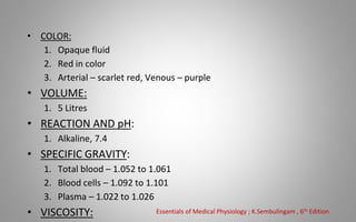
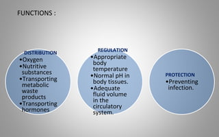
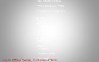
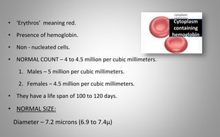
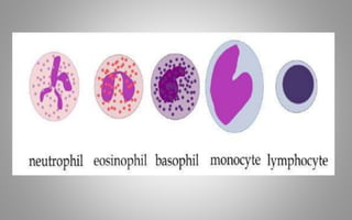
Presented by:-
DR. ROHIT PATIL
MDS I
BLOOD : COMPONENTS, DISORDERS AND
IMPLICATIONS IN PROSTHODONTICS
CONTENTS
• Introduction
• Composition of blood
• Red blood cell and its disorders
• White blood cell and its disorders
• Platelets and its disorders
• Coagulation disorders
• History taking
• Conclusion
• References
• William Harvey father of physiology discovered blood
circulated through the body in 1628.
• Blood is fluid connective tissue present in circulatory system
• It carries oxygen from lungs to all parts of the body and
carbon dioxide from all parts of the body to the lungs.
INTRODUCTION
• COLOR:
1. Opaque fluid
2. Red in color
3. Arterial – scarlet red, Venous – purple
• VOLUME:
1. 5 Litres
• REACTION AND pH:
1. Alkaline, 7.4
• SPECIFIC GRAVITY:
1. Total blood – 1.052 to 1.061
2. Blood cells – 1.092 to 1.101
3. Plasma – 1.022 to 1.026
• VISCOSITY: Essentials of Medical Physiology ; K.Sembulingam , 6th Edition
FUNCTIONS :
DISTRIBUTION
•Oxygen
•Nutritive
substances
•Transporting
metabolic
waste
products
•Transporting
hormones
REGULATION
•Appropriate
body
temperature
•Normal pH in
body tissues.
•Adequate
fluid volume
in the
circulatory
system.
PROTECTION
•Preventing
infection.
•Red blood cells (RBCs)
•White Blood Cells (WBCs)
•Platelets or Thrombocytes
•PLASMA
•Solids
•(7% - 8%)
•Organic substances
•Inorganic substances
•Water
•(92% - 93%)
•Gases
•Oxygen
•Carbon Dioxide
•Nitrogen
Essentials of Medical Physiology ; K.Sembulingam , 6th Edition
RED BLOOD CELL AND ITS
DISORDERS
• ‘Erythros’ meaning red.
• Presence of hemoglobin.
• Non - nucleated cells.
• NORMAL COUNT – 4 to 4.5 million per cubic millimeters.
1. Males – 5 million per cubic millimeters.
2. Females – 4.5 million per cubic millimeters.
• They have a life span of 100 to 120 days.
• NORMAL SIZE:
Diameter – 7.2 microns (6.9 to 7.4μ)
Cytoplasm
containing
hemoglobin
RED BLOOD CELL DISORDERS
• Erythrocytoses
• Polycythemia vera
• Anemia
✔Iron deficiency anemia
✔Anemia owing to hemolysis
✔Sickle cell anemia
✔Erythroblastosis fetalis
✔Thalassemia
✔Pernicious anemia
✔Aplastic anemia
ERYTHROCYTOSES
• A conditions with an increase in circulating red blood cells (RBCs),
characterized by a increased hemoglobin level.
• 2 types- relative and absolute
• Relative polycythemia: Occur as a result of loss of fluid with
hemoconcentration of cells.
•Absolute polycythemia: Increase in the number of circulating
RBC of the hemoglobin.
•Primary polycythemia
•Secondary polycythemia
• Chronic stem cell disorder with an insidious onset
characterized as a panhyperplastic, malignant and
neoplastic marrow disorder.
• Absolute increase in the number of circulating RBC and in
the total blood volume because of uncontrolled RBC
production.
• Accompanied by increase in WBC and platelet production.
POLYCYTHEMIA VERA
Oral Manifestations
• Erythema (red-purple
color) of mucosa.
• Glossitis.
• Erythematous &
edematous gingiva.
• Spontaneous gingival
bleeding.
• Pruritis
• Vertigo
• Gastrointestinal pain
• Headache
• Paresthesias, fatigue,
weakness
• Visual disturbances, tinnitus,
Clinical
Manifestations
ANEMIA
• Anemia refers to reduction in
1. Red blood cell count
2. Hemoglobin content
3. Packed cell volume
• It can also be defined as a lowered ability of
the blood to carry oxygen.
Etiologic classification of Anemia:
1. Loss of blood:
• Acute posthemorragic anemia.
• Chronic posthemorrhagic anemia.
2.Excessive destruction of RBC:
A.Extra corpuscular causes: Antibodies, Infection(malaria), Splenic
sequestration, Drugs, chemicals and physical agents.
B.Intracorpuscular haemolytic disease :Hereditary,Disorder of glycolysis,
abnormalities in RBC membrane,Abnormalities in synthesis of globin,lead
poisoning.
3. Impaired blood production resulting from deficiency of
substances essential for erythropoiesis:
a. Iron deficiency
b. Deficiency of vitamin B12 ,folic acid and Protein deficiency
4. Inadequate production of mature erythrocytes :
a) Deficiency of erythroblasts
b) Pure red cell aplasia
c) Infiltration of bone marrow- Leukemia, lymphoma, Multiple
myeloma
d) Endocrine abnormality- myxedema
e) Chronic renal disease
f) Chronic inflammatory disease
g) Cirrohisis of liver
IRON DEFICIENCY ANEMIA
• Iron deficiency is defined as a reduction in total body
iron to an extent that iron stores are fully exhausted and
some degree of tissue iron deficiency is present.
• Females are mostly affected.
Etiology
• Chronic blood loss
• Inadequate dietary intake
• Faulty iron absorption
• Increased requirements in pregnancy.
Clinical Manifestations
• Chronic fatigue
• Pallor of the conjunctiva, lips, and oral
mucosa;
• Brittle nails with spooning, cracking,
• Splitting of nail beds, koilonychia
• Palmar creases
• Palpitations
• Shortness of breath, numbness
• Bone pain
Oral Manifestations
• Angular cheilitis
• Glossitis with different degrees of
atrophy
of fungiform and filliform papillae
• Pale oral mucosa
• Oral candidiasis
• Recurrent aphthous stomatitis
• Erythematous mucositis
• Burning mouth
Plummer-Vinson Syndrome/ Paterson-Kelly syndrome
• Rare syndrome , middle-aged white women.
• Classic triad : Dysphagia,
Iron deficiency anemia
Upper esophageal webs or strictures.
Etiopathogenesis :
• Iron deficiency.
• Malnutrition
• Genetic predisposition and Autoimmune processes.
Treatment: Iron supplementation.
ANEMIA OWING TO HEMOLYSIS
• Normal RBC life span - 90 to 120 days.
Hemolytic diseases result in anemia if the bone marrow is not
able to replenish adequately the prematurely destroyed
RBCs.
• Either inherited or acquired.
3 mechanism for accelerated destruction of RBCs:
1. Molecular defect inside the red cell.
2. Abnormality in membrane structure and function.
3. Environmental factor- mechanical trauma.
Oral Manifestations
• Pallor or jaundice of oral
mucosa
• Paresthesia of mucosa
• Hyperplastic marrow spaces in
the mandible,maxilla and facial
bones
• Acute back pain
• Renal failure
• Fatigue
• Loss of stamina
• Breathlessness
• Tachycardia
• Hemoglobinuria
Clinical
Manifestations
SICKLE CELL DISEASE/SICKLE CELL ANEMIA
• Hereditary type of chronic hemolytic anemia transmitted as a
mendalian dominant, nongender linked characteristic.
• Exclusively in blacks and in whites of Mediterranean origin.
• A concordance exists between the prevalence of malaria and
HbS
HbA is genetically altered to produce HbS
Substituition of valine for glutamine at the sixth position of the
β globin chain
• Erythrocytes have their normal biconcave discoid shape
distorted, generally presenting a sickle-like shape.
• Reduces both their plasticity and lifetime from the normal
120 days average down to 14 days.
• This results in the underlying anemia and
hypertrophic bone marrow.
• In heterozygote- 40% of hemoglobin is HbS.
• In homozygote- nearly all hemoglobin is HbS.
• Common in females, before the age of 30 years
• Cerebrovascular accidents/ strokes
• Aplastic crises leading to severe anemia
• chronic leg ulcers
• Hematuria
• Aseptic osteonecrosis
• Retinitis leading to blindness
• Splenic sequestration
• Renal failure
• Acute chest syndrome - fever, cough, sputum production,
dyspnea,
or hypoxia.
Clinical Manifestations
Oral Manifestations
• Significant bone change in dental
radiograph
• Mild to severe generalized osteoporosis
• Loss of trabeculation of the jaw bone
• Enamel hypomineralization
• Increased overjet and overbite
• Pallor of the oral mucosa
• Delayed eruption of the teeth
• Pulpal necrosis
Smooth tongue
ERYTHROBLASTOSIS FETALIS
• Congenital hemolytic anemia due to Rh incompatibility
results from the destruction of fetal blood brought about
by a reaction between maternal and fetal blood factors.
• Rh factor, named after the rhesus monkey, was
discovered by Landsteiner and wiener in 1940 as a factor
in human RBC that would react with rabbit antiserum
produced by administration of RBC from the rhesus
monkey.
• If father is Rh- positive and mother is Rh- negative → fetus
inherits parental factor, which may act as an antigen to the
mother and immunize her with resultant antibody formation.
Clinical features
• Some infants are stillborn.
• Anemia with pallor
• Jaundice
• Compensatory erythropoiesis
• Fetal hydrops
• Deposition of blood pigments in
the enamel and dentin
• Ground sections- positive test for
bilirubin
• Intrinsic stains
• Enamel hypoplasia
• Rh hump
Oral manifestation
THALASSEMIAS
Thalassemia is a group of genetic disorders of hemoglobin
synthesis characterized by a disturbance of either alpha (α) or beta
(β) hemoglobin chain production.
• In heterozygotes the disease is mild and is called as
Thalassemia minor or thalassemia trait.
• Represent both α and β thalassemia.
• In homozygote, severe form, called Thalassemia major or β -
thalassemia/ Cooley's anemia.
• Production of β chain is markedly decreased or absent.
Clinical Manifestations
• Yellowish pallor of the skin
• Fever, chills, malaise
• Generalized weakness
• Splenomegaly and hepatomegaly
• RODENT FACIES- due to prominence of
the cheeks, protrusion of the maxillary
anterior teeth, depression of the bridge
of the nose.
• Unusual prominance of
the premaxilla
• Anemic pallor
observed
Oral
manifestation
• It is adult form of anemia that is associated with gastric atrophy
and a loss of intrinsic factor production in gastric secretions.
• Rare congenital autosomal recessive form.
• Autoimmune disease resulting from autoantibodies directed
against intrinsic factor (a substance needed to absorb vitamin
B12 from the gastrointestinal tract) and gastric parietal cells.
• Vitamin B12 → erythrocyte – maturing factor.
Megaloblastic (Pernicious) Anemia and Vitamin B12
(Cobalamin) Deficiency
Oral Manifestations
• Burning sensation in the tongue, lips, buccal
mucosa, and other mucosal sites.
• The tongue is generally inflammed often
described as ‘beefy red’in color.
• Hunter’s glossitis or Moeller’s glossitis.
• Fiery red appearance of the tongue may undergo
periods of remission, recurrent attacks are
common.
• Dysphagia and taste alterations have been
reported.
Aplastic Anemia
• Aplastic anemia (AA) is a rare blood dyscrasia in which
peripheral blood pancytopenia results from reduced or
absent blood cell production in the bone marrow and
normal hematopoietic tissue in the bone marrow has been
replaced by fatty marrow.
• Environmental exposures, such as to drugs, viruses, and
toxins, are thought to trigger the aberrant immune
response in some patients, but most cases are classified as
idiopathic
Clinical Manifestations
• Pancytopenia
• Anemia
• Leukopenia, particularly
neutropenia, can result in
fever and infection.
• Preceded by infections by
hepatitis viruses, EBV, HIV
parvovirus, mycobacterial
infections.
• Hemorrhage
• Candidiasis
• Viral infections
• Gingival bleedings
Oral
Manifestations
PROSTHODONTIC IMPLICATIONS OF RBC DISORDERS
Anemia :
• In edentulous and partially edentulous patients, the inefficiency
in mastication may lead to nutritional deficiency and eventually,
anemia.
• The length of appointment in anemic patients should be less as
there may be irritability, weakness and early fatigue.
• Cheilosis (fissures at the corners of the mouth) sign of advanced
tissue iron deficiency.
Singh N. Systemic Diseases Concern to Prosthodontist. Int J Oral Health Med Res 2015;2(2):89-93.
• The abnormal bleeding in anemic patients, due to hemorrhage
causes difficulty in placement of implants.
• The increased edema increases the risk of postoperative infection.
• In majority of anemic patients, implant procedures are not
contraindicated.
• However preoperative and postoperative antibiotics should be
administered.
Singh N. Systemic Diseases Concern to Prosthodontist. Int J Oral Health Med Res 2015;2(2):89-93.
SICKLE CELL ANEMIA :
•Preventive therapy
•Ideal approach
•Decrease possibility of oral infections
•Home Fluoride therapy
•Shorten time of Dental Procedures
•Schedule morning appointments
•Avoid elective surgery
•Pre-prosthetic Surgery
•Analgesics
•Avoid Steroidal anti-inflammatory drugs
•May trigger Acute Chest Syndrome
These patients are on long term prophylactic antibiotic therapy and are hence prone to
fungal infections such as candidiasis.
R. Sams et al 1990, Managing The Dental Patient With Sickle Cell Anemia: A Review Of The Literature
WHITE BLOOD CELLS AND ITS
DISORDERS
• White blood cells(WBCs),also called leukoytes or leucocytes, are
the cells of the immune system that are involved in protecting
the body against both infectious disease and foreign invaders.
• All white blood cells are produced and derived from multipotent
cells in the bone marrow known as hematopoietic stem
cells.
• Leukocytes are found throughout the body, including the
blood and lymphatic system.
• 2 types: granulocytes and agranulocytes
WBC Disorders
• Leukocytosis
• Leukopenia
• Agranulocytosis
• Neutropenia
• Leukamia
LEUKOCYTOSIS
• Defined as abnormal increase in the number of circulating WBCs.
• Considered to be a manifestation of the reaction of the body to a
pathologic situation.
• It may also occur after exercise, convulsions such as epilepsy,
emotional stress, pregnancy, anesthesia, and epinephrine
administration.
• There are five principal types of leukocytosis:
1. Neutrophilia (the most common form)
2. Lymphocytosis
3. Monocytosis
4. Eosinophilia
5. Basophilia
LEUKOPENIA
• Leukopenia is a decrease in the number of white blood cells
(leukocytes) found in the blood, which places individuals at
increased risk of infection.
CAUSES:
• Infections:Bacterial, Viral and Rickettsial, Protozoal.
• Hemopoietic disorders: Gaucher’s disease, Pernicious anemia,
Aplastic anemia, Chronic hypochromic anemia, Agranuocytosis
• Chemical agents: Mustards, Benzene, Urethane.
• X-ray radiations
• Anaphylactic shock
• Liver cirrhosis
Agranulocytosis (Neutropenia/Granulocytopenia)
• Serious disease involving the WBC and is characterized by
decrease in the number of circulating granulocytes.
• The terms agranulocytosis, neutropenia, and
granulocytopenia are commonly used interchangeably for a
reduced quantity of leukocytes.
Clinical features
• Occur at any age- particularly
among adults.
• Women are more affected.
• High fever, chills, sore throat.
• Malaise, weakness.
• Skin appears pale and anemic
• Presence of infections.
• Regional lymphadenitis.
• Complication-
Generalized sepsis.
• Necrotizing ulceration of the
oral cavity, tonsils and
pharynx particularly gingiva
and palate.
• Necrotic ulcers are covered
by gray or even black
membrane.
• No purulent discharge are
noted.
• Excessive salivation.
• Oral surgical procedures are
contraindicated.
Oral
manifestations
CYCLIC NEUTROPENIA
• Cyclic neutropenia is a rare hematologic disorder, characterized by
repetitive episodes of fever, mouth ulcers, and infections
attributable to recurrent severe neutropenia.
• Characterized by periodic or cyclic diminution in circulating PMNs
as a result of bone marrow maturation arrest.
• Neutropenia recurs with a regular periodicity of 21 days, persists for
3 to 5 days, and is characterized by infectious events that are
usually less severe than in severe chronic neutropenia.
• Autosomal dominant cyclic neutropenia is caused by a mutation of
the gene for neutrophil elastase, ELA2, located at 19p13.3
Clinical features
• Occurs at any age, infants
or young adults.
• Symptoms are milder
Fever, malaise, sore
throat, stomatitis.
• Regional
lymphadenopathy.
• Headache, arthritis
Cutaneous infection,
conjunctivitis
• Severe gingivitis
• Stomatitis with
Ulceration
• Isolated painful
ulcers- lasts for 10-
14 days , heals
with scarring.
• With return of the
neutrophil count to
normal, gingiva
appears normal.
Oral manifestations
LEUKEMIA
• Leukemia is a disease characterized by the progressive
overproduction of WBCs which usually appear in the
circulating blood in an immature form.
• True malignant neoplasm- proliferation of WBC or their
precursors occurs in such as uncoordinated and
independent fashion.
• Leukemic cells multiply at the expense of normal
hematopoietic cell lines, resulting in marrow failure,
altered blood cell counts, and, when untreated, death
Leukemia is classified into:
• Lymphoid (lymphoblastic, lymphocytic) leukemia- involving
the lymphocytic series.
• Myeloid (myelogenous) leukemia- involving progenitor cells
that gives rise to terminally differentiated cells of the myeloid
series (erythrocytes, granulocytes, monocytes, platelets).
Classification may be modified to
indicate the course of the disease :
Acute –survival is less than 6 months
Subacute- survival is between acute and
chronic
Chronic- survival of over 1 year
Oral manifestations
• Gingivitis, gingival hyperplasia.
• Hemorrhage, petechiae and ulceration of the mucosa.
• Rapid loosening of the tooth due to necrosis of the PDL.
• Destruction of the alveolar bone.
• Oral mucositis, exfoliative cheilitis.
• Infection with herpes and candida.
PROSTHODONTIC IMPLICATIONS OF LEUKOCYTE DISORDERS
• Leukopenia is the reduction of circulating WBC’s to less than
5000/mm3. The common cause of Leukopenia is viral infections.
• WBC disorders cause delayed wound healing which can affect the
socket healing after extractions.
• During tooth preparation procedure special care should be taken
so as to avoid injury to the adjacent gingiva.
Singh N. Systemic Diseases Concern to Prosthodontist. Int J Oral Health Med Res 2015;2(2):89-93.
• For most implant procedures the first few months are critical
for long-term success. Delayed healing may increase the risk
of secondary infection.
• Severe bleeding in these patients also complicates the
implant surgery.
• Most implant procedures are contraindicated for the patient
with acute or chronic leukemia.
PLATELET AND ITS
DISORDERS
• Platelets or thrombocytes are small colorless, nonnucleated
and moderately refractive bodies.
• Considered to be fragments of cytoplasm.
• Spherical or rod shaped, becomes oval or disc shaped when
inactivated.
Properties:
1. Adhesiveness
2. Aggregation
3. Agglutination
Platelets
Normal count and disorders
• Normal platelet count- 2,00,000-4,00,000/cu mm of
blood.
• Platelet disorders may be divided into two categories by
etiology— congenital and acquired—
• Two additional categories by type—
thrombocytopenias and thrombocytopathies.
• Occur when platelet quantity is reduced and are
caused by one of three mechanisms:
1. Decreased production in the bone marrow.
2. Increased sequestration in the spleen or
3. Accelerated destruction.
THROMBOCYTOPENIAS
•Characterized by dysfunctional platelets (thrombocytes),
which result in prolonged bleeding time, defective clot
formation, and a tendency to hemorrhage.
•May result from defects in anyof the three critical
platelet reactions:
1. Adhesion
2. Aggregation or
3. Granule release.
THROMBOCYTOPATHIE
S
Purpura
• Purpura is defined as a purplish discoloration of the skin
and mucous membrane due to spontaneous
extravasation of blood.
•Classification:
• Nonthrombocytic purpura
• Thrombocytic purpua
a) Primary or essential purpura
b) Secondary or symptomatic purpura
Nonthrombocytopenic purpura
• Heterogeneous group of disease.
• Not mediated through changes in blood platelets.
• Due to alterations in the capillaries themselves that results in many
instances in increased permeability.
Thrombocytic purpura
• Patient develops focal hemorrhages in to various tissues and organs,
including skin and mucous membranes.
2 basic forms-
• Primary- unknown etiology
• Secondary- known etiology ,abnormal reduction in the number of
circulating blood platelets.
Idiopathic purpura/ Primary thrombocytopenia
• Autoimmune disorder in which person becomes
immunized and develops antibodies against his/her own
platelet.
• An antiplatelet globulin which results in a decrease in the
number of circulating platelets when administered to
normal patients.
• Acute form- children, oftenfollowing certain
viral infections.
• Chronic type- adults.
Clinical features:
• Spontaneous appearance of purpuric or
hemorrhagic lesions of the skin which vary
in size – tiny red pinpoint petechiae to large
purplish ecchymoses.
• Massive hemartomas
• Bruising tendency
• Epistaxis
• Hematuria
• Malena
• Complications- intracranial hemorrhage,
hemiplagia.
• Severe and profuse gingival bleeding.
• Hemorrhage may be spontaneous.
• Petechiae- palate.
• Ecchymosis.
Oral
manifestations
THROMBOCYTHEMIA/
THROMBOCYTOSIS
• Condition characterized by an increase in the number of
circulating blood platelets.
• 2 types:
• Primary- unknown etiology
• Secondary- occur after traumatic injury,
inflammatory
conditions, surgical procedures, parturition.
- may be due to the overproduction of proinflammatory
cytokines such as IL-1, IL-6, IL-11, that occurs in chronic
inflammatory, infective, and malignant states.
• No gender or age
predilection is seen.
• Bleeding tendency in
spite of the fact that
their platelet
count is elevated.
• Epistaxis.
• Bleeding into-
Genitourinary tract and
CNS.
• Spontaneous
gingival bleeding.
• Excessive and
prolonged bleeding
after extraction.
Clinical
features
Oral
manifestations
PROSTHODONTIC IMPLICATIONS OF PLATELET DISORDERS
• Hemosiderin (iron deposits) and other blood degradation products may
cause brown deposits on surface of teeth due to chronic bleeding.
• The minimum blood platelet level before dental surgical procedures is
approximately 50,000/μL.
• Extensive surgery may require > 100,000/μL.
• Replacement therapy may be required if the count is below this level.
A. Gupta et al, 2007, Bleeding Disorders of Importance in Dental Care and Related Patient Management
• Usually, platelet transfusion is carried out 30 minutes before surgery.
• In patients with platelet levels below 100,000/μL, prolonged oozing
may occur, but local measures are usually sufficient to control the
bleeding.
• In cases of idiopathic thrombocytopenic purpura, an acquired platelet
disorder, oral systemic steroids may be prescribed 7–10 days before
surgery to increase the platelet count to safe levels.
• Patients with Glanzmann thrombasthenia, an autosomal recessive
disorder causing a defect in platelet aggregation, are given platelet
infusion before surgery.
Lockhart PB et al; 2003, Dental management considerations for the patient with an acquired coagulopathy.
• For fixed prosthodontic procedures,
– Use rubber dam isolation to avoid tissue lacerations.
– avoid creating ecchymoses and hematomas with high
speed evacuators or saliva ejectors.
67
COAGULATION
DISORDERS
• Blood disease characterized by prolonged
coagulation time and
hemorrhagic tendencies.
• Hereditary disease, defect being carried by x-chromosome,
• Transmitted as a gender-linked Mendelian recessive trait.
• Occurs only in males, transmitted through an unaffected daughter to
a grandson.
Etiology
• Hemophilia A- Plasma Thromboplastinogen (AHG factor VIII)
• Hemophilia B- Plasma Thromboplastin component (PTC factor IX)
• Hemophilia C- Plasma Thromboplastin antecedent (PTA factor XI)
HEMOPHILIA
Prosthodontic Implications in Hemophilia
• Complete dentures and partial dentures are well tolerated in hemophilic
patients.
• Maintain meticulous oral hygiene because clasp can trap food debris,
resulting in gingivitis and subsequent hemorrhage.
• Drugs precaution—Aspirin and other NSAIDs are avoided in patients with
bleeding disorders owing to inhibition of their platelet function.
• Anesthesia—local anesthesia is contraindicated in severe hemophiliac
patients with prior replacement therapy. If to be given, it should be
intrapulpal anesthesia, intraligamentary (periodontal) and papillary
injection.
70
• hematomas, hemarthroses,
hematuria.
• gastrointestinal bleeding, and
bleeding from lacerations.
• head trauma or spontaneous
intracranial bleeding.
• Joint synovitis, hemophilic
arthropathies.
• Intramuscular bleed and
pseudotumors.
• Gingival Hemorrhage-
massive and prolonged.
• Pseudotumor.
Clinical signs Oral
manifestations
von Willebrand’s Disease
• vWD, a unique disorder that was described originally by
Erik von Willebrand in 1926, can result from inherited
defects in the concentration, structure, or function of von
Willebrand’s factor (vWF).
• It promotes its function in two ways:
(1) By supporting platelet adhesion to the injured vessel wall
under conditions of high shear forces .
(2) By its carrier function for factor VIIIc in plasma.
• Transmitted as an autosomal dominant trait.
vWD is classified into four primary categories.
1. Type 1 (85% of all vWD) includes
partial quantitative deficiency.
2. Type 2 (10–15% of all vWD) includes qualitative defects.
3. Type 3 (rare) includes virtually complete deficiency of vWF.
4. pseudo- or platelet-type vWD, and it is
a primary platelet disorder that mimics vWD.
Clinical features
• Usually mild and include mucosal bleeding.
• soft tissue hemorrhage, menorrhagia hemarthrosis.
DISSEMINATED INTRAVASCULAR COAGULATION
(DIC)
• Acquired bleeding disorder which is generally acute but
may be chronic in onset in certain instances.
• The acute DIC is clinically severe with depletion of multiple
clotting factors like fibrinogen, prothrombin, factor V and
factor VIII and platelets are also reduced in number.
74
History Taking
• The initial recognition of a bleeding disorder may occur in a dental
practice.
• Proper dental and medical evaluation of a patient is necessary before
treatment.
• Any clinically significant bleeding episodes must be noted during
history-taking:
– bleeding continuing past 12 hours.
– bleeding for which patient calls or returns to dental clinic or seeks medical
care.
– bleeding causing a hematoma or ecchymosis within soft tissue requires
blood product support.
– history of nasal or oral bleeding .
75
• Majority of dental bleeding episodes are minor and do not
require special precautions or treatment.
• Many bleeding disorders run in families so a careful family
history for bleeding disorders is needed.
• Medications with haemostatic effect (Anticoagulants, Heparin,
Aspirin).
• If a bleeding disorder is suspected, refer the patients for
primary care to physician for laboratory testing, including blood
counts and coagulation studies.
76
Conclusion
• A wide array of disorders of blood and hemostasis encountered in
internal medicine has manifestations in the oral cavity and the facial
region.
• It is important that all clinicians are aware of the physiopathology
and oral manifestations of blood disorders .
• Dental surgeons should carefully obtain the patient’s clinical history
and information about particular features so that they can plan any
dental treatment such that it is appropriate to the patient’s
limitations and needs.
• Proper diagnosis is essential to initiate the correct treatment.
References
1. Guyton and Hall, textbook of medical physiology, 11thedition.
2. K. sembulingam, Essentials of medical physiology, 3rd edition.
3. Burket’s oral medicine, 11th edition.
4. Shafer’s textbook of oral pathology, 6th edition.
5. Oral and maxillofacial pathology, 3rd edition, Neville
6. Singh N. Systemic Diseases Concern to Prosthodontist. Int J Oral Health Med Res
2015;2(2):89-93.
7. A. Gupta et al, 2007, Bleeding Disorders of Importance in Dental Care and Related Patient
Management
8. Lockhart PB et al; 2003, Dental management considerations for the patient with an
acquired coagulopathy.
9. R. Sams et al 1990, Managing The Dental Patient With Sickle Cell Anemia: A Review Of The
Literature
Thank you…

SEMINAR 6BLOOD DISORDERS .pptx

  • 1.
    Presented by:- DR. ROHITPATIL MDS I BLOOD : COMPONENTS, DISORDERS AND IMPLICATIONS IN PROSTHODONTICS
  • 2.
    CONTENTS • Introduction • Compositionof blood • Red blood cell and its disorders • White blood cell and its disorders • Platelets and its disorders • Coagulation disorders • History taking • Conclusion • References
  • 3.
    • William Harveyfather of physiology discovered blood circulated through the body in 1628. • Blood is fluid connective tissue present in circulatory system • It carries oxygen from lungs to all parts of the body and carbon dioxide from all parts of the body to the lungs. INTRODUCTION
  • 4.
    • COLOR: 1. Opaquefluid 2. Red in color 3. Arterial – scarlet red, Venous – purple • VOLUME: 1. 5 Litres • REACTION AND pH: 1. Alkaline, 7.4 • SPECIFIC GRAVITY: 1. Total blood – 1.052 to 1.061 2. Blood cells – 1.092 to 1.101 3. Plasma – 1.022 to 1.026 • VISCOSITY: Essentials of Medical Physiology ; K.Sembulingam , 6th Edition
  • 5.
  • 6.
    •Red blood cells(RBCs) •White Blood Cells (WBCs) •Platelets or Thrombocytes •PLASMA •Solids •(7% - 8%) •Organic substances •Inorganic substances •Water •(92% - 93%) •Gases •Oxygen •Carbon Dioxide •Nitrogen Essentials of Medical Physiology ; K.Sembulingam , 6th Edition
  • 7.
    RED BLOOD CELLAND ITS DISORDERS
  • 8.
    • ‘Erythros’ meaningred. • Presence of hemoglobin. • Non - nucleated cells. • NORMAL COUNT – 4 to 4.5 million per cubic millimeters. 1. Males – 5 million per cubic millimeters. 2. Females – 4.5 million per cubic millimeters. • They have a life span of 100 to 120 days. • NORMAL SIZE: Diameter – 7.2 microns (6.9 to 7.4μ) Cytoplasm containing hemoglobin
  • 9.
    RED BLOOD CELLDISORDERS • Erythrocytoses • Polycythemia vera • Anemia ✔Iron deficiency anemia ✔Anemia owing to hemolysis ✔Sickle cell anemia ✔Erythroblastosis fetalis ✔Thalassemia ✔Pernicious anemia ✔Aplastic anemia
  • 10.
    ERYTHROCYTOSES • A conditionswith an increase in circulating red blood cells (RBCs), characterized by a increased hemoglobin level. • 2 types- relative and absolute • Relative polycythemia: Occur as a result of loss of fluid with hemoconcentration of cells. •Absolute polycythemia: Increase in the number of circulating RBC of the hemoglobin. •Primary polycythemia •Secondary polycythemia
  • 11.
    • Chronic stemcell disorder with an insidious onset characterized as a panhyperplastic, malignant and neoplastic marrow disorder. • Absolute increase in the number of circulating RBC and in the total blood volume because of uncontrolled RBC production. • Accompanied by increase in WBC and platelet production. POLYCYTHEMIA VERA
  • 12.
    Oral Manifestations • Erythema(red-purple color) of mucosa. • Glossitis. • Erythematous & edematous gingiva. • Spontaneous gingival bleeding. • Pruritis • Vertigo • Gastrointestinal pain • Headache • Paresthesias, fatigue, weakness • Visual disturbances, tinnitus, Clinical Manifestations
  • 13.
    ANEMIA • Anemia refersto reduction in 1. Red blood cell count 2. Hemoglobin content 3. Packed cell volume • It can also be defined as a lowered ability of the blood to carry oxygen.
  • 14.
    Etiologic classification ofAnemia: 1. Loss of blood: • Acute posthemorragic anemia. • Chronic posthemorrhagic anemia. 2.Excessive destruction of RBC: A.Extra corpuscular causes: Antibodies, Infection(malaria), Splenic sequestration, Drugs, chemicals and physical agents. B.Intracorpuscular haemolytic disease :Hereditary,Disorder of glycolysis, abnormalities in RBC membrane,Abnormalities in synthesis of globin,lead poisoning.
  • 15.
    3. Impaired bloodproduction resulting from deficiency of substances essential for erythropoiesis: a. Iron deficiency b. Deficiency of vitamin B12 ,folic acid and Protein deficiency 4. Inadequate production of mature erythrocytes : a) Deficiency of erythroblasts b) Pure red cell aplasia c) Infiltration of bone marrow- Leukemia, lymphoma, Multiple myeloma d) Endocrine abnormality- myxedema e) Chronic renal disease f) Chronic inflammatory disease g) Cirrohisis of liver
  • 16.
    IRON DEFICIENCY ANEMIA •Iron deficiency is defined as a reduction in total body iron to an extent that iron stores are fully exhausted and some degree of tissue iron deficiency is present. • Females are mostly affected. Etiology • Chronic blood loss • Inadequate dietary intake • Faulty iron absorption • Increased requirements in pregnancy.
  • 17.
    Clinical Manifestations • Chronicfatigue • Pallor of the conjunctiva, lips, and oral mucosa; • Brittle nails with spooning, cracking, • Splitting of nail beds, koilonychia • Palmar creases • Palpitations • Shortness of breath, numbness • Bone pain
  • 18.
    Oral Manifestations • Angularcheilitis • Glossitis with different degrees of atrophy of fungiform and filliform papillae • Pale oral mucosa • Oral candidiasis • Recurrent aphthous stomatitis • Erythematous mucositis • Burning mouth
  • 19.
    Plummer-Vinson Syndrome/ Paterson-Kellysyndrome • Rare syndrome , middle-aged white women. • Classic triad : Dysphagia, Iron deficiency anemia Upper esophageal webs or strictures. Etiopathogenesis : • Iron deficiency. • Malnutrition • Genetic predisposition and Autoimmune processes. Treatment: Iron supplementation.
  • 20.
    ANEMIA OWING TOHEMOLYSIS • Normal RBC life span - 90 to 120 days. Hemolytic diseases result in anemia if the bone marrow is not able to replenish adequately the prematurely destroyed RBCs. • Either inherited or acquired. 3 mechanism for accelerated destruction of RBCs: 1. Molecular defect inside the red cell. 2. Abnormality in membrane structure and function. 3. Environmental factor- mechanical trauma.
  • 21.
    Oral Manifestations • Palloror jaundice of oral mucosa • Paresthesia of mucosa • Hyperplastic marrow spaces in the mandible,maxilla and facial bones • Acute back pain • Renal failure • Fatigue • Loss of stamina • Breathlessness • Tachycardia • Hemoglobinuria Clinical Manifestations
  • 22.
    SICKLE CELL DISEASE/SICKLECELL ANEMIA • Hereditary type of chronic hemolytic anemia transmitted as a mendalian dominant, nongender linked characteristic. • Exclusively in blacks and in whites of Mediterranean origin. • A concordance exists between the prevalence of malaria and HbS HbA is genetically altered to produce HbS Substituition of valine for glutamine at the sixth position of the β globin chain
  • 23.
    • Erythrocytes havetheir normal biconcave discoid shape distorted, generally presenting a sickle-like shape. • Reduces both their plasticity and lifetime from the normal 120 days average down to 14 days. • This results in the underlying anemia and hypertrophic bone marrow. • In heterozygote- 40% of hemoglobin is HbS. • In homozygote- nearly all hemoglobin is HbS.
  • 24.
    • Common infemales, before the age of 30 years • Cerebrovascular accidents/ strokes • Aplastic crises leading to severe anemia • chronic leg ulcers • Hematuria • Aseptic osteonecrosis • Retinitis leading to blindness • Splenic sequestration • Renal failure • Acute chest syndrome - fever, cough, sputum production, dyspnea, or hypoxia. Clinical Manifestations
  • 25.
    Oral Manifestations • Significantbone change in dental radiograph • Mild to severe generalized osteoporosis • Loss of trabeculation of the jaw bone • Enamel hypomineralization • Increased overjet and overbite • Pallor of the oral mucosa • Delayed eruption of the teeth • Pulpal necrosis Smooth tongue
  • 26.
    ERYTHROBLASTOSIS FETALIS • Congenitalhemolytic anemia due to Rh incompatibility results from the destruction of fetal blood brought about by a reaction between maternal and fetal blood factors. • Rh factor, named after the rhesus monkey, was discovered by Landsteiner and wiener in 1940 as a factor in human RBC that would react with rabbit antiserum produced by administration of RBC from the rhesus monkey.
  • 27.
    • If fatheris Rh- positive and mother is Rh- negative → fetus inherits parental factor, which may act as an antigen to the mother and immunize her with resultant antibody formation.
  • 28.
    Clinical features • Someinfants are stillborn. • Anemia with pallor • Jaundice • Compensatory erythropoiesis • Fetal hydrops • Deposition of blood pigments in the enamel and dentin • Ground sections- positive test for bilirubin • Intrinsic stains • Enamel hypoplasia • Rh hump Oral manifestation
  • 29.
    THALASSEMIAS Thalassemia is agroup of genetic disorders of hemoglobin synthesis characterized by a disturbance of either alpha (α) or beta (β) hemoglobin chain production. • In heterozygotes the disease is mild and is called as Thalassemia minor or thalassemia trait. • Represent both α and β thalassemia. • In homozygote, severe form, called Thalassemia major or β - thalassemia/ Cooley's anemia. • Production of β chain is markedly decreased or absent.
  • 30.
    Clinical Manifestations • Yellowishpallor of the skin • Fever, chills, malaise • Generalized weakness • Splenomegaly and hepatomegaly • RODENT FACIES- due to prominence of the cheeks, protrusion of the maxillary anterior teeth, depression of the bridge of the nose. • Unusual prominance of the premaxilla • Anemic pallor observed Oral manifestation
  • 31.
    • It isadult form of anemia that is associated with gastric atrophy and a loss of intrinsic factor production in gastric secretions. • Rare congenital autosomal recessive form. • Autoimmune disease resulting from autoantibodies directed against intrinsic factor (a substance needed to absorb vitamin B12 from the gastrointestinal tract) and gastric parietal cells. • Vitamin B12 → erythrocyte – maturing factor. Megaloblastic (Pernicious) Anemia and Vitamin B12 (Cobalamin) Deficiency
  • 32.
    Oral Manifestations • Burningsensation in the tongue, lips, buccal mucosa, and other mucosal sites. • The tongue is generally inflammed often described as ‘beefy red’in color. • Hunter’s glossitis or Moeller’s glossitis. • Fiery red appearance of the tongue may undergo periods of remission, recurrent attacks are common. • Dysphagia and taste alterations have been reported.
  • 33.
    Aplastic Anemia • Aplasticanemia (AA) is a rare blood dyscrasia in which peripheral blood pancytopenia results from reduced or absent blood cell production in the bone marrow and normal hematopoietic tissue in the bone marrow has been replaced by fatty marrow. • Environmental exposures, such as to drugs, viruses, and toxins, are thought to trigger the aberrant immune response in some patients, but most cases are classified as idiopathic
  • 34.
    Clinical Manifestations • Pancytopenia •Anemia • Leukopenia, particularly neutropenia, can result in fever and infection. • Preceded by infections by hepatitis viruses, EBV, HIV parvovirus, mycobacterial infections. • Hemorrhage • Candidiasis • Viral infections • Gingival bleedings Oral Manifestations
  • 35.
    PROSTHODONTIC IMPLICATIONS OFRBC DISORDERS Anemia : • In edentulous and partially edentulous patients, the inefficiency in mastication may lead to nutritional deficiency and eventually, anemia. • The length of appointment in anemic patients should be less as there may be irritability, weakness and early fatigue. • Cheilosis (fissures at the corners of the mouth) sign of advanced tissue iron deficiency. Singh N. Systemic Diseases Concern to Prosthodontist. Int J Oral Health Med Res 2015;2(2):89-93.
  • 36.
    • The abnormalbleeding in anemic patients, due to hemorrhage causes difficulty in placement of implants. • The increased edema increases the risk of postoperative infection. • In majority of anemic patients, implant procedures are not contraindicated. • However preoperative and postoperative antibiotics should be administered. Singh N. Systemic Diseases Concern to Prosthodontist. Int J Oral Health Med Res 2015;2(2):89-93.
  • 37.
    SICKLE CELL ANEMIA: •Preventive therapy •Ideal approach •Decrease possibility of oral infections •Home Fluoride therapy •Shorten time of Dental Procedures •Schedule morning appointments •Avoid elective surgery •Pre-prosthetic Surgery •Analgesics •Avoid Steroidal anti-inflammatory drugs •May trigger Acute Chest Syndrome These patients are on long term prophylactic antibiotic therapy and are hence prone to fungal infections such as candidiasis. R. Sams et al 1990, Managing The Dental Patient With Sickle Cell Anemia: A Review Of The Literature
  • 38.
    WHITE BLOOD CELLSAND ITS DISORDERS
  • 39.
    • White bloodcells(WBCs),also called leukoytes or leucocytes, are the cells of the immune system that are involved in protecting the body against both infectious disease and foreign invaders. • All white blood cells are produced and derived from multipotent cells in the bone marrow known as hematopoietic stem cells. • Leukocytes are found throughout the body, including the blood and lymphatic system. • 2 types: granulocytes and agranulocytes
  • 41.
    WBC Disorders • Leukocytosis •Leukopenia • Agranulocytosis • Neutropenia • Leukamia
  • 42.
    LEUKOCYTOSIS • Defined asabnormal increase in the number of circulating WBCs. • Considered to be a manifestation of the reaction of the body to a pathologic situation. • It may also occur after exercise, convulsions such as epilepsy, emotional stress, pregnancy, anesthesia, and epinephrine administration. • There are five principal types of leukocytosis: 1. Neutrophilia (the most common form) 2. Lymphocytosis 3. Monocytosis 4. Eosinophilia 5. Basophilia
  • 43.
    LEUKOPENIA • Leukopenia isa decrease in the number of white blood cells (leukocytes) found in the blood, which places individuals at increased risk of infection. CAUSES: • Infections:Bacterial, Viral and Rickettsial, Protozoal. • Hemopoietic disorders: Gaucher’s disease, Pernicious anemia, Aplastic anemia, Chronic hypochromic anemia, Agranuocytosis • Chemical agents: Mustards, Benzene, Urethane. • X-ray radiations • Anaphylactic shock • Liver cirrhosis
  • 44.
    Agranulocytosis (Neutropenia/Granulocytopenia) • Seriousdisease involving the WBC and is characterized by decrease in the number of circulating granulocytes. • The terms agranulocytosis, neutropenia, and granulocytopenia are commonly used interchangeably for a reduced quantity of leukocytes.
  • 45.
    Clinical features • Occurat any age- particularly among adults. • Women are more affected. • High fever, chills, sore throat. • Malaise, weakness. • Skin appears pale and anemic • Presence of infections. • Regional lymphadenitis. • Complication- Generalized sepsis. • Necrotizing ulceration of the oral cavity, tonsils and pharynx particularly gingiva and palate. • Necrotic ulcers are covered by gray or even black membrane. • No purulent discharge are noted. • Excessive salivation. • Oral surgical procedures are contraindicated. Oral manifestations
  • 46.
    CYCLIC NEUTROPENIA • Cyclicneutropenia is a rare hematologic disorder, characterized by repetitive episodes of fever, mouth ulcers, and infections attributable to recurrent severe neutropenia. • Characterized by periodic or cyclic diminution in circulating PMNs as a result of bone marrow maturation arrest. • Neutropenia recurs with a regular periodicity of 21 days, persists for 3 to 5 days, and is characterized by infectious events that are usually less severe than in severe chronic neutropenia. • Autosomal dominant cyclic neutropenia is caused by a mutation of the gene for neutrophil elastase, ELA2, located at 19p13.3
  • 47.
    Clinical features • Occursat any age, infants or young adults. • Symptoms are milder Fever, malaise, sore throat, stomatitis. • Regional lymphadenopathy. • Headache, arthritis Cutaneous infection, conjunctivitis • Severe gingivitis • Stomatitis with Ulceration • Isolated painful ulcers- lasts for 10- 14 days , heals with scarring. • With return of the neutrophil count to normal, gingiva appears normal. Oral manifestations
  • 48.
    LEUKEMIA • Leukemia isa disease characterized by the progressive overproduction of WBCs which usually appear in the circulating blood in an immature form. • True malignant neoplasm- proliferation of WBC or their precursors occurs in such as uncoordinated and independent fashion. • Leukemic cells multiply at the expense of normal hematopoietic cell lines, resulting in marrow failure, altered blood cell counts, and, when untreated, death
  • 49.
    Leukemia is classifiedinto: • Lymphoid (lymphoblastic, lymphocytic) leukemia- involving the lymphocytic series. • Myeloid (myelogenous) leukemia- involving progenitor cells that gives rise to terminally differentiated cells of the myeloid series (erythrocytes, granulocytes, monocytes, platelets). Classification may be modified to indicate the course of the disease : Acute –survival is less than 6 months Subacute- survival is between acute and chronic Chronic- survival of over 1 year
  • 50.
    Oral manifestations • Gingivitis,gingival hyperplasia. • Hemorrhage, petechiae and ulceration of the mucosa. • Rapid loosening of the tooth due to necrosis of the PDL. • Destruction of the alveolar bone. • Oral mucositis, exfoliative cheilitis. • Infection with herpes and candida.
  • 51.
    PROSTHODONTIC IMPLICATIONS OFLEUKOCYTE DISORDERS • Leukopenia is the reduction of circulating WBC’s to less than 5000/mm3. The common cause of Leukopenia is viral infections. • WBC disorders cause delayed wound healing which can affect the socket healing after extractions. • During tooth preparation procedure special care should be taken so as to avoid injury to the adjacent gingiva. Singh N. Systemic Diseases Concern to Prosthodontist. Int J Oral Health Med Res 2015;2(2):89-93.
  • 52.
    • For mostimplant procedures the first few months are critical for long-term success. Delayed healing may increase the risk of secondary infection. • Severe bleeding in these patients also complicates the implant surgery. • Most implant procedures are contraindicated for the patient with acute or chronic leukemia.
  • 53.
  • 54.
    • Platelets orthrombocytes are small colorless, nonnucleated and moderately refractive bodies. • Considered to be fragments of cytoplasm. • Spherical or rod shaped, becomes oval or disc shaped when inactivated. Properties: 1. Adhesiveness 2. Aggregation 3. Agglutination Platelets
  • 55.
    Normal count anddisorders • Normal platelet count- 2,00,000-4,00,000/cu mm of blood. • Platelet disorders may be divided into two categories by etiology— congenital and acquired— • Two additional categories by type— thrombocytopenias and thrombocytopathies.
  • 56.
    • Occur whenplatelet quantity is reduced and are caused by one of three mechanisms: 1. Decreased production in the bone marrow. 2. Increased sequestration in the spleen or 3. Accelerated destruction. THROMBOCYTOPENIAS
  • 57.
    •Characterized by dysfunctionalplatelets (thrombocytes), which result in prolonged bleeding time, defective clot formation, and a tendency to hemorrhage. •May result from defects in anyof the three critical platelet reactions: 1. Adhesion 2. Aggregation or 3. Granule release. THROMBOCYTOPATHIE S
  • 58.
    Purpura • Purpura isdefined as a purplish discoloration of the skin and mucous membrane due to spontaneous extravasation of blood. •Classification: • Nonthrombocytic purpura • Thrombocytic purpua a) Primary or essential purpura b) Secondary or symptomatic purpura
  • 59.
    Nonthrombocytopenic purpura • Heterogeneousgroup of disease. • Not mediated through changes in blood platelets. • Due to alterations in the capillaries themselves that results in many instances in increased permeability. Thrombocytic purpura • Patient develops focal hemorrhages in to various tissues and organs, including skin and mucous membranes. 2 basic forms- • Primary- unknown etiology • Secondary- known etiology ,abnormal reduction in the number of circulating blood platelets.
  • 60.
    Idiopathic purpura/ Primarythrombocytopenia • Autoimmune disorder in which person becomes immunized and develops antibodies against his/her own platelet. • An antiplatelet globulin which results in a decrease in the number of circulating platelets when administered to normal patients. • Acute form- children, oftenfollowing certain viral infections. • Chronic type- adults.
  • 61.
    Clinical features: • Spontaneousappearance of purpuric or hemorrhagic lesions of the skin which vary in size – tiny red pinpoint petechiae to large purplish ecchymoses. • Massive hemartomas • Bruising tendency • Epistaxis • Hematuria • Malena • Complications- intracranial hemorrhage, hemiplagia.
  • 62.
    • Severe andprofuse gingival bleeding. • Hemorrhage may be spontaneous. • Petechiae- palate. • Ecchymosis. Oral manifestations
  • 63.
    THROMBOCYTHEMIA/ THROMBOCYTOSIS • Condition characterizedby an increase in the number of circulating blood platelets. • 2 types: • Primary- unknown etiology • Secondary- occur after traumatic injury, inflammatory conditions, surgical procedures, parturition. - may be due to the overproduction of proinflammatory cytokines such as IL-1, IL-6, IL-11, that occurs in chronic inflammatory, infective, and malignant states.
  • 64.
    • No genderor age predilection is seen. • Bleeding tendency in spite of the fact that their platelet count is elevated. • Epistaxis. • Bleeding into- Genitourinary tract and CNS. • Spontaneous gingival bleeding. • Excessive and prolonged bleeding after extraction. Clinical features Oral manifestations
  • 65.
    PROSTHODONTIC IMPLICATIONS OFPLATELET DISORDERS • Hemosiderin (iron deposits) and other blood degradation products may cause brown deposits on surface of teeth due to chronic bleeding. • The minimum blood platelet level before dental surgical procedures is approximately 50,000/μL. • Extensive surgery may require > 100,000/μL. • Replacement therapy may be required if the count is below this level. A. Gupta et al, 2007, Bleeding Disorders of Importance in Dental Care and Related Patient Management
  • 66.
    • Usually, platelettransfusion is carried out 30 minutes before surgery. • In patients with platelet levels below 100,000/μL, prolonged oozing may occur, but local measures are usually sufficient to control the bleeding. • In cases of idiopathic thrombocytopenic purpura, an acquired platelet disorder, oral systemic steroids may be prescribed 7–10 days before surgery to increase the platelet count to safe levels. • Patients with Glanzmann thrombasthenia, an autosomal recessive disorder causing a defect in platelet aggregation, are given platelet infusion before surgery. Lockhart PB et al; 2003, Dental management considerations for the patient with an acquired coagulopathy.
  • 67.
    • For fixedprosthodontic procedures, – Use rubber dam isolation to avoid tissue lacerations. – avoid creating ecchymoses and hematomas with high speed evacuators or saliva ejectors. 67
  • 68.
  • 69.
    • Blood diseasecharacterized by prolonged coagulation time and hemorrhagic tendencies. • Hereditary disease, defect being carried by x-chromosome, • Transmitted as a gender-linked Mendelian recessive trait. • Occurs only in males, transmitted through an unaffected daughter to a grandson. Etiology • Hemophilia A- Plasma Thromboplastinogen (AHG factor VIII) • Hemophilia B- Plasma Thromboplastin component (PTC factor IX) • Hemophilia C- Plasma Thromboplastin antecedent (PTA factor XI) HEMOPHILIA
  • 70.
    Prosthodontic Implications inHemophilia • Complete dentures and partial dentures are well tolerated in hemophilic patients. • Maintain meticulous oral hygiene because clasp can trap food debris, resulting in gingivitis and subsequent hemorrhage. • Drugs precaution—Aspirin and other NSAIDs are avoided in patients with bleeding disorders owing to inhibition of their platelet function. • Anesthesia—local anesthesia is contraindicated in severe hemophiliac patients with prior replacement therapy. If to be given, it should be intrapulpal anesthesia, intraligamentary (periodontal) and papillary injection. 70
  • 71.
    • hematomas, hemarthroses, hematuria. •gastrointestinal bleeding, and bleeding from lacerations. • head trauma or spontaneous intracranial bleeding. • Joint synovitis, hemophilic arthropathies. • Intramuscular bleed and pseudotumors. • Gingival Hemorrhage- massive and prolonged. • Pseudotumor. Clinical signs Oral manifestations
  • 72.
    von Willebrand’s Disease •vWD, a unique disorder that was described originally by Erik von Willebrand in 1926, can result from inherited defects in the concentration, structure, or function of von Willebrand’s factor (vWF). • It promotes its function in two ways: (1) By supporting platelet adhesion to the injured vessel wall under conditions of high shear forces . (2) By its carrier function for factor VIIIc in plasma. • Transmitted as an autosomal dominant trait.
  • 73.
    vWD is classifiedinto four primary categories. 1. Type 1 (85% of all vWD) includes partial quantitative deficiency. 2. Type 2 (10–15% of all vWD) includes qualitative defects. 3. Type 3 (rare) includes virtually complete deficiency of vWF. 4. pseudo- or platelet-type vWD, and it is a primary platelet disorder that mimics vWD. Clinical features • Usually mild and include mucosal bleeding. • soft tissue hemorrhage, menorrhagia hemarthrosis.
  • 74.
    DISSEMINATED INTRAVASCULAR COAGULATION (DIC) •Acquired bleeding disorder which is generally acute but may be chronic in onset in certain instances. • The acute DIC is clinically severe with depletion of multiple clotting factors like fibrinogen, prothrombin, factor V and factor VIII and platelets are also reduced in number. 74
  • 75.
    History Taking • Theinitial recognition of a bleeding disorder may occur in a dental practice. • Proper dental and medical evaluation of a patient is necessary before treatment. • Any clinically significant bleeding episodes must be noted during history-taking: – bleeding continuing past 12 hours. – bleeding for which patient calls or returns to dental clinic or seeks medical care. – bleeding causing a hematoma or ecchymosis within soft tissue requires blood product support. – history of nasal or oral bleeding . 75
  • 76.
    • Majority ofdental bleeding episodes are minor and do not require special precautions or treatment. • Many bleeding disorders run in families so a careful family history for bleeding disorders is needed. • Medications with haemostatic effect (Anticoagulants, Heparin, Aspirin). • If a bleeding disorder is suspected, refer the patients for primary care to physician for laboratory testing, including blood counts and coagulation studies. 76
  • 77.
    Conclusion • A widearray of disorders of blood and hemostasis encountered in internal medicine has manifestations in the oral cavity and the facial region. • It is important that all clinicians are aware of the physiopathology and oral manifestations of blood disorders . • Dental surgeons should carefully obtain the patient’s clinical history and information about particular features so that they can plan any dental treatment such that it is appropriate to the patient’s limitations and needs. • Proper diagnosis is essential to initiate the correct treatment.
  • 78.
    References 1. Guyton andHall, textbook of medical physiology, 11thedition. 2. K. sembulingam, Essentials of medical physiology, 3rd edition. 3. Burket’s oral medicine, 11th edition. 4. Shafer’s textbook of oral pathology, 6th edition. 5. Oral and maxillofacial pathology, 3rd edition, Neville 6. Singh N. Systemic Diseases Concern to Prosthodontist. Int J Oral Health Med Res 2015;2(2):89-93. 7. A. Gupta et al, 2007, Bleeding Disorders of Importance in Dental Care and Related Patient Management 8. Lockhart PB et al; 2003, Dental management considerations for the patient with an acquired coagulopathy. 9. R. Sams et al 1990, Managing The Dental Patient With Sickle Cell Anemia: A Review Of The Literature
  • 79.